NEET PG Exam  >  NEET PG Test  >  Medicine  >  Test: Thalassemia and Sickle Cell Anemia - NEET PG MCQ

Thalassemia and Sickle Cell Anemia - Free MCQ Practice Test with solutions,


MCQ Practice Test & Solutions: Test: Thalassemia and Sickle Cell Anemia (17 Questions)

You can prepare effectively for NEET PG Medicine with this dedicated MCQ Practice Test (available with solutions) on the important topic of "Test: Thalassemia and Sickle Cell Anemia". These 17 questions have been designed by the experts with the latest curriculum of NEET PG 2026, to help you master the concept.

Test Highlights:

  • - Format: Multiple Choice Questions (MCQ)
  • - Duration: 30 minutes
  • - Number of Questions: 17

Sign up on EduRev for free to attempt this test and track your preparation progress.

Test: Thalassemia and Sickle Cell Anemia - Question 1

Pancytopenia may occur in the following except: (Recent Pattern 2014-15)

Detailed Solution: Question 1

Causes of pancytopenia encompass:

  • Reduced bone marrow function
  • Aplasia
  • Acute leukaemia, myelodysplasia, myeloma
  • Infiltration by lymphoma or solid tumours
  • Tuberculosis
  • Megaloblastic anaemia
  • Paroxysmal nocturnal haemoglobinuria
  • Myelofibrosis
  • Haemophagocytic syndrome

Test: Thalassemia and Sickle Cell Anemia - Question 2

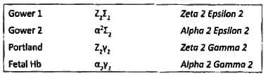
Hemoglobin with zeta 2 and gamma 2 chains are seen in which of the following: (Recent Pattern 2014-15)

Detailed Solution: Question 2

Test: Thalassemia and Sickle Cell Anemia - Question 3

The most appropriate drug used for chelation therapy in beta thalassemia major is: (Recent Pattern 2014-15)

Detailed Solution: Question 3

Drug of choice for acute iron poisoning is desferrioxamine; however, it must be given parenterally. It is ineffective when taken orally. In the case of beta thalassemia major, excess iron can accumulate due to frequent blood transfusions and significant haemolysis.
  • The chelating agent needs to be administered over an extended period in this scenario.
  • Consequently, oral Deferiprone is the preferred option in this situation.

Test: Thalassemia and Sickle Cell Anemia - Question 4

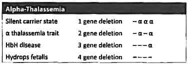
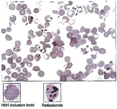
HbH is characterized by: (Recent Pattern 2014-15)

Detailed Solution: Question 4

Test: Thalassemia and Sickle Cell Anemia - Question 5

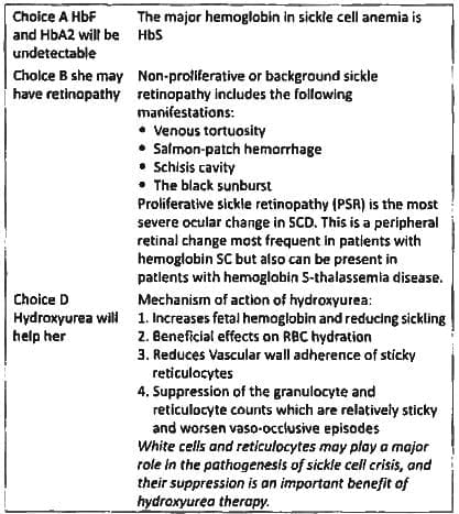
Which of the following is not seen in a chronic case of Sickle cell anemia: (AI 1996)

Detailed Solution: Question 5

Sickle cell anaemia is marked by hyposplenism or autosplenectomy, rather than splenomegaly.

Test: Thalassemia and Sickle Cell Anemia - Question 6

All are true for sickle cell anemia, except (AIIMS May 94)

Detailed Solution: Question 6

"Sickle cell anemia is associated with granulocytosis (leukocytosis) and not leukopenia".

Test: Thalassemia and Sickle Cell Anemia - Question 7

Golf ball inclusion bodies in RBCs are seen in? (Recent Question 2016-17)

Detailed Solution: Question 7

Golf ball inclusion bodies are called as HbH bodies and are seen in alpha thalassemia. The peripheral smear stained with supravital stain brilliant cresyl blue shows multiple inclusions in an evenly distributed panel.

Reticulocytes are differentiated from these RBCs with inclusions as they have uneven reticulin deposits.
Heinz bodies are larger, single inclusions and not so numerous as Howell jolly bodies.

Test: Thalassemia and Sickle Cell Anemia - Question 8

A 25-year-old lady came with anemia jaundice and recurrent joint pains. All of the following are true except: (APPG 2015)

Detailed Solution: Question 8

  • Anemia with jaundice points to hemolytic anemia and presence of joint pain indicates sickling leading to vaso-occlusive crisis.
  • Bone pain occurs as gnawing, progressive pain, most commonly in the humerus, tibia, and femur and less commonly in the facial bones.
  • Periarticular pain and joint effusion, often associated with a sickle cell crisis, are considered a result of ischemia and infarction of the synovium and adjacent bone and bone marrow 
  • Patients can present with acute chest syndrome and not pulmonary syndrome.
  • Acute chest syndrome is a distinctive manifestation characterized by chest pain, tachypnea, fever, cough, and arterial oxygen desaturation. It can mimic pneumonia, pulmonary emboli, bone marrow infarction and embolism, myocardial ischemia, or in situ lung infarction.

Acute chest syndrome is a distinctive manifestation characterized by chest pain, tachypnea, fever, cough, and arterial oxygen desaturation. It can mimic pneumonia, pulmonary emboli, bone marrow infarction and embolism, myocardial ischemia, or in situ lung infarction. Acute chest syndrome is thought to reflect in situ sickling within the lung, producing pain and temporary pulmonary dysfunction.

Test: Thalassemia and Sickle Cell Anemia - Question 9

The abnormality in X-ray skull shown can be seen in the following conditions except: (APPG 2015)

Detailed Solution: Question 9

The X- Skull shows presence of Lytic lesions
Differential diagnosis of lytic lesion on X ray Skull:

  1. Multiple myeloma
  2. Hyperparathyroidism
  3. Histiocytosis-X
  4. Metastasis (breast, thyroid, lung and kidney cancer)
  5. TB
  6. Osteomylelitis
  7. CNS Epidermoid cyst
  8. Hemangioma
  9. Fibrous dysplasia

Test: Thalassemia and Sickle Cell Anemia - Question 10

Persistent priapism is due to: (Recent Pattern 2014-15)

Detailed Solution: Question 10

Acute vaso-occlusion may also cause stroke due to sinus thrombosis in priapism.
Priapism is defined as erection lasting for > 4 hours. Low flow priapism may be due to any of the following:

  • An excessive release of neurotransmitters
  • Blockage of draining venules (e.g., mechanical interference in sickle cell crisis, leukemia, or excessive use of intravenous parenteral lipids)
  • Paralysis of the intrinsic detumescence mechanism
  • Prolonged relaxation of the intracavernous smooth muscles (most often caused by the use of exogenous smooth-muscle relaxants such as injectable intra-cavernosal prostaglandin El)

Prolonged low flow priapism leads to a painful ischemic state, which can cause fibrosis of the corporeal smooth muscle and cavernosal artery thrombosis. The degree of ischemia is a function of the number of emissary veins involved and the duration of occlusion. Light-microscopy studies conducted early on demonstrated that corporeal tissue becomes thickened, edematous, and flbrotic after days of priapism.

Test: Thalassemia and Sickle Cell Anemia - Question 11

Regarding to Thalassemia minor the following is incorrect: (Recent Pattern 2014-15)

Detailed Solution: Question 11

  • Beta thalassemia minor: Is a thalassemia trait, these patients have a modest anemia with hematocrit between 28% and 40%.
  • The MCV ranges from 55 to 75 fL, and the red blood cell count is normal or increased.
  • The peripheral blood smear is mildly abnormal, with hypo chromia, microcytosis, and target cells. In contrast to alpha thalassemia, basophilic stippling slightly elevated.
  • Hemoglobin electrophoresis (using quantitative techniques) may show an elevation of hemoglobin A2 to 4 8% & occasional elevations of hemoglobin present is hemoglobin.

Test: Thalassemia and Sickle Cell Anemia - Question 12

Basic defect in HbS is: (Recent Pattern 2014-15)

Detailed Solution: Question 12

  • When HbS is deoxygenated the sticky patch present on its surface binds to the complementary patch on another deoxygenated HbS molecule.
  • “Binding of a number of deoxygenated HbS in this way leads to formation of long fibrous polymers of HbS."
  • This stiffens and distort the red cells producing rigid misshaped erythrocytes.
  • A molecule of hemoglobin S (HbS) contains two normal a globin chains and two mutant a globin chains in which glutamate at position 6 has been substituted with valine.
  • This substitution replaces the polar glutamine residue with a nonpolar valine.
  • This change decreases the solubility of the hemoglobin when deoxygenated.
  • The replacement of glutamate by valine generates a sticky patch on the surface of HbS.
  • The sticky patch is present on both oxygenated and deoxygenated HbS but deoxygenated HbS also contains a complementary site for the sticky patch.
  • Such stckle cells frequently block the flow of blood in the narrow capillaries, (The mean diameter of red blood cells is 7.5 m while that of micro vasculature is 3 - 4 m so instead of squeezing through the microvasculature like HbA containing RBCs, sickled RBC s have decreased ability to deform and so have difficulty moving through small vessels).

Test: Thalassemia and Sickle Cell Anemia - Question 13

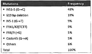
In Beta thalassemia, the most common gene mutation is: (Recent Pattern 2014-15)

Detailed Solution: Question 13

The most common mutation causing a thalassemia is intron /inversion

  • Thalassemias are autosomal recessive disorder.
  • β thalassemias are caused due to reduced synthesis of beta chain of hemoglobin,
  • β thalassemias are the consequence of various point mutations on the globin gene dusters.
  • Most of the β thalassemias are caused by point mutations affecting one or few bases.
  • Five or six specific mutations usually accounts for more than 90% of cases of β thalassemias.

The most common mutation causing b thalassemia is intron / inversion
Also know:
Synthesis of alpha chain is controlled by 2 gene clusters on → chromosome 16
Synthesis of beta chain is controlled by 2 gene dusters on → chromosome 11
Thalassemia mutations in India

Test: Thalassemia and Sickle Cell Anemia - Question 14

Fetal hemoglobin achieves adult values by: (Recent Pattern 2014-15)

Detailed Solution: Question 14

  • At approximately 20 weeks of fetal development, the site of erythropoiesis begins to switch from the liver and spleen to the bone marrow, where progenitors show increased expression of adult globins, a and β.
  • Hemoglobin A may constitute 5% of β-family globin expression during this time. Beginning at the thirtieth week and proceeding to the time of birth, a significant switch from fetal to adult erythropoiesis takes place, such that at the time of birth, fetal hemoglobin constitutes approximately 80% of the total hemoglobin.
  • Over the next 25 to 30 weeks after birth, fetal hemoglobin concentration decreases by approximately 10% every 2 weeks until it reaches its normal adult level of less than 2% by 30 weeks of age 

Test: Thalassemia and Sickle Cell Anemia - Question 15

Sickle cell anemia is usually associated with all, except: (Recent Pattern 2014-15)

Detailed Solution: Question 15

  • Reticulocytosis is a feature of all hemolytic anemia. The reticulocyte count is used to estimate the degree of effective erythro-poiesis,which can be reported as absolute reticulocyte count or as a reticulocyte percent-age. The (Ref: range of the reticulocyte percentage in adults is 0.5% - 1.5%
  • Sickle cell anemia is an autosomal recessive disorder in which an abnormal hemoglobin leads to chronic hemolytic anemia with numerous clinical consequences.
  • A single DNA base change leads to an amino acid substitution of valine for glutamine in the sixth position on the a-globin chain.
  • The abnormal beta chain is designated as p and the tetramer of a2a2 is designated hemoglobin S.
  • When in the deoxy-form, hemoglobin S forms polymers that damage the red blood cell membrane. Both polymer formation & early membrane damage are reversible.
  • However, red blood cells that have undergone repeated sickling are damaged beyond repair & become irreversibly sickled.
  • The rate of sickling is influenced by a number of factors, most importantly by the concentration of hemoglobin S in the individual red blood cell. Red cell dehydration makes the cell quite vulnerable to sickling.
  • Sickling is also strongly influenced by the presence of other hemoglobins within the cell.
  • Hemoglobin F cannot participate in polymer formation.

Test: Thalassemia and Sickle Cell Anemia - Question 16

X-ray skull characteristically shows “Hair standing on end” appearance in one of the following disease: (Recent Pattern 2014-15)

Detailed Solution: Question 16

The hair on end sign refers to a radiographic appearance on a skull which results from a periosteal reaction manifesting as perpendicular trabeculations interspersed by radiolucent marrow hyperplasia along the skull vault. It is classically described with plain skull radiographs although can also be appreciated on CT or MRI.
Mnemonic: H.l -N.E.S.T

  • Herditary spherocytosis
  • Iron deficiency Anemia
  • Neuroblastoma
  • Enzyme deficiency (e.g. G-6-P deficiency causing Hemolytic Anemia)
  • Sickle cell disease
  • Thalassemia major

Test: Thalassemia and Sickle Cell Anemia - Question 17

Auto-splenectomy is associated with: (Recent Pattern 2014-15)

Detailed Solution: Question 17

SLE patients with thrombocytosis and anti-phospholipid antibody syndrome are at risk of developing auto-splenectomy. Patients with sickle cell disease often suffer from autosplenectomy as a result of splenic destruction by the numerous infarcts associated with sickle cell crises during childhood. Indeed, the presence of a palpable spleen in a patient with sickle cell disease after age five suggests a coexisting hemoglobinopathy, e.g„ thalassemia or hemoglobin C. In addition, patients who receive splenic irradiation for a neoplastic or autoimmune disease are also functionally hyposplenia.

52 docs|64 tests
Information about Test: Thalassemia and Sickle Cell Anemia Page
In this test you can find the Exam questions for Test: Thalassemia and Sickle Cell Anemia solved & explained in the simplest way possible. Besides giving Questions and answers for Test: Thalassemia and Sickle Cell Anemia, EduRev gives you an ample number of Online tests for practice
52 docs|64 tests
Download as PDF