NEET Exam  >  NEET Tests  >  Daily Test for NEET Preparation  >  Test: Electrocardiograph (ECG) (August 19) - NEET MCQ

Test: Electrocardiograph (ECG) (August 19) - NEET MCQ


Test Description

10 Questions MCQ Test Daily Test for NEET Preparation - Test: Electrocardiograph (ECG) (August 19)

Test: Electrocardiograph (ECG) (August 19) for NEET 2024 is part of Daily Test for NEET Preparation preparation. The Test: Electrocardiograph (ECG) (August 19) questions and answers have been prepared according to the NEET exam syllabus.The Test: Electrocardiograph (ECG) (August 19) MCQs are made for NEET 2024 Exam. Find important definitions, questions, notes, meanings, examples, exercises, MCQs and online tests for Test: Electrocardiograph (ECG) (August 19) below.
Solutions of Test: Electrocardiograph (ECG) (August 19) questions in English are available as part of our Daily Test for NEET Preparation for NEET & Test: Electrocardiograph (ECG) (August 19) solutions in Hindi for Daily Test for NEET Preparation course. Download more important topics, notes, lectures and mock test series for NEET Exam by signing up for free. Attempt Test: Electrocardiograph (ECG) (August 19) | 10 questions in 10 minutes | Mock test for NEET preparation | Free important questions MCQ to study Daily Test for NEET Preparation for NEET Exam | Download free PDF with solutions
Test: Electrocardiograph (ECG) (August 19) - Question 1

In a standard ECG which one of the following alphabets is the correct representation of the respective activity of the human heart?

Detailed Solution for Test: Electrocardiograph (ECG) (August 19) - Question 1

In a standard ECG, the P wave is a small upward wave that represents depolarisation of atria which leads to contraction of both atria.

Test: Electrocardiograph (ECG) (August 19) - Question 2

Read the following statements and select the correct option
Statement 1: The 4-chambered heart of Birds is superior to the 4-chambered heart of Crocodiles.
Statement 2: Crocodilian heart retains both systemic arches that join, causing the mixing of blood in the dorsal aorta while the avian heart has lost the left systemic arch.

Detailed Solution for Test: Electrocardiograph (ECG) (August 19) - Question 2
  • The 4-chambered heart of birds is not superior to the 4-chambered heart of crocodiles. Both have heart as a muscle that pumps blood. Crocodile's heart has two aortic arches helping it to breath underwater, which makes it's heart more complex than bird's heart.
  • Crocodile has two aortic arches, the two aortic arches connect with each other with the help of foramen of penizza as soon as they leave the heart but merge completely in the lower parts of the body away from the heart.
1 Crore+ students have signed up on EduRev. Have you? Download the App
Test: Electrocardiograph (ECG) (August 19) - Question 3

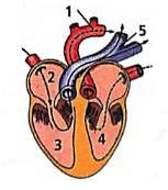
In the given figure of the heart which of the labelled part (1,2,3,4,5) carries oxygenated blood?
Detailed Solution for Test: Electrocardiograph (ECG) (August 19) - Question 3

The labelled parts '1' and '4' are aorta and left ventricle respectively, which carry oxygenated blood. Left ventricle receives oxygenated blood from left auricle which received it from pulmonary veins and this oxygenaed blood then moves into the aorta to be supplied to the whole body.

Test: Electrocardiograph (ECG) (August 19) - Question 4

Which of the following sequences is truly a systemic circulation pathway?

Detailed Solution for Test: Electrocardiograph (ECG) (August 19) - Question 4

Systemic circulation is the movement of the blood between heart and rest of the body (tissues) and the back to the heart. The oxygenated blood moves from the left auricle into the left ventricle. From here the blood moves into the aorta which ultimately divides into arteries that carry oxygenated blood to various tissues in the body. From the tissues deoxygenated blood is carried by the veins into the right atrium via superior vena cava and inferior vena cava.

Test: Electrocardiograph (ECG) (August 19) - Question 5

What is the nature of blood passing through blood vessels A, B, C and D respectively?

Detailed Solution for Test: Electrocardiograph (ECG) (August 19) - Question 5

'A' is vena cava that carries deoxygenated blood
'B' is aorta that carries oxygenated blood
'C' is pulmonary artery that carries deoxygenated blood
'D' is pulmonary vein that carries oxygenated blood.

Test: Electrocardiograph (ECG) (August 19) - Question 6

Which of the following statements is correct?

Detailed Solution for Test: Electrocardiograph (ECG) (August 19) - Question 6

In a standard ECG, a patient is connected to the machine with three electrical leads, one to each wrist and one to the left ankle.

Test: Electrocardiograph (ECG) (August 19) - Question 7

Choose the schematic diagram which properly respresents pulmonary circulation in humans.

Detailed Solution for Test: Electrocardiograph (ECG) (August 19) - Question 7

Pulmonary circulation is the movement of blood between heart and lungs. During this pathway deoxygenated blood entering the right atrium, moves into the right ventricle From here it moves through the pulmonary arch into the lungs for oxygenation. Then from lungs oxygenated blood moves into the left atrium through pulmonary veins.

Test: Electrocardiograph (ECG) (August 19) - Question 8

In ECG, P-R interval corresponds to

Detailed Solution for Test: Electrocardiograph (ECG) (August 19) - Question 8

In ECG, P-R interval is the time from the beginning of the P wave to the beginning of the ORS complex. It corresponds to the time lag from the onset of atrial depolarisation to the onset of ventricular depolarisation. Most of the delay occurs in the AV node.

Test: Electrocardiograph (ECG) (August 19) - Question 9

Which of the following is the diagrammatic representation of standard electrocardiogram(ECG)?

Detailed Solution for Test: Electrocardiograph (ECG) (August 19) - Question 9

A normal electrogram (ECG) is composed of a ‘P’ wave, a QRS wave (complex), and a T wave. The ‘P’ wave is a small upward wave that represents electrical excitation or atrial depolarization which leads to contraction of both the atria. QRS wave (complex) begins after a fraction of a second of the P wave. It begins as a small downward deflection (Q) and continues as a larger upright (R) and triangular wave, ending as a downward wave (S) at its base. It represents ventricular depolarization. The ‘T’ wave is dome-shaped which represents ventricular repolarization. The potential generated by the recovery of the ventricle from the depolarization state is called the repolarization wave. The end of the ‘T’ wave marks the end of the systole.

Test: Electrocardiograph (ECG) (August 19) - Question 10

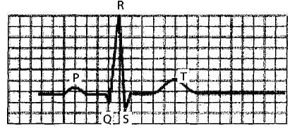
The given figure is the ECG of a normal human. Which one of its components is correctly interpreted below 
Detailed Solution for Test: Electrocardiograph (ECG) (August 19) - Question 10

The QRS complex represents the depolarisation of the ventricles, which initiates the ventricular contraction. By counting the number of QRS complexes that occur in a given time period, One can determine the rate of heart beat of an individual.

12 docs|366 tests
Information about Test: Electrocardiograph (ECG) (August 19) Page
In this test you can find the Exam questions for Test: Electrocardiograph (ECG) (August 19) solved & explained in the simplest way possible. Besides giving Questions and answers for Test: Electrocardiograph (ECG) (August 19), EduRev gives you an ample number of Online tests for practice

Top Courses for NEET

Download as PDF

Top Courses for NEET