Amoeba Proteus - Class 11 PDF Download
GENERAL CHARACTERISTICS OF AMOEBA PROTEUS
 Amoeba Proteus
Classification
Phylum -
- Protozoa
- Unicellular or Acellular
- organism.
Class -
- Sarcodin
- Locomotion by pseudopodia
- Pseudopodia finger like (lobose) called lobopodiaBody is naked uninulceate ormultinucleate.
Genus - Amoeba Psuedopodia distinct; uninulceate body; contractile vacuole for osmoregulation.
Species - Proteu
Introduction
(i) Protozoa name proposed by Goldfuss.
(ii) Amoeba was discovered by Russel Von Rosenhoff in 1755.
Rossenhoff called it “The Little Proteus”.
(iii) Word Amoeba is originated from Greek word-
Amoeba = Change
Proteus = Greek Sea god, who is believed to be capable of changing shape.
(iv) Detailed study of Amoeba proteus is done by H.I. Herschfield
(v) Amoeba name was proposed by B.D.S. Vinscent.
(vi) Detail description of Amoeba was done by Leidy (1879).
(vii) 'Amoeba of man' is written by Dobel.Distribution
(i) Majority of species of Amoeba are found in fresh water. Some are also found in moist soil.
(ii) Some species of Amoeba are marine, which are as follows :
(a) A. verrucosa
(b) A. striata
(iii) It is found in ponds, lakes, ditches etc where there is plenty of organic matter. It feeds on algae, bacteria etc.
Species
(i) Amoeba proteus.
(ii) Amoeba verrucosa.
(a) In Amoeba verrucosa pellicle is found.
(b) In Amoeba verrucosa single pseudopodia is found.
(iii) In Amoeba discoides
(a) Amoeba discoides is fresh water Amoeba.
(b) Amoeba discoides is most closely related with Amoeba proteus.
(c) In it disc type nucleus is found. Pellicle is present.
– A. terricula.
– A. striata. It has pellicle.
– A. dubia.
– A.diploidea.
– A. radiosa
Culture
The process of Amoeba culture is called Hay infusion method.
Size
0.2 - 0.6 mm is an average size of Amoeba.
Size of Amoeba proteus - 0.6 mm.
Amoeba proteus can be seen through naked eye.
Note :
(i) Largest Amoeba is Pelomyxa (chaos chaos) and the smallest Amoeba is A. verrucosa.
(ii) Immortality of Amoeba was described by Hertman. He said that Amoeba do not die by natural death because its body is not divided into somatoplasm and germplasm.
2. STRUCTURE

Shape
Shape of Amoeba is not fixed.
Reason : Because of appearance and disappearance of pseudopodia. Because of absence of pellicle (Main cause).
Note : Pellicle is found in following species of Amoeba -
(i) Amoeba striata
(ii) Amoeba verrucosa
(iii) Amoeba discoidea
Pseudopodia
(i) At a time, numerous pseudopodia get formed in Amoeba proteus so it is called polypodial.
(ii) Pseudopodia perform two important functions-
(a) It helps in locomotion so, pseudopodia is a locomotory organ.
(b) It helps in ingestion of food.
(iii) At anterior end of pseudopodia hyaline cap is found.
(iv) Uroid is older pseudopodia and look like wrinkles of body for sometime at posterior end of the body.
(v) Peudopodia of Amoeba is called Lobopodia.
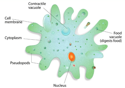
Body
It has 3 main parts.
- Plasmalemma
- Nucleus
- Cytoplasm
Plasmalemma
(i) No cell wall is found.
(ii) It is 1 to 2 ? thick
(iii) Regeneration capacity of plasmalemma is extremely good so if it get injured it get regenerated very quickly and prevent any loss of water.
(iv) It does not get wet because of lipid bilayer.
Lipid + Protein + Lipid
(v) Extremely small microvilli are found on its surface. Scientist believe that microvilli have adhesivecapacity and it helps in attachment of Amoeba. An adhesive layer of glycolipids & glycoproteins ispresent at outer surface called as Glycocalyx.
(vi) Microvilli is composed of mucoprotein
(vii) It is firm and elastic hence it neither allows protoplasm to flow away nor hinder in growth and formation of pseudopodia.
(viii)It is capable of invagination into the body and thus both pinocytosis and phagocytosis occurs.
(ix) Osmosis and diffusion takes place through plasmalemma.
Nucleus
(i) Amoeba proteus is mononucleated.
(ii) Within nucleus number of nucleolus varies from 3.
(iii) Nucleus remain present in the centre of the body.
(iv) Nucleus of young Amoeba is biconcave in shape.
(v) Nucleus of adult Amoeba is biconvex in shape.
(vi) Just below the nuclear membrane in the nucleus net like structure is found which resembles like Honey bee comb. so, the net is called Honey comb lattice.
(vii) Honey comb lattice maintain the shape of the nucleus.
(viii) Nucleus is massive type. (Nucleus in which chromatin substance is more in comparison to nucleoplasm is called massive type of nucleus)
(ix) In the nucleus chromatin substances are found in form of granules called chromidia.
(x) In living Amoeba nucleus cannot be seen because light get refracted from the nucleus.
Note : Chromidia is called chromosomes of Amoeba. 500-600 in number.
(xii)Nucleus remain surrounded by porous double nuclear membrane or karyotheca.
Exceptions :
(i) A. pelomyxa or A. chaos-chaos - Multi nucleated. 400 to 1000 nuclei are found.
(ii) A. diploidea - binucleated.
Function of Nucleus
Nucleus controls metabolism and reproduction of Amoeba.
Note : Vesicular nucleus- Nucleus in which more nucleoplasm is found, in comparison to chromatin substance is called vesicular nucleus.
Cytoplasm
Cytoplasm of Amoeba differentiated into ectoplasm and endoplasm.
(A) Ectoplasm
(i) Outer cortical layer of cytoplasm.
(ii) Found just below plasmalemma.
(iii) Also called ectosarc.
(iv) It is thin, transparent, tough, non granular and contractile layer.
(v) It is composed of two layers, which are as follows :
(a) Inner sticky, granular-jelly like layer.
(b) Outer hyaline layer which is clear, glossy and fluid layer. This layer becomes thin when in contact with substratum and becomes thick at tips of pseudopodia forming hyaline cap.
(B) Endoplasm
(i) It is fluid like, granular and semitransparent.
(ii) According to Mast, endoplasm is differential into two parts.
(a) Outer - Solid plasma gel
(b) Inner - Liquid plasma sol
Sol and gel are reversible.
(iii) Conversion of gel into sol is called- Solation.
(iv) Conversion of sol into gel is called - Gelation.
(v) Endoplasm shows streaming movement called cyclosis.
(vi) pH of endoplasm is 7.4.
Vacuoles
In Amoeba three types of vacuoles are found which are as follows-
- Contractile vacuole
- Food vacuole
- Water vacuole
- Contractile vacuole
(i) Found within endoplasm.
(ii) Only (1) in number.
(iii) Formed because of fusion of numerous small vacuoles.
(iv) It appears like a bubble and remain enclosed within a delicate and elastic condensation membrane.
Position :
(i) Found in between nucleus and posterior end of the Amoeba.
(ii) Numerous mitochondria remain scattered around contractile vacuole.
Function :
Osmoregulation : Environment around the Amoeba is hypotonic as a result water from outside rushes within Amoeba and contractile vacuole expels the water outside of the body.
For osmoregulation contractile vacuole undergoes diastole and systole.
Note :
Systole - Filling of water within contractile vacoule is called systole.
Diastole - When contractile vacoule get filled with water it get ruptured near to plasmalemma and water comes out. After getting ruptured contractile vacoule disappears.
During systole and diastole energy is obtained from mitochondria.
Amoeba spends maximum energy in water regulation
(i) So thousand of mitochondria are found around the contractile vacuole.
(ii) Function of contractile vacuole is like kidney in higher animal.
(iii) Marine form do not have contractile vacuole due to non requirement of water.
Note :
(i) When fresh water Amoeba kept in sea water, contractile vacuole disappear.
(ii) When marine Amoeba is kept in fresh water contractile vacuole get appeared.
(iii) When Amoeba is kept in distilled water contractile vacuole becomes more functional.
(iv) When Amoeba is kept from marine water to distilled water, Amoeba will get swelled, contractile vacouleget formed.
Food vacuole
(i) Numerous in number.
(ii) Size varies on type of food ingested.
(iii) Moves freely in endoplasm.
(iv) Temporary structure.
(v) It is wall of food vacuole which is composed of broken pieces of plasmalemma.
(vi) Food vacuole reappears at its usual place as a minute vacuole and start growing.
(vii) Food get digested within food vacuole.
Water vacuole
(i) Remain filled with water.
(ii) Small in size.
(iii) Numerous in number.
(iv) It is non-contractile structure.
Cell organelles
Following are the cell organelles which are found in Amoeba.
Mitochondria
(i) Oval and rod like
(ii) Five lakhs in number
(iii) Provides energy.
Golgi body
(i) Related with secretion.
(ii) Golgi body are found 2 or 3 in number in Amoeba.
Ribosome
Ribosome like particles either found attached with E.R. or Scattered freely in the endoplasm.
Lysosome
Round vesicles containing enzyme.
Endoplasmic reticulum
Found in the form of net.
Note : In Endoplasmic reticulum no cisternae is found.
Bi-Urate and Tri-Urate Crystals
(i) Biurates and Triurates are crystals found in endoplasm of Amoeba.
(ii) Biurates are (bipyramidal) and triurates are (tripyramidal)
(iii) It posses carbonyl diurea.
(iv) Its excretion is not known.
(v) Some scientists believe that it is excreted during reproduction.
Stored food :
In Amoeba food remain stored in form of glycogen granules and oil drops.
3. NUTRITION
(i) In Amoeba nutrition is holozoic.
(ii) Amoeba is omnivorous.
Note :
(i) Amoeba takes bacteria, diatoms, algae, and other protozoans as food.
(ii) Amoeba can selectively choose food.
(iii) Amoeba can even distinguish organic food particles from inorganic sand particle.
Nutrition of Amoeba includes
Ingestion
Digestion
Absorption and assimilation
Egestion
Ingestion
(i) Rhumbler in 1930 at first described the process of ingestion in Amoeba.
(ii) No mouth is found for ingestion.
(iii) In Amoeba 4 ways of ingestion are found.
Import
(i) This is passive way of ingestion, Amoeba has to make no effort.
Invagination
(i) In this process of ingestion plasmalemma and ectoplasm suddenly invaginates at the point of contact of food particle.
(ii) As a result of invagination a tube get formed taking the particle into the endoplasm later on tube disappear within the endoplasm.
Circumfluence
When Amoeba comes in contact of less active substance (food) like-algae whole cytoplasm flows around the food encircles it in a food cup and takes it within endoplasm.
Circumvallation
(i) In this process of ingestion motile and active food like ciliate and flagellate is ingested.
(ii) Prey get surrounded by several pseudopodia as such. Pseudopodia soon fuses together and prey comes within endoplasm and get surrounded by food vacuole.
Note :
(i) Biggest food vacuole get produced during circumvallation.
(ii) Ingestion of solid food is called phagocytosis.
(iii) Ingestion of liquid food is called pinocytosis. (cell drinking)
(iv) Pinocytic vesicles or pinosome get formed by invagination of plasmalemma.
Food particle Food vacuole
(A) Import (B)Food particleInvagination
(A) tube (B)
Invagination tube Food vacuole
(C) (D) (E)
Invagination (A to E)
(v) During ingestion some amount of water always remain present.
(vi) Food and water mainly forms food vacuole in endoplasm.
Digestion
(i) In Amoeba digestion mainly takes place in food vacuole.
(ii) Food vacuole is also called gastric vacuole/gastrioles.
(iii) In Amoeba digestion is intracellular.
(iv) Digestive enzymes are formed in cytoplasm and gets accumulated in lysosome.
(v) Lysosomes attaches with food vacuole and transfers enzyme.
(vi) Medium within food vacuole is acidic at first which becomes alkaline later on.
(vii) Medium within food vacuole remain acidic for 45 minute only.
(viii)No digestion of nutrients occur in acidic medium & in this phase the fluid of vacuoles contains HCl,hence it is acidic.
Proteases, trypsin and peptidase Protein
Lipase Fats
Amylase Carbohydrate.
Note :
Presence of amylolytic enzyme is doubtful.
Assimilation and Absorption
(i) Digestion of food get completed in 15 to 30 hours.
(ii) During digestion food vacuole circulates during cyclosis.
(iii) After completion of digestion food vacuole shrinks and digested food get well distributed within cytoplasm.
(iv) Absorbed food immediately get involved in metabolic reactions.
Egestion
(i) Egestion may take at any place.
(ii) During egestion, food vacuole with undigested food stops moving and becomes stagnant. As it comes in contact of plasmalemma, plasmalemma bursts. and food vacuole comes out.
(iii) Plasmalemma immediately heals up to prevent any loss of cytoplasm.
Respiration :
Through body surface by diffusion. Oxygen required is obtained from water.
Excretion
Main excretory substances is ammonia. Excretion takes place by diffusion.
4. REPRODUCTION
Reproduction in Amoeba is always asexual and take place by various methods which are as follows-
- Binary fission
- Sporulation
- Multiple fission
- Encystment
- Regeneration

Binary Fission
(i) Takes place in favourable condition.
(ii) In this period temperature of water should remain optimum, and plenty of food should be present.
(iii) Get completed in 30 minutes.
(iv) This is the commonest and simplest method of reproduction in Amoeba. In this the whole body divides like an ordinary cell in to two daughter Amoeba by mitosis Hence it is termed binary fission.
(v) Size of the two daughter Amoeba is slightly more than half of the size of the parent.
(vi) Duration in between two binary fission is 24 hours.
(vii) During mitosis in Amoeba nuclear membrane does not disappear such type of mitosis is called
Cryptomitosis.
In binary fission of Amoeba the different phases of mitosis are as follows :
- Disintegrating nucleus
- Daughter nuclei
- Chromatin
- Spores
- Sporulation
(i) Amoeba undergoes sporulation in unfavourable condition.
(ii) At first nuclear membrane get disintegrated and chromatin body get released in the block of 2 or 3 in to the cytoplasm.
(iii) Each chromatin block become a small daughter nucleus by which it acquire a nuclear membrane around it.
(iv) Each daughter nucleus get surrounded by cytoplasm.
(v) Thus about 200 minute spore like daughter Amoeba are formed in the body of parent Amoeba.
(vi) The peripheral layer of each daughter Amoeba changes into tough and resistant spore membrane as a result spore get formed.
(vii) The spore sink down, at the bottom of the pond on return of favourable condition the spore wall rupture
(viii) Thus minute Amoeba emerges out from each spore and rapidly grows into an adult Amoeba.
Encystment and Multiple Fission
(i) In this type of reproduction Amoeba withdraws its pseudopodia and becomes spherical and secretesa highly resistant 3 layered cyst around itself.
(ii) Nucleus undergoes repeated mitotic division forming about 500 to 600 minute daughter nuclei.
(iii) On return of favourable condition the cyst absorbs water and ruptures to release the amoebule which soon grow into adult Amoeba.
Encystment helps in protection and survival as well as in dispersal of Amoeba.
Cystwall Daughter nuclei Pseudopodiospores Young Amoeba
Regeneration
(i) Discovered by - A.Trembley in Amoeba.
(ii) Amoeba posses excellent regenerative capacity. If cut into numerous pieces with nucleus each piecedevelop into separate Amoeba.
Conjugation
Temporary closeness between two Amoeba after some time they become separate again, this form of reproduction is called as Rejuvenation.
5. LOCOMOTION
(i) Locomotion in Amoeba takes place by pseudopodia.
(ii) Amoeba is polypodial animal, means, numerous pseudopodia get formed at a time.
(iii) In Amoeba monolocomotion is found it means, during locomotion Amoeba uses only one pseudopodia.
(iv) Locomotion speed - 0.02 - 0.03 mm/minute.
(v) Locomotion includes 3- steps :
A. Attachment with Substratum
For attachment of Amoeba, with substratum, microvilli, glycocalyx and ions present in water (Na+, Mg+,K+ Br¯) helps.
B. Sol and gel interconversion
Pseudopodia get formed because of sol and gel inter conversion.
C. Contractile force
Because of contractile force in the body of Amoeba locomotion takes place.
Locomotion Theories
Sol and Gel Theory or Pressure Theory or Ectoplasmic Theory
(i) This theory was proposed by Hyman (1917).
(ii) This theory was supported and explained in detail by Pantin (1923) & Mast (1925)
(iii) Most accepted theory.
(iv) According to this theory formation of pseudopodia depends on solation and gelation; solation andgelation takes place because of change in viscosity.
(v) This theory is also called change of viscosity theory
Plasmagel changing intoplasmasol (solation)
Uroid
Pseudopodium Plasmagel
Ectoplasm Plasmasol
Plasmalemma
Plasmasol changing into
plasmagel (Gelation)
Endoplasma
6. BEHAVIOUR
(i) No specialized sensory organelles is found.
(ii) No nervous system is found.
(iii) Shows reactions or response towards environmental changes.
(iv) By locomotory behaviour Amoeba respond to various stimuli.
(a) Positive response- If Amoeba continue to advance towards the source of stimulation it is called positive response.
(b) Negative response- If Amoeba move away from the source of stimulation it is called negative response.
(c) Response to stimuli is called taxis
FAQs on Amoeba Proteus - Class 11
| 1. What is Amoeba Proteus and why is it important in the study of cells? |  |
| 2. What are the key features of Amoeba Proteus? |  |
| 3. How does Amoeba Proteus obtain its nutrition? |  |
| 4. What are the ecological roles of Amoeba Proteus? |  |
| 5. How can Amoeba Proteus be used in medical research? |  |


