MCAT Exam > MCAT Notes > Biology for MCAT > Signal propagation: The Movement of Signals Between Neurons
Signal propagation: The Movement of Signals Between Neurons | Biology for MCAT PDF Download
| Table of contents |

|
| Introduction |

|
| Signal Transmission within Neurons |

|
| Signal Transmission between Neurons |

|
| Signal Integration and Threshold Activation |

|
| Understanding the Complexity |

|
Introduction
The human brain is a remarkable organ that constantly engages in electrochemical activity. With around 100 billion neurons firing off 5-50 messages, known as action potentials, per second, the brain enables us to process our environment, control our muscles, and maintain our balance. It is responsible for intricate tasks such as responding to touch, avoiding danger, and reacting to stimuli like bright lights. To accomplish these actions effectively, the brain relies on a rapid and efficient process of signal propagation, which occurs in two crucial steps: within individual cells (action potential) and between cells (neurotransmitters).

Signal Transmission within Neurons
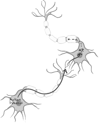
Neurons, the building blocks of the nervous system, are organized in networks that facilitate the transmission of information between the body and the brain. The key components of signal transmission within neurons can be summarized as follows:
- Action Potentials: Information is transmitted as packets of messages called action potentials. These action potentials travel along a single neuron cell through an electrochemical cascade, resulting in a net inward flow of positively charged ions into the axon.
- Cell Body and Axon: Action potentials are triggered at the cell body, travel down the axon, and ultimately reach the axon terminal.
- Axon Terminal and Neurotransmitters: The axon terminal contains vesicles filled with neurotransmitters, which are chemical messengers responsible for transmitting signals between neurons.

Signal Transmission between Neurons
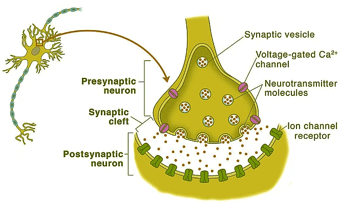
The critical juncture where signals are transmitted between neurons is known as the synapse. The synapse encompasses the machinery required for information transfer, including the pre-synaptic and post-synaptic membranes. While we will focus on the synapse between two neurons, it is essential to acknowledge that a single neuron can influence multiple post-synaptic neurons, receiving inputs from neighboring cells.
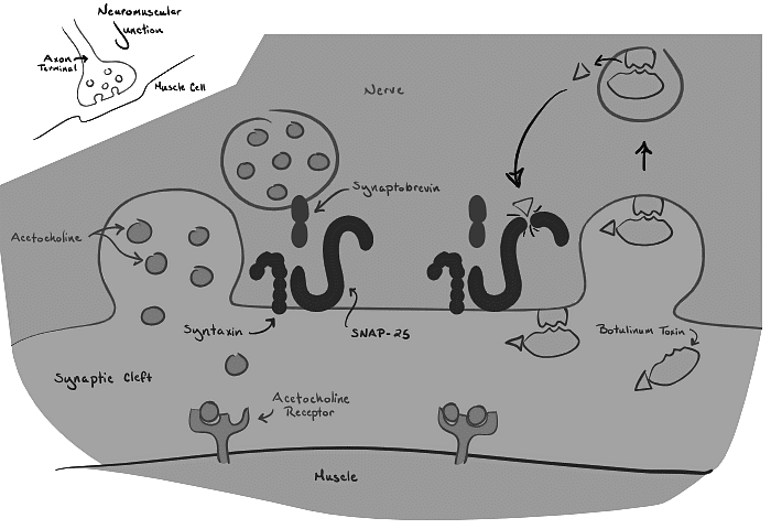
- Pre-Synaptic Cell: At the axon terminal of neurons, neurotransmitters are stored in membrane-bound vesicles. Membrane proteins on the vesicles interact with membrane proteins at the axon terminal to anchor the vesicles in place. The protein complexin acts as a brake, preventing vesicles from fusing into the membrane and releasing their contents. However, the protein synaptotagmin, triggered by the influx of positive ions during the action potential, can bind and release complexin in the presence of calcium. This mechanism allows the vesicles to fuse with the cell membrane and release their contents into the synaptic cleft.
- Neurotransmitters: Neurotransmitters play a crucial role in communication between cells. Synapses can be either excitatory or inhibitory, depending on the neurotransmitter released. Excitatory neurotransmitters propagate the signal by triggering more action potentials, while inhibitory signals work to counteract the signal. Glutamate, the major excitatory neurotransmitter, facilitates signal propagation, while GABA (gamma-aminobutyric acid) serves as the major inhibitory neurotransmitter. Various other neurotransmitters, including dopamine, serotonin, adrenaline, and histamine, have specific functions in different regions of the brain and the body.
- Post-Synaptic Cell: The post-synaptic cell, consisting of dendrites and the cell body, is the recipient of the neurotransmitter signal. Dendrites are specialized projections designed to increase surface area and enhance information reception. Neurotransmitter receptors on the post-synaptic terminal, primarily ligand-gated ion channels, open when they bind to specific neurotransmitters. The type of channel opened determines whether the neurotransmitter is excitatory or inhibitory. Excitatory neurotransmitters allow positive ions to enter, depolarizing the cell body, while inhibitory neurotransmitters open channels that enable the entry of negative ions, hyperpolarizing the cell body.

Signal Integration and Threshold Activation
While a single vesicle of neurotransmitter is insufficient to depolarize the cell body and trigger an action potential in the post-synaptic cell, the brain compensates for this by employing neuron networks. Multiple signals are sent to a single cell, and repeated firing of a neuron may be necessary to relay the message effectively. Moreover, competition among neurons can occur, with a post-synaptic neuron receiving both excitatory glutamate signals and inhibitory GABA signals. The post-synaptic cell will transmit the message if it receives enough excitatory input to reach the depolarization threshold, which subsequently opens voltage-gated ion channels in its axon.
Understanding the Complexity
While the basic signaling method outlined here provides a fundamental understanding of signal propagation, it is essential to acknowledge the nuanced variations that can occur. For instance, cells in the basal ganglia release both GABA and Substance P simultaneously, leading to intricate command patterns or the activation of a broader network of cells.
Case Study: Botulism
To gain a deeper understanding of the signal propagation process, it is helpful to examine what happens when it malfunctions. Botulism, caused by the bacteria Clostridium botulinum, involves the secretion of botulinum toxin. This toxin destroys the membrane and vesicle proteins responsible for neurotransmitter release at the neuromuscular junction, leading to muscle weakness and fatigue. The same botulinum toxin is used in cosmetic procedures such as Botox.
Conclusion
Signal propagation is a remarkable process that enables the transmission of information within and between neurons. From the initiation of action potentials to the release and binding of neurotransmitters, every step contributes to the efficient functioning of the brain. Understanding the intricate mechanisms involved in signal propagation enhances our comprehension of how the brain processes and responds to the world around us.
The document Signal propagation: The Movement of Signals Between Neurons | Biology for MCAT is a part of the MCAT Course Biology for MCAT.
All you need of MCAT at this link: MCAT
|
233 videos|16 docs|32 tests
|
Related Searches


