Mitochondria, Plastid, Ribosome, Cytoskeleton & Nucleus


This content covers critical membrane-bound and non-membrane-bound organelles essential for cellular metabolism, protein synthesis, structural support, and genetic control. These structures are fundamental to understanding cellular organization and function, with high relevance in cell biology questions focusing on structure-function relationships, comparative analysis, and biochemical processes.
1. Mitochondria
1.1 Basic Characteristics
- Visibility: Not easily visible under light microscope unless specifically stained
- Number per cell: Variable, depends on physiological activity of the cell
- Shape: Sausage-shaped or cylindrical with considerable variability
- Size: Diameter 0.2-1.0 µm (average 0.5 µm); Length 1.0-4.1 µm
- Nickname: Power houses of the cell (produce ATP through aerobic respiration)
1.2 Structural Organization
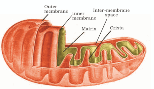
Mitochondria are double membrane-bound structures with distinct compartmentalization:
- Outer membrane: Forms continuous limiting boundary of the organelle; smooth and permeable
- Inner membrane: Forms numerous infoldings called cristae (singular: crista) that project towards the matrix
- Cristae function: Increase surface area for ATP synthesis reactions
- Inter-membrane space: Compartment between outer and inner membranes (outer compartment)
- Matrix: Dense homogeneous substance filling the inner compartment
1.3 Components of Matrix
- DNA: Single circular DNA molecule (prokaryotic type)
- RNA: Few RNA molecules present
- Ribosomes: 70S ribosomes (prokaryotic type)
- Protein synthesis machinery: Components required for synthesis of proteins
- Enzymes: Both membranes have specific enzymes associated with mitochondrial function
1.4 Functional Significance
- Main function: Sites of aerobic respiration
- Energy production: Produce cellular energy in the form of ATP
- Reproduction: Divide by fission (independent of cell division)
- Semi-autonomous organelle: Has own DNA, RNA, and ribosomes
2. Plastids
2.1 General Features
- Distribution: Found in all plant cells and euglenoides
- Visibility: Easily observed under microscope (large organelles)
- Pigments: Bear specific pigments that impart specific colors to plants
- Classification basis: Type of pigments present
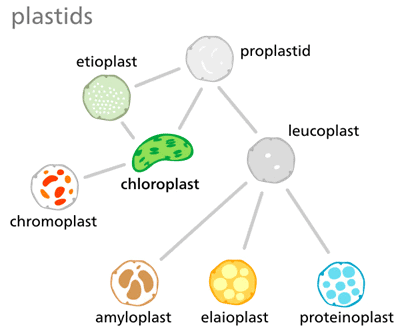
2.2 Types of Plastids
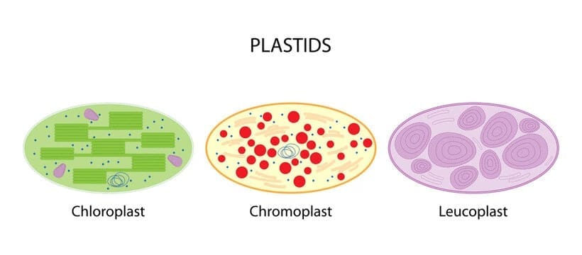
2.2.1 Chloroplasts
- Pigments: Contain chlorophyll and carotenoid pigments
- Function: Trap light energy essential for photosynthesis
- Location: Majority found in mesophyll cells of leaves
- Shape: Lens-shaped, oval, spherical, discoid, or ribbon-like
- Size: Length 5-10 µm; Width 2-4 µm
- Number: Variable from 1 per cell (Chlamydomonas) to 20-40 per cell (mesophyll)
2.2.2 Chromoplasts
- Pigments: Fat-soluble carotenoid pigments (carotene, xanthophylls)
- Color: Impart yellow, orange, or red color to plant parts
- Function: Provide color to flowers and fruits
2.2.3 Leucoplasts
Colorless plastids of varied shapes and sizes with stored nutrients:
- Amyloplasts: Store carbohydrates (starch); Example: potato tubers
- Elaioplasts: Store oils and fats
- Aleuroplasts: Store proteins
2.3 Chloroplast Structure (Detailed)
2.3.1 Membrane System
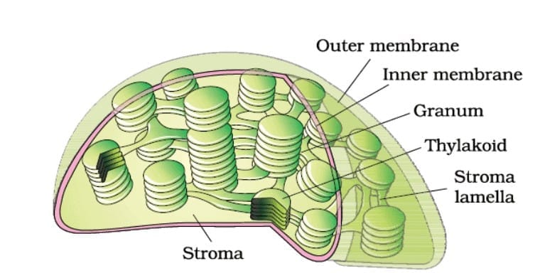
- Double membrane-bound: Like mitochondria
- Outer membrane: Relatively more permeable
- Inner membrane: Relatively less permeable
- Stroma: Space limited by inner membrane; contains enzymes for carbohydrate and protein synthesis
2.3.2 Thylakoid System
- Thylakoids: Organized flattened membranous sacs present in stroma
- Grana (singular: granum): Stacks of thylakoids arranged like piles of coins
- Intergranal thylakoids: Thylakoids present between grana
- Stroma lamellae: Flat membranous tubules connecting thylakoids of different grana
- Lumen: Space enclosed by thylakoid membrane
- Chlorophyll location: Present in the thylakoids
2.3.3 Genetic System
- DNA: Small, double-stranded circular DNA molecules
- Ribosomes: 70S ribosomes (smaller than cytoplasmic 80S ribosomes)
- Semi-autonomous: Can synthesize some of their own proteins
3. Ribosomes
3.1 Discovery and Basic Features
- Discovered by: George Palade (1953)
- Observation: First observed as dense particles under electron microscope
- Composition: Composed of ribonucleic acid (RNA) and proteins
- Membrane: Not surrounded by any membrane (non-membrane-bound)
- Function: Sites of protein synthesis
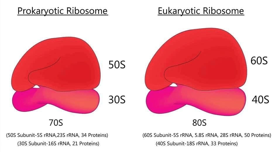
3.2 Types Based on Sedimentation Coefficient
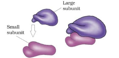
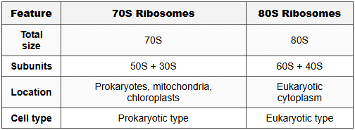
3.2.1 Eukaryotic Ribosomes (80S)
- Total size: 80S
- Larger subunit: 60S
- Smaller subunit: 40S
- Location: Cytoplasm of eukaryotic cells
3.2.2 Prokaryotic Ribosomes (70S)
- Total size: 70S
- Larger subunit: 50S
- Smaller subunit: 30S
- Location: Prokaryotic cells, mitochondria, chloroplasts
3.3 Important Terminology
- S (Svedberg's Unit): Stands for sedimentation coefficient
- Significance of S: Indirectly a measure of density and size
- Structural organization: Both 70S and 80S composed of two subunits (larger and smaller)
Trap Alert: The S values are NOT additive (60S + 40S ≠ 100S but = 80S). This is because S represents sedimentation coefficient which depends on shape, density, and mass, not just mass alone.
4. Cytoskeleton
4.1 Definition and Composition
- Definition: An elaborate network of filamentous proteinaceous structures in the cytoplasm
- Components:Three types of structures collectively form cytoskeleton:
- Microtubules (largest diameter)
- Microfilaments (smallest diameter)
- Intermediate filaments (medium diameter)
- Distribution: Present in both plant and animal cells
4.2 Functions of Cytoskeleton
- Mechanical support: Provides structural framework to the cell
- Motility: Helps in cell movement and intracellular transport
- Shape maintenance: Maintains the shape of the cell
- Additional roles: Involved in cell division, vesicle transport, organelle positioning
5. Cilia and Flagella
5.1 Basic Characteristics
- Structure: Hair-like outgrowths of the cell membrane
- Cilia (singular: cilium): Small structures that work like oars
- Flagella (singular: flagellum): Comparatively longer structures
- Function of cilia: Cause movement of either the cell or surrounding fluid
- Function of flagella: Responsible for cell movement
Trap Alert: Prokaryotic bacteria also possess flagella but these are structurally different from eukaryotic flagella. Prokaryotic flagella lack the 9+2 arrangement.
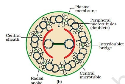
5.2 Ultra-structure of Eukaryotic Cilia/Flagella
5.2.1 External Structure
- Covering: Covered with plasma membrane (extension of cell membrane)
- Core: Called axoneme
5.2.2 Axoneme Organization (9+2 Array)
- Microtubule arrangement: 9+2 array (most important structural feature)
- Peripheral microtubules: Nine doublets of radially arranged microtubules
- Central microtubules: A pair (two) of centrally located microtubules
- Orientation: Microtubules run parallel to the long axis
5.2.3 Associated Structures
- Central sheath: Encloses the two central tubules
- Bridges: Connect the two central tubules
- Radial spokes: Nine radial spokes connect central sheath to one tubule of each peripheral doublet
- Linkers (interdoublet bridges): Interconnect the peripheral doublets
- Basal bodies: Centriole-like structures from which cilium and flagellum emerge
6. Centrosome and Centrioles
6.1 Centrosome Structure
- Definition: Organelle usually containing two cylindrical structures called centrioles
- Pericentriolar material: Amorphous material surrounding the centrioles
- Centriole orientation: Both centrioles lie perpendicular to each other
- Distribution: Typically present in animal cells
6.2 Centriole Structure
- Organization: Each has organization like a cartwheel
- Peripheral fibrils: Made up of nine evenly spaced peripheral fibrils
- Triplet structure: Each peripheral fibril is a triplet of microtubules
- Linkage: Adjacent triplets are also linked
- Hub: Central part of proximal region is proteinaceous, called the hub
- Radial spokes: Connect hub with peripheral triplets (made of protein)
- Protein composition: Fibrils made of tubulin protein
6.3 Functions of Centrioles
- Basal body formation: Form the basal body of cilia or flagella
- Spindle fiber formation: Give rise to spindle fibers during cell division in animal cells
- Spindle apparatus: Essential for chromosome separation during cell division
7. Nucleus
7.1 Historical Background
- First described by: Robert Brown (1831)
- Chromatin naming: Material stained by basic dyes named chromatin by Flemming
- Interphase nucleus: Nucleus of a cell when it is not dividing
7.2 Components of Nucleus
- Nuclear envelope: Double membrane boundary
- Chromatin: Highly extended nucleoprotein fibers (in interphase)
- Nuclear matrix/Nucleoplasm: Ground substance of nucleus
- Nucleolus (plural: nucleoli): One or more spherical bodies
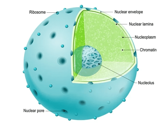
7.3 Nuclear Envelope Structure
7.3.1 Membrane Organization
- Double membrane: Consists of two parallel membranes
- Perinuclear space: Space between the two membranes (10 to 50 nm)
- Barrier function: Forms barrier between nuclear materials and cytoplasm
- Outer membrane: Usually continuous with endoplasmic reticulum; bears ribosomes on it
- Inner membrane: Smooth, without ribosomes
7.3.2 Nuclear Pores
- Formation: Formed by fusion of two membranes at numerous places
- Function: Passages for bidirectional movement of RNA and protein molecules
- Transport direction: Movement occurs between nucleus and cytoplasm in both directions
7.4 Nuclear Number Variations
- Normally: Only one nucleus per cell
- Multinucleate cells: Some cells have more than one nucleus (e.g., Paramecium has two nuclei - macronucleus and micronucleus)
- Anucleate cells:Some mature cells lack nucleus:
- Erythrocytes (RBCs) of many mammals
- Sieve tube cells of vascular plants
Trap Alert: Anucleate cells like mature RBCs and sieve tube cells are still considered living cells despite lacking a nucleus, as they perform metabolic activities for a limited period.
7.5 Nucleoplasm (Nuclear Matrix)
- Contents: Contains nucleolus and chromatin
- Composition: Semi-fluid matrix containing various nuclear components
- Function: Site for nucleic acid synthesis and processing
7.6 Nucleolus Structure and Function
- Shape: Spherical structures present in nucleoplasm
- Membrane: Not membrane-bound (content continuous with nucleoplasm)
- Main function: Site for active ribosomal RNA (rRNA) synthesis
- Number and size correlation: Larger and more numerous nucleoli present in cells actively carrying out protein synthesis
- Composition: Contains RNA, DNA (nucleolar organizer regions), and proteins
7.7 Chromatin and Chromosomes
7.7.1 Chromatin Structure
- Appearance in interphase: Loose and indistinct network of nucleoprotein fibers
- Composition: Contains DNA and proteins (histones, non-histone proteins) and RNA
- DNA length: Single human cell has approximately two meter long thread of DNA
- Chromosome distribution: Distributed among 46 chromosomes (23 pairs) in humans
7.7.2 Proteins Associated with Chromatin
- Histone proteins: Basic proteins providing structural support
- Non-histone proteins: Regulatory and enzymatic proteins
- RNA: Various RNA molecules associated with chromatin
7.7.3 Chromosome Structure (During Cell Division)
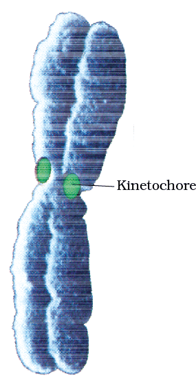
Primary constriction (Centromere):
- Essential component of every chromosome
- Holds two chromatids of a chromosome together
- Kinetochores: Disc-shaped structures present on sides of centromere
- Function of kinetochores: Attachment site for spindle fibers during cell division
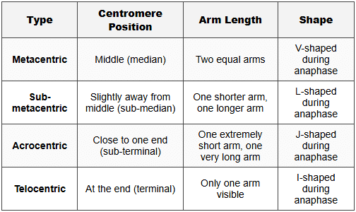
7.8 Types of Chromosomes Based on Centromere Position
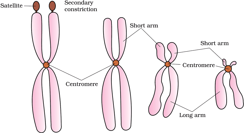
7.9 Secondary Constriction and Satellite
- Secondary constriction: Non-staining constrictions at constant locations on some chromosomes
- Satellite: Small fragment-like appearance created by secondary constriction
- Chromosomes with satellites: Called SAT chromosomes (Satellite chromosomes)
- Function: Secondary constrictions are sites of nucleolar organizer regions (NOR) where rRNA genes are located
8. Microbodies
8.1 Basic Features
- Structure: Membrane-bound minute vesicles
- Contents: Contain various enzymes
- Distribution: Present in both plant and animal cells
- Size: Small vesicular structures
- Examples: Peroxisomes, glyoxysomes
9. Comparative Analysis of Organelles
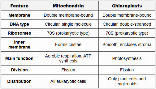
9.1 Semi-Autonomous Organelles Comparison
9.2 Ribosome Types Comparison
Understanding these organelles is crucial for comprehending cellular metabolism, energy production, protein synthesis, and genetic control. Each structure demonstrates remarkable specialization for its specific function while maintaining coordinated cellular activities. The semi-autonomous nature of mitochondria and chloroplasts, with their own genetic systems, supports the endosymbiotic theory of their evolutionary origin.
FAQs on Mitochondria, Plastid, Ribosome, Cytoskeleton & Nucleus
| 1. What is the function of the cytoskeleton in a cell? |  |
| 2. How do cilia and flagella differ in terms of structure and function? | |
| 3. What is the role of the centrosome and centrioles in cell division? | |
| 4. What is the primary function of the nucleus in a cell? | |
| 5. How are chromosomes organized within the nucleus during cell division? | |


