Oogenesis - Human Reproduction - Class 12 PDF Download
OOGENESIS
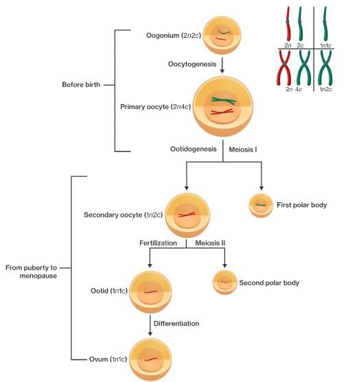
The process of formation of a mature female gamete is called oogenesis which is markedly different from spermatogenesis Oogenesis is initiated during the embryonic stage
FORMATION OF PRIMARY FOLLICLE
The formation of ovum takes place in the follicles which are initially formed as cluster of oogonia which converted into primary oocyte surrounding by some granulosa cells as primary follicles. All such follicles are formed only once when the foetus is 25 weeks old in her mother’s womb. The primary oocytes already remain halted in the diplotene stage of its first meiotic division.
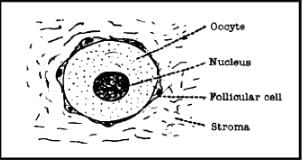
The flattened follicular cells now become columnar. Follicles upto this stage of development are called primary follicle.(having primary oocyte and granulosa cell) till puberty in ovary primary
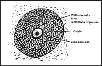
The follicular cells proliferate now to form several layers of cells to form the membrane granulosa now they are known as secondary follicle. A membrane called the zona pellucida, now appears between the follicular cells and the oocyte.
Tertiary Follicle: A cavity appears within the membrane granulosa. It is called the antrum. With the appearance of this cavity, the follicle is formed (follicle means a small sac).
As the follicle expands, the stromal cells surrounding the membrane granulosa become condensed to form a covering called the theca Interna. The cells of theca interna (Thecal cells) afterwards secrete a hormone called oestrogen.
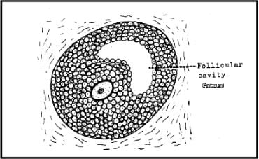
The cavity of the follicle rapidly increases in size and gets filled with a fluid called liquor folliculi. Than meiosis I get completed and primary oocyte of Tertiary follicle get converted into secondary oocyte and first Polar body(FPB). In humans (and most vertebrates), the first polar body does not undergo meiosis II, whereas the secondary oocyte proceeds as far as the metaphase stage of meiosis II.
However, it then stops advancing any further, it awaits the arrival of the spermatozoa for completion of second meiotic division. Entry of the sperm restarts the cell cycle breaking down MPF (M-phase promoting factor and turning on the APC (Anaphase promoting complex).Completion of meiosis II converts the secondary oocyte into a fertilised egg or zygote (and also a second polar body).
The ovarian follicle is now fully formed and is now called the Graafian follicle.
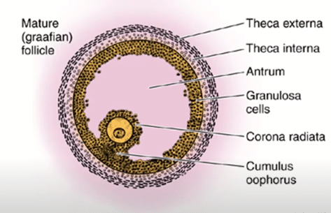 Fig: Graafian follicle
After 14 days of menstrual cycle (on 14th day when cycle is ideally for 28 days) Graafian follicle is ruptured & egg is released.
After ovulation the ruptured Graafian follicle is called corpus luteum. Soon after ovulation, the granulosa cells of Graafian follicle proliferate & these cells look yellow due to accumulation of pigment called Lutein. These cells are called lutein cells. Before ovulation the follicle was a vascular but soon after ovulation blood vessels grow & corpus luteum becomes filled with blood. Lutein cells synthesize the progesterone hormone.
If fertilization occurs in fallopian tube, the corpus luteum then becomes stable for next nine months. If fertilization does not occur then the corpus luteum starts degenerating after about 9 days of its formation. The degeneration is completed by 14 days to form corpus albicans, which gradually disappears. Progesterone hormone maintains pregnancy and repairs the wall of uterus to make its surface adhesive to help in implantation.
The total number of follicles in the two ovaries of a normal young adult woman is about four lakhs. However most of them undergo regression. This is termed as follicular atresia. This is responsible for limited number of gamete production in females. Generally, only one ovum is liberated in each menstrual cycle, by alternate ovaries. Only about 450 ova are produced by a human female over the entire span of her reproductive life which lasts till about 40-50 years of age.
Due to action of estrogens and progesterone, the endometrium of uterus is prepared for implantation. By the 6th to 7th day, embryo is implanted into endometrium (most commonly at the fundus).
EGG OF MAMMALS
Mammalian eggs have very less amount of yolk, so the eggs are oligolecithal and isolecithal or microlecithal and homolecithal.
The egg has 2 egg-membranes
(i) Zona pellucida - This is a transparent membrane like covering and is a primary membrane secreted by the ovum/oocyte itself.
(ii) Corona radiata - This is a layer of follicular cells" and these cells are attached to the surface of egg through " hyaluronic acid" This is a secondary membrane, which is secreted by the ovary. These eggs don't have tertiary membrane. Mammalian eggs are approx 0.1 mm in size.
FAQs on Oogenesis - Human Reproduction - Class 12
| 1. What is oogenesis and how does it relate to human reproduction? |  |
| 2. What are the stages of oogenesis in humans? |  |
| 3. How does oogenesis differ from spermatogenesis? |  |
| 4. What factors regulate oogenesis in the human body? |  |
| 5. How does oogenesis contribute to genetic diversity in humans? |  |


