Class 9 Exam > Class 9 Notes > Lab Manuals for Class 9 > Lab Manual: Slide of Onion Peel and Cheek Cells
Lab Manual: Slide of Onion Peel and Cheek Cells | Lab Manuals for Class 9 PDF Download
Objective
To prepare stained temporary mounts of
- onion peel and
- human cheek cells and to record observations and draw their labelled diagrams.
Theory
- Plant cell to be studied in lab: Onion peel
- The cells are very clearly visible as compartments with prominent nucleus in it.
- The cell walls are very distinctly seen under the microscope.
- The big vacuoles are also seen in each cell.
- The cells can be stained very easily using safranin solution.
- Animal cell to be studied in lab: Cheek cell
- The cells are flat and irregular in shape, with no cell wall.
- The nucleus can be seen very prominently in the center of the cell.
- The nucleus of the cell is stained easily with the methylene blue stain.
- The cell membrane and the cytoplasm can also be studied through these cells.
- Functions of the Organelles:
(i) Nucleus: Contains nucleic acid, DNA (genetic material) , which controls cell activities
(ii) Cytoplasm: Most chemical reactions take place here; controlled by enzymes
(iii) Cell membrane: Controls substances that move in and out of cell. It is partially permeable.
(iv) Cell wall : It is made up of cellulose, strengthens and protects the cell and gives definite shape to the cell.
(v) Chloroplasts: Contain chlorophyll, which absorbs light energy and is a site for photosynthesis.
(vi) Mitochondrion: Site of respiration. It is known as the powerhouse of the cell.
(vii) Vacuole: Large, and filled with cell sap to help keep the cell turgid. Acts as a store of water, sugars, in some cases, of waste products that the cell needs to excrete.
To prepare stained temporary mount of onion peel.
Materials Required
Onion, slides, coverslips, watch glass, petridish, forceps, needles, dropper, glycerine, blotting paper, blade/knife, safranin solution and a microscope.
Procedure
- Take a medium sized onion, cut its outer surface with knife.
- Use forceps to remove the peel of onion.
- With the help of needle separate the small portion of epidermis (peel)
- Keep dilute safranin solution in a watch glass.
- Put this small peel in this watch glass with brush and allow it to stain for 3-5 minutes.
- Transfer the stained peel to another watch glass that contains distilled water in it, to remove extra stain.
- Take a clean dry slide and place two drops of water/glycerine on the centre of the slide.
- Transfer the stained peel with needle and brush on the middle of the slide, if the peel curls straighten it and flatten it with brush and needle, do this gently.
- With the help of blade cut the peel into a square shape.
- Take a dry and clean coverslip and gently place it on the slide with the help of needle such that no air bubbles enter in it.
- Gently press the coverslip with needle for even spreading of glycerine.
- Remove the extra stain and water with the help of blotting paper.
 Cell Structure of onion peel (Plant cell)
- Clean the sides of the coverslip with dry blotting paper and place it under the lens of the microscope and record your observations.
(a) Take a piece of onion bulb (b) Scrab the scale backwards
 (c) Pull the transparent film of onion peel
(d) Stain the peel taken (e) Place the onion peel on the slide (avoid overstaining)
 (f) Put a drop of glycerine
 (g) Place a coverslip with the help of needle (avoid air bubbles)
Observations
The cells under observation are the plant cells. It consists of cell wall and large vacuoles. The nucleus is very prominent and is clearly visible.
Inference
Plant cell shows the following:
- It consists of cell wall.
- The nucleus is prominent and present at the periphery of cytoplasm.
- Large vacuoles are seen at the centre of the cell.
- A lightly stained cytoplasm is present in the cell.
Precautions
- Use dilute stain for staining.
- Avoid the formation of air-bubbles while placing the coverslip on the slide.
- Take very thin peel of onion to get a single layer of cells, no overlapping of cells should be seen.
- Use dry and clean slide, wipe out extra stain or water present on the sides of the slide.
To prepare stained, temporary mount of human cheek cells.
Materials Required
Slide, coverslip, watch glass, methylene blue stain, blotting paper, toothpick, needle, dropper, brush, microscope and glycerine.
Procedure
- Make a dilute methylene blue solution in a watch glass.
- Keep a clean slide with a drop of distilled water at the middle of the slide.
- Take a clean/unused toothpick and scrap the inner wall of your mouth/cheek gently to obtain the epithelial animal tissue, (use the blunt side of toothpick)
- Transfer the scrap on the middle of the glass slide and put a drop of methylene blue solution on it, to stain the cells.
- After 2-3 minutes place the coverslip gently on the cheek cell with the help of needle and avoid the air bubble. (A drop of glycerine can be spread on the cheek cells, it is optional)
- With the help of blotting paper remove the extra stain/water present on the slide.
- Place the slide under microscope and observe it.
Observations
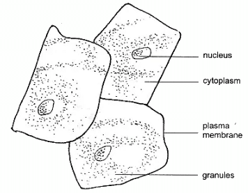
- Cells with irregular shapes are seen.
- A prominent nucleus is seen in the middle of the cell.
- A thin membrane called plasma-membrane is visible at the boundary of each cell.
- The cells do not show any intercellular space.
- No big vacuoles and cell wall is seen.
 Human Cheek Cells
Inference
The cells observed under the microscope do not have cell wall and big vacuoles, these are the cells of animal.
Precautions
- Use unused/new toothpick for scraping of cheek cells.
- Placing of coverslip should be done carefully to avoid air bubbles.
- Avoid overstaining.
- Use clean/dry mounted slide while placing it under the lens of the microscope.
- Avoid overlapping of the cells.
The document Lab Manual: Slide of Onion Peel and Cheek Cells | Lab Manuals for Class 9 is a part of the Class 9 Course Lab Manuals for Class 9.
All you need of Class 9 at this link: Class 9
|
15 videos|98 docs
|
FAQs on Lab Manual: Slide of Onion Peel and Cheek Cells - Lab Manuals for Class 9
| 1. What is the purpose of examining onion peel and cheek cells in a lab? |  |
Ans. The purpose of examining onion peel and cheek cells in a lab is to understand the structure and function of plant and animal cells. By observing these cells under a microscope, students can learn about the different organelles present in each type of cell and how they contribute to the overall functioning of the organism.
| 2. How can onion peel cells be obtained for examination? |  |
Ans. Onion peel cells can be obtained for examination by carefully peeling off a thin layer from the innermost part of an onion bulb. This layer, known as the epidermis, consists of a single layer of cells that can be easily separated and placed on a microscope slide for observation.
| 3. What are the characteristics of onion peel cells? |  |
Ans. Onion peel cells are plant cells that possess several distinct characteristics. They have a rectangular shape with distinct cell walls, vacuoles, and nuclei. The cytoplasm of onion peel cells usually contains a high concentration of starch grains, which can be observed as small, dark spots within the cell.
| 4. How do cheek cells differ from onion peel cells? |  |
Ans. Cheek cells, also known as buccal cells, are animal cells that differ from onion peel cells in several ways. Unlike onion peel cells, cheek cells do not possess a cell wall. They are round or irregular in shape and do not contain chloroplasts. Cheek cells also have a larger nucleus compared to onion peel cells.
| 5. What can the examination of onion peel and cheek cells tell us about their respective organisms? |  |
Ans. The examination of onion peel and cheek cells can provide insights into the structural and functional differences between plant and animal cells. It allows us to understand how these cells adapt to their respective environments and perform specific functions. Additionally, studying these cells can help us appreciate the complexity and diversity of life forms on Earth.

|
Explore Courses for Class 9 exam
|

|
Signup for Free!
Signup to see your scores go up within 7 days! Learn & Practice with 1000+ FREE Notes, Videos & Tests.
Related Searches
 (b) Scrab the scale backwards
 (c) Pull the transparent film of onion peel
 (e) Place the onion peel on the slide (avoid overstaining)
 (f) Put a drop of glycerine
 (g) Place a coverslip with the help of needle (avoid air bubbles)


