Most Important Diagrams Class 11 Biology (Zoology) for NEET 2026 Biology Exam Preparation


Introduction
For NEET Biology preparation, Class 11 Zoology diagrams are extremely important. These diagrams cover topics from the animal kingdom to circulatory, excretory systems, and neural control. They have been organized chapter-wise and can be found in the following list. As diagram-based questions are a part of the NEET Biology section, it is essential to have an in-depth understanding of them in order to succeed. In this article we will see various important diagrams.
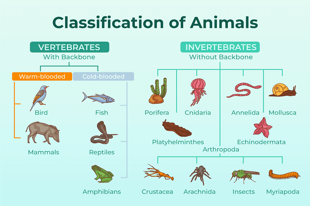
Animal Kingdom
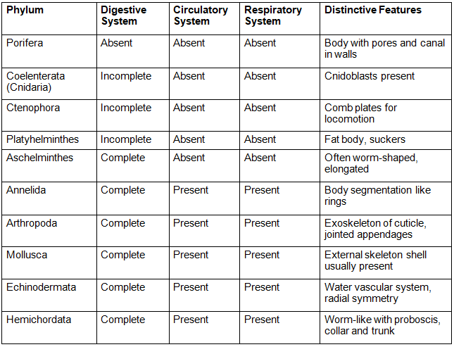
The animal kingdom can be classified into two main groups - those with bones (chordates) and those without bones (non-chordates). The basis of these classifications is the arrangement of cells, body symmetry, patterns of digestive system, nature of coelom, pattern of reproductive and circulatory systems.
- Non-chordates include phyla Porifera, Cnidaria, Aschelminthes, Platyhelminthes, Annelida, Arthropoda, Mollusca, Hemichordata and Echinodermata.
- Chordates include classes Cyclostomata, Chondrichthyes, Osteichthyes, Amphibia, Reptilia, Aves and Mammalia.
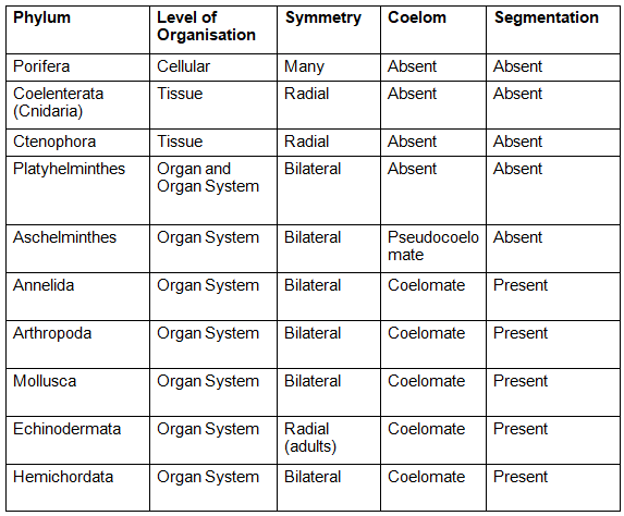
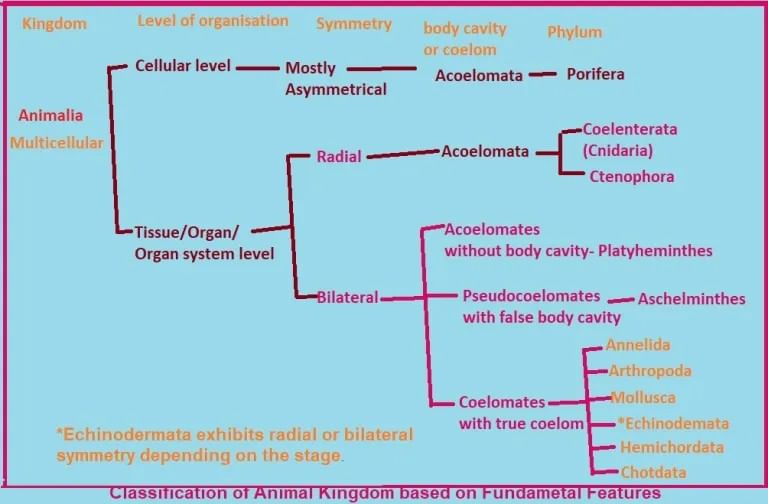
Salient Features of Different Phyla in the Animal Kingdom
Structural Organisations in Animals
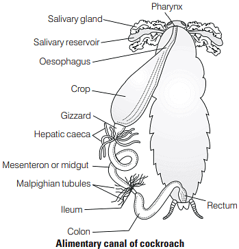
The diagrams in this chapter show the shapes and forms of animals such as frogs, cockroaches and earthworms.
Alimentary Canal of Cockroach
The morphology of the cockroach is divided into three main parts: the head, thorax and abdomen. In addition, its digestive system within the body cavity can be anatomically split into three components - the foregut, midgut and hindgut. The diagram below illustrates the cockroach's digestive system with labeled parts, such as the Malpighian tubules, Hepatic caeca, Gizzard and Crop, which are important for NEET objectives.
Open circulatory system of cockroach
Cockroaches possess a thirteen-chamber heart and an open circulatory system. It is important to understand the anatomy of the heart and the accompanying sinuses when studying the subject. Oxygenated blood enters each chamber through a slit known as an Ostia.
Biomolecules
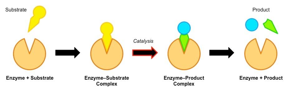
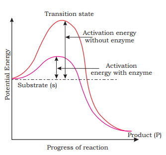
This chapter covers macromolecules such as proteins, nucleic acids, and polysaccharides, as well as enzymes and their roles. Enzymes are proteins that act as catalysts, speeding up biochemical reactions in cells.
Mechanism of Enzyme Action
Enzymes are proteins which interact with substrates to form a transient complex known as an ES complex. This complex reduces the amount of energy needed to initiate a reaction, as illustrated by a graph. After the reaction has taken place, the ES complex breaks down into the enzyme and the product. Factors like pH, temperature, concentration of substrates, inhibitors, and allosteric regulators can affect enzyme activity. The only exception to enzymes being proteins is the class of RNA catalysts known as ribozymes, which take their name from ribonucleic acid enzymes.
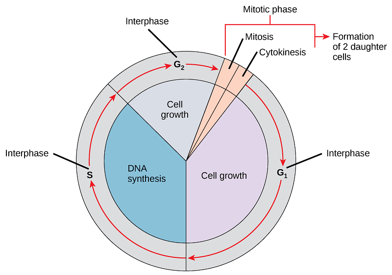
Cell Cycle and Cell Division
The cell cycle is composed of two distinct parts: Interphase and M phase (Mitosis phase). Interphase is further divided into 3 phases: G1 phase (Gap 1), S phase (Synthesis), and G2 phase (Gap 2). It is important to remember the sequence of these phases in order to understand the process of cell division.
Digestion and Absorption
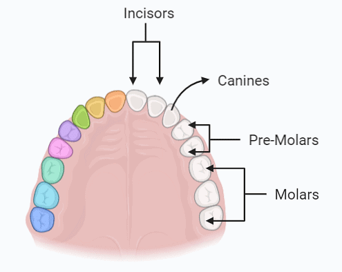
The digestive system starts with the mouth, which takes us to the oral cavity. This cavity is composed of the tongue and various kinds of teeth. The dental formula is used to portray the number and kind of teeth in each half of both the upper and lower jaws.
Milk Teeth or Primary Dentition
Premolars are absent in the primary dentition of humans.
I 2/2 C 1/1 M 2/2 = 10
10 (upper jaw) + 10 (lower jaw) = 20
Permanent Dentition
I 2/2 C 1/1 Pm 2/2 M 3/3 = 16
16 (upper jaw) + 16 (lower jaw) = 32
Here, I represent incisors, C - canine, Pm - premolars and M - molars.
Breathing and Exchange of Gases
This chapter focuses on the organs of respiration, the processes of breathing, and the exchange of gases. Consequently, diagrams such as the structure of the lungs, the process of inspiration and expiration, the alveolus, the oxygen transport curve, and the exchange of gases are featured.
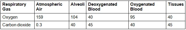
Transport of Gases between Tissue and Alveoli
The alveoli are made up of thin cells known as simple squamous epithelium, which is the main location for gas exchange. The epithelial cells of alveoli are of two types, type I and type II, with the latter secreting a lipoprotein (surfactant) to reduce surface tension. Gases are exchanged between the surrounding tissue and blood due to a difference in their concentrations or pressure gradient, with oxygen and carbon dioxide diffusing from areas of higher to lower concentration. The oxygen partial pressure (pO2) of the alveoli (104 mm Hg) is higher than that of deoxygenated blood (40 mm Hg), while the partial pressure of carbon dioxide (pCO2) in alveoli (40 mm Hg) is lower than that of blood from the tissue (45 mm Hg).
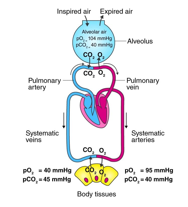 Transport of Gases
The diagram illustrates the differences between systemic veins, which transport deoxygenated blood, and systemic arteries, which transport oxygenated blood. The table below shows the comparison of the partial pressure of carbon dioxide and oxygen (in mmHg) in the systemic veins and arteries to the atmospheric air.
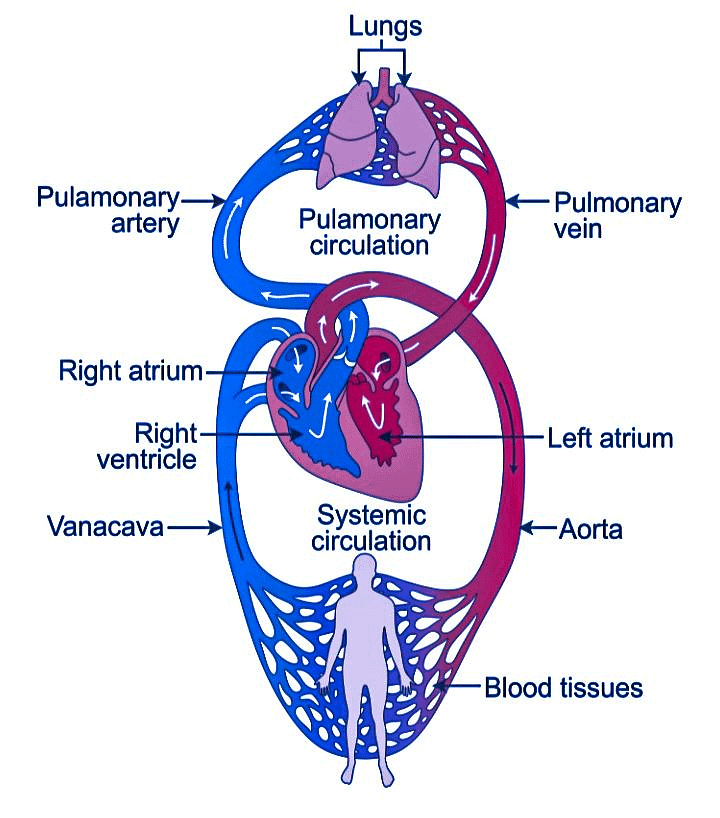
Body Fluid and Circulation
This section covers different bodily fluids, such as blood and lymph, as well as the circulatory system. It is essential to brush up on diagrams of blood cells, the structure of the heart, and a flow chart of double circulation, which could appear in an exam.
Systemic circulation
- The left ventricles send oxygenated blood to the aorta.
- The oxygenated blood is sent to the capillaries for distribution.
- The veins and venules collect the carbon dioxide-rich, deoxygenated blood from the various parts of the body.
- This deoxygenated blood is returned to the superior vena cava and then carried to the right atrium.
- From there, the blood is pumped into the right ventricle for pulmonary circulation.
Pulmonary circulation
- The pulmonary artery receives the blood from the right ventricle and transports it to the lungs, where it is oxygenated.
- The oxygenated blood is brought back to the left atrium via the pulmonary vein, which is then sent to the left ventricles.
- The left ventricles then pump the oxygenated blood to the aorta, allowing it to be circulated throughout the body.
Electrocardiograph
An electrocardiograph is a device used to measure the electrical activity of the heart during a cardiac cycle and produce a graph, known as an electrocardiogram, to visually represent this activity.
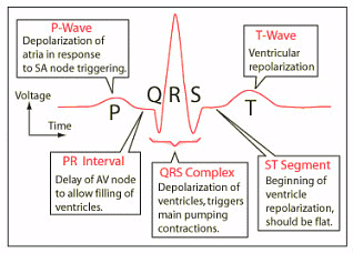
The P wave indicates electrical activation of the atria, while the QRS complex is an indication of the ventricles contracting. The Q wave marks the onset of systole and is followed by an R wave with a higher amplitude, and then a downward-pointing S wave. The T wave shows the ventricles returning to their resting state from an excited state.
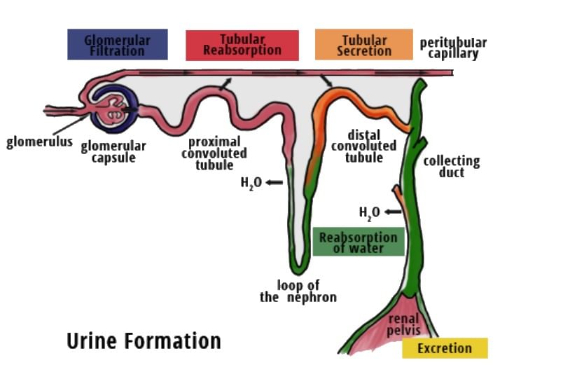
Excretory Products and their Elimination
The human body contains a set of organs for the purpose of excretion, which are the kidneys, bladder, ureters and urethra. This chapter outlines the anatomy of the urinary system, showing the location of the kidneys and the internal structure of a nephron.
Urine Formation: Reabsorption and Secretion
The nephron is comprised of two components, the renal corpuscle and the renal tubule. The renal corpuscle, also known as the Malpighian body, is made up of the glomerulus and Bowman's capsule. The renal tubule is a tubular structure that consists of a proximal convoluted tubule (PCT), Henle's loop, distal convoluted tubule (DCT), and collecting duct. The renal corpuscle, PCT, and DCT are located in the cortex region, while Henle's loop is primarily found in the medulla.
- The Proximal Convoluted Tubule (PCT) is lined with simple cuboidal epithelium, complete with microvilli and brush borders to maximize surface area for absorption. Approximately 70-80% of reabsorption occurs in this area.
- The Ascending Limb of Henle's Loop carries out only minimal reabsorption. This loop is essential in sustaining a high osmolarity in the fluid, with the ascending limb being impermeable to water, and the descending limb permeable.
- The Distal Convoluted Tubule (DCT) is responsible for the conditional reabsorption of water and sodium. Subsequently, the Collecting Duct is responsible for the reabsorption of a large quantity of water to produce a highly concentrated urine.
Locomotion and Movement
This chapter contains diagrams related to muscles, muscle fibers, and contractile proteins. The most significant of these is the diagram that illustrates the sliding filament theory, which explains that muscle contraction is caused by the movement of thin filaments over thick filaments.
- The central nervous system stimulates muscle contraction by sending a signal through motor neurons that triggers the release of neurotransmitters.
- Once the acetylcholine reaches the synaptic cleft, it binds to the muscle fiber receptors and leads to the depolarization of the sarcolemma. This causes an action potential to move through the muscle fibers and activate the release of calcium ions into the sarcoplasm.
- The calcium ions then attach to the troponin subunit, leading to structural changes that make the myosin-binding sites on the actin filaments visible.
- The myosin head contains a binding site for ATP and also has an ATPase activity. This ATPase activity is responsible for ATP hydrolysis which allows the energized myosin head to bind to the active binding sites on actin forming a cross bridge.
ATP = ADP + Pi + Energy - The actin filaments and the Z-lines attached to them are pulled inward toward the center of the A-band, resulting in a shortening or contraction of the sarcomere. Meanwhile, ADP and Pi are released from the myosin, allowing it to return to its relaxed state. A new ATP binds and breaks the cross-bridge.
- This process is then repeated, leading to further muscle contraction. This continues until the calcium ions are pumped back to the sarcoplasmic cisternae, which masks the actin filaments and allows the Z-lines to return to their original, relaxed position.
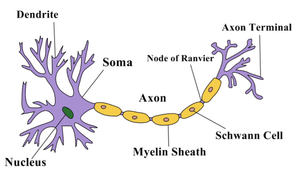
Neural Control and Coordination
The last chapter of class 11 contains several important diagrams including a sagittal section of the brain, the structure of a neuron, the structure of an ear, an eye, a schematic representation of a reflex action, and a diagram of an axon terminal and synapses.
Structure of a Neuron
Neurons are microscopic structures in the nervous system composed of dendrites, a cell body and an axon. The cell body consists of particles known as Nissl's granules. Dendrites, which are branch-like projections from the cell body, also contain Nissl's granules. Axons are long, tube-like structures that transmit messages away from the cell body to the junction between neurons and muscles, called synapses. Axons can be either myelinated, with a myelin sheath formed by Schwann cells, and gaps between the myelin sheaths called nodes of Ranvier, or unmyelinated, with Schwann cells but without a myelin sheath.
Nerve signals are transmitted from one neuron to another by way of synapses. The axon of the neuron ends in a bulb-like structure called a synaptic knob that has synaptic vesicles containing neurotransmitters. A labelled diagram is provided to illustrate the synapse.
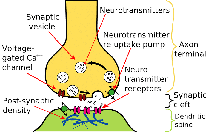
Axon Terminal and Synapse
The axon terminal of a pre-synaptic neuron and the membrane of a post-synaptic neuron come together to form a synapse, which may be separated by a gap known as the synaptic cleft. When an action potential is triggered, the synaptic vesicles present in the axon terminal release neurotransmitters into the synaptic cleft. These neurotransmitters then bind to the specific receptors on the postsynaptic membrane.
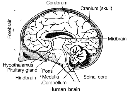
Sagittal section of the Human Brain
The human brain is securely held in place within the skull and is covered by three layers of cranial meninges, the outer layer (dura mater), the middle thin layer (arachnoid) and the inner layer (pia mater). This organ can be divided into three sections: the forebrain (which includes the cerebrum, thalamus and hypothalamus), the midbrain (which sits between the forebrain's hypothalamus and hindbrain's pons) and the hindbrain (which is composed of the pons, cerebellum and medulla). The two halves of the cerebrum are linked by a bundle of nerve fibers called the corpus callosum.
Eye Structure
The human eyeball is composed of 3 layers - sclera (external layer), retina (inner layer) and choroid (middle layer).
- The front part of the sclera is called the cornea.
- Moving forward, the choroid layer forms a thicker ciliary body which is surrounded by an opaque and pigmented structure known as the iris. Its opening is referred to as the pupil.
- The eyeball contains a transparent lens held in place by suspensory ligaments. The area between the lens and cornea is known as the aqueous chamber and is filled with aqueous fluid.
- The innermost layer of the eye is called the retina and contains two photoreceptor cells, rods and cones.
- Between the lens and the retina is the vitreous chamber which is filled with vitreous humor.
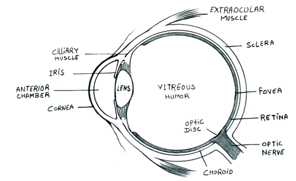 Structure of Human Eye
Ear Structure
The ear is divided into three parts - the outer ear, middle ear and inner ear. The outer ear includes the external auditory canal and the pinna, which ends at the eardrum.
The middle ear is made up of three ossicles - malleus, stapes and incus - and it is connected to the throat through the Eustachian tube.
The inner ear is known as the labyrinth and consists of two parts, a membranous labyrinth and a bony portion. The membranous labyrinth is filled with endolymph and surrounded by perilymph. It also features a coiled region known as the cochlea.
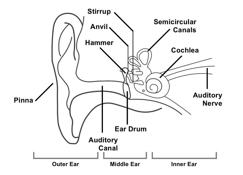 Human Ear
FAQs on Most Important Diagrams Class 11 Biology (Zoology) for NEET 2026 Biology Exam Preparation
| 1. Which are the most important diagrams I need to memorize for NEET biology zoology? |  |
| 2. How do I draw the human digestive system diagram correctly for NEET exam? | |
| 3. What's the difference between diagrams of plant and animal cells in Class 11 biology? | |
| 4. Which diagrams of tissue types appear most frequently in NEET questions? | |
| 5. How should I label the neuron diagram to score full marks in NEET biology? | |

