Important Diagrams: Body Fluids and Circulation | Biology Class 11 - NEET PDF Download
| Table of contents |

|
| 1. Formed Elements |

|
| 2. Human Heart |

|
| 3. Electrocardiogram (ECG) |

|
| 4. Double Circulation |

|
| Diagrams Based Questions NEET |

|
1. Formed Elements
Erythrocytes (Red Blood Cells - RBCs):Erythrocytes are the most numerous cells in blood, averaging 5 to 5.5 million per mm³ in healthy adults and are primarily responsible for oxygen transport due to their hemoglobin content. Lacking nuclei, these biconcave cells have a lifespan of 120 days and are processed in the spleen.
Leucocytes (White Blood Cells - WBCs):Leucocytes, devoid of hemoglobin and thus colorless, range from 6,000 to 8,000 per mm³ of blood and include granulocytes (neutrophils, eosinophils, basophils) and agranulocytes (lymphocytes, monocytes). These cells play crucial roles in immune defense, inflammation, and allergic reactions, with neutrophils being the most prevalent.
Platelets (Thrombocytes):Platelets, small fragments from megakaryocytes, number between 150,000 to 350,000 per mm³ of blood and are vital for blood clotting; a decrease in their count can lead to significant clotting disorders and excessive blood loss.
 Diagrammatic representation of formed elements in blood
2. Human Heart
The heart is a mesodermally derived organ located in the thoracic cavity between the lungs, slightly tilted to the left. It is protected by the pericardium, a double-walled membranous sac, which encloses the pericardial fluid. The heart has four chambers: two upper chambers (atria) and two lower chambers (ventricles). These chambers are separated by muscular walls: the interatrial and interventricular septa. Valves, like the tricuspid and bicuspid, ensure one-way blood flow between the chambers and major arteries.The heart is composed of cardiac muscles, and its walls are thicker in the ventricles. Specialized nodal tissue, such as the sinoatrial node (SAN) and atrioventricular node (AVN), helps in initiating and maintaining the heart's rhythmic contractions. The SAN, known as the pacemaker, generates action potentials that cause the heart to beat at an average rate of 72 beats per minute.
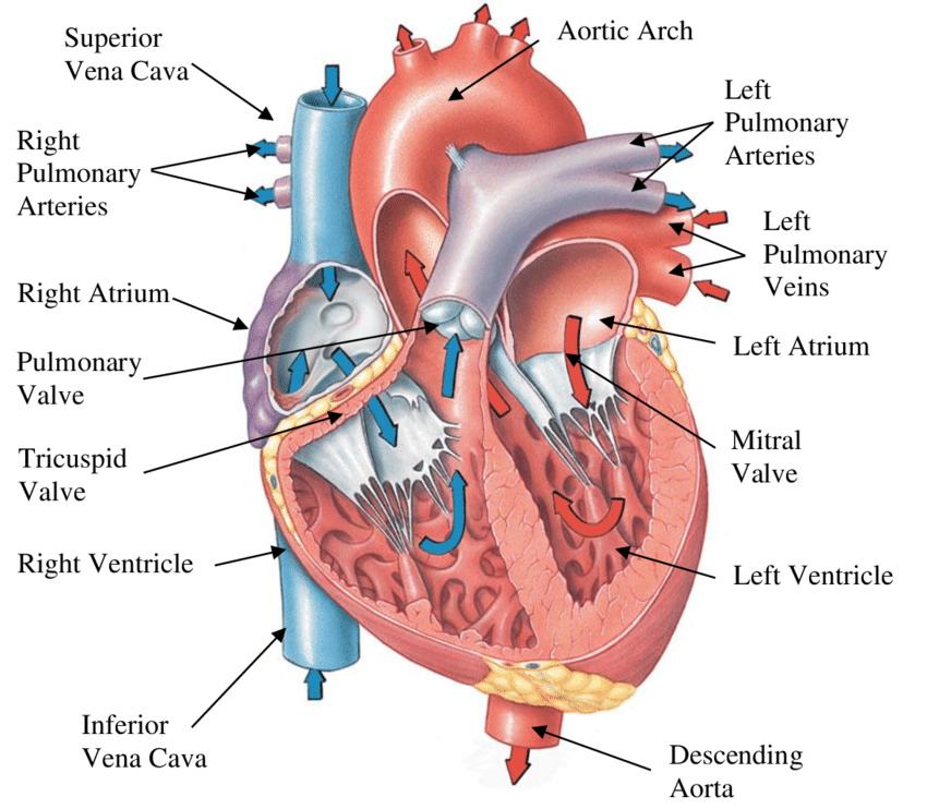 Section of Human Heart
3. Electrocardiogram (ECG)
- P-wave: Represents atrial depolarization, leading to atrial contraction.
- QRS complex: Represents ventricular depolarization, initiating ventricular contraction and marking the start of systole.
- T-wave: Represents ventricular repolarization, marking the end of systole.
- Heartbeat rate: Determined by counting the number of QRS complexes over a given time.
- Clinical significance: Deviations in ECG shape may indicate abnormalities or disease.
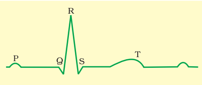 Diagrammatic presentation of a standard ECG
4. Double Circulation
Blood vessels: Arteries and veins have three layers:
- Tunica intima (inner squamous endothelium),
- Tunica media (smooth muscle, thinner in veins),
- Tunica externa (outer connective tissue with collagen).
Pulmonary circulation: Deoxygenated blood is pumped from the right ventricle to the lungs via the pulmonary artery. Oxygenated blood returns to the left atrium via pulmonary veins.
Systemic circulation: Oxygenated blood from the left ventricle travels through arteries to tissues, and deoxygenated blood returns to the right atrium via veins.
Hepatic portal system: Connects the digestive tract and liver, carrying blood from the intestines to the liver.
Coronary system: Dedicated circulation for the heart muscles.
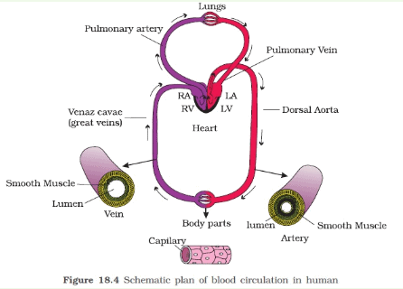 Schematic plan of blood circulation in human
Diagrams Based Questions NEET
Q1: Following are the stages of pathway for conduction of an action potential through the heart (NEET 2024)
A. AV bundle
B. Purkinje fibres
C. AV node
D. Bundle branches
E. SA node
Choose the correct sequence of pathway from the options given below
(a) E-C-A-D-B
(b) A-E-C-B-D
(c) B-D-E-C-A
(d) E-A-D-B-C
Ans: (a)
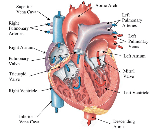 The electrical pathway for the conduction of an action potential through the heart is a precisely coordinated process, essential for maintaining the heart's rhythmic beating. To understand this conduction pathway, it’s important to know the roles of the specific components involved:
SA node (Sinoatrial node): Often referred to as the pacemaker of the heart. It initiates the electrical impulse, causing the atria to contract.
AV node (Atrioventricular node): Receives the impulse from the SA node and provides a slight delay, allowing the ventricles time to fill with blood before they contract.
AV bundle (Bundle of His): Transfers the electrical impulse from the AV node to the bundle branches.
Bundle branches: Conducts the impulses through the interventricular septum.
Purkinje fibers: Distribute the electrical impulse throughout the ventricles, stimulating them to contract uniformly and powerfully.
The correct sequence for the pathway of an action potential through the heart follows a route designed to efficiently coordinate the heartbeat starting from the initiation of the action potential to the consequential contraction of the heart muscles. The sequence is:
SA node (E): The pacemaker where the electrical activity originates.
AV node (C): Where the impulse is delayed slightly.
AV bundle (A): Conducts the impulse from the AV node to the bundle branches.
Bundle branches (D): Leads the impulse to the Purkinje fibers.
Purkinje fibers (B): Distributes the impulse throughout the ventricles.
By examining the options provided with the above understanding:
Option A: E-C-A-D-B
Option B: A-E-C-B-D
Option C: B-D-E-C-A
Option D: E-A-D-B-C
Option A (E-C-A-D-B) correctly reflects the order in which the electrical impulse travels through the cardiac conduction system, beginning with the SA node and ending in the Purkinje fibers. Hence, Option A is the correct answer.
Q2: Match List I with List II : (NEET 2024)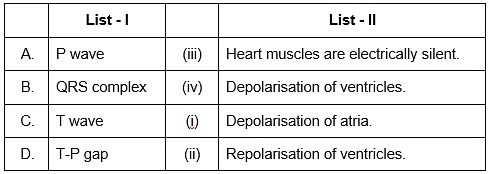 Choose the correct answer from the options given below :
(a) A-I, B-III, C-IV, D-II
(b) A-III, B-II, C-IV, D-I
(c) A-II, B-III, C-I, D-IV
(d) A-IV, B-II, C-I, D-III
Ans: (b)
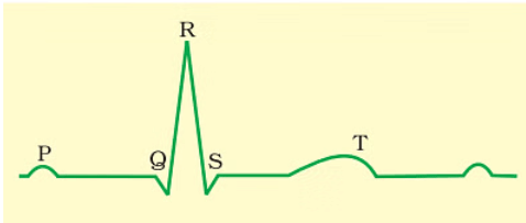
Sol: The correct answer is option no. (B) as
A. P wave - III. Depolarisation of atria.
B. QRS complex - II. Depolarisation of ventricles.
C. T wave - IV. Repolarisation of ventricles.
D. T-P gap - I. Heart muscles are electrically silent.
|
150 videos|399 docs|136 tests
|
FAQs on Important Diagrams: Body Fluids and Circulation - Biology Class 11 - NEET
| 1. What are the main formed elements of blood and their functions? |  |
| 2. What is the structure and functional significance of the human heart? |  |
| 3. How does an electrocardiogram (ECG) work and what does it measure? |  |
| 4. What is double circulation and why is it important for humans? |  |
| 5. What are the key diagrams to study for body fluids and circulation in NEET? |  |


