Human Digestive System | Famous Books for UPSC Exam (Summary & Tests) PDF Download
Introduction
The human digestive system is responsible for converting biomacromolecules found in food, such as carbohydrates and proteins, into simpler substances that our body can utilize. Rather than being used in their original form, these biomacromolecules undergo a process of breakdown and conversion within the digestive system. For instance, carbohydrates are broken down into simple sugars like glucose, fats are converted into fatty acids and glycerol, and proteins are transformed into amino acids. This transformation of complex food substances into easily absorbable forms is referred to as digestion.
Alimentary Canal
- The food passes through a continuous canal called alimentary canal. The canal can be divided into various compartments: (1) the buccal cavity, (2) foodpipe or oesophagus, (3) stomach, (4) small intestine, (5) large intestine ending in the rectum and (6) the anus.
- The activities of the gastro-intestinal tract [alimentary canal] are under neural and hormonal control for proper coordination of different parts.
- The sight, smell and/or the presence of food in the oral cavity can stimulate the secretion of saliva.
- Gastric and intestinal secretions are also, similarly, stimulated by neural signals.
- The muscular activities of different parts of the alimentary canal can also be moderated by neural mechanisms.
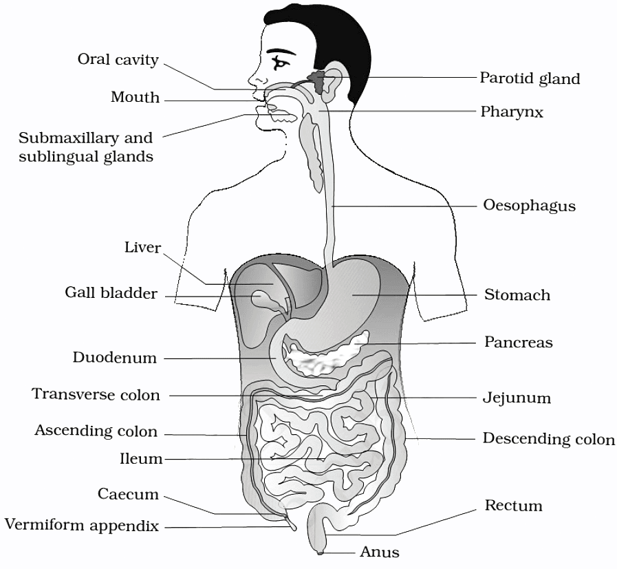
Buccal Cavity or Oral Cavity – Teeth, Tongue, Saliva
- The buccal cavity, also known as the oral cavity, is involved in the initial stages of food intake. This process, known as ingestion, occurs through the mouth. The mouth serves as an entry point to the buccal cavity or oral cavity.
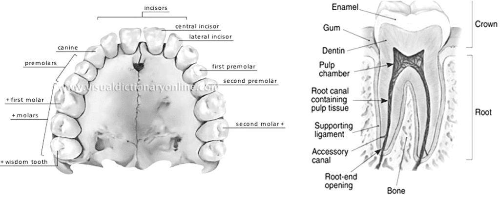
- Within the oral cavity, several components are present, including a collection of teeth and a muscular tongue. Each tooth is securely anchored in a socket within the jaw bone. In most mammals, including humans, two sets of teeth are formed throughout their lifetime. Initially, a temporary set of teeth, known as milk or deciduous teeth, are replaced by a permanent set of teeth, also referred to as adult teeth.
- An adult human typically possesses 32 permanent teeth, which can be classified into four different types: incisors (I), canines (C), premolars (PM), and molars (M). The arrangement of these teeth in each half of the upper and lower jaw follows a specific pattern, represented by a dental formula. In humans, this formula is typically expressed as 2123/2123, indicating two incisors, one canine, two premolars, and three molars on each side.
- The teeth are equipped with a hard chewing surface composed of enamel, which happens to be the hardest substance in the human body and contains a high concentration of minerals. This robust structure aids in the process of mastication, or chewing, of food.
- Within our mouth, we have salivary glands that produce and release saliva. This saliva serves an important role in breaking down starch into sugars. The saliva that is secreted into the oral cavity contains electrolytes such as sodium (Na+), potassium (K+), chloride (Cl-), and bicarbonate ions (HCO3-), along with enzymes known as salivary amylase and lysozyme.
- The chemical process of digestion begins in the oral cavity through the hydrolytic action of the salivary amylase enzyme. This enzyme specifically targets carbohydrates and breaks them down. Approximately 30 percent of the starch present in the food we consume is hydrolyzed by this enzyme, with its optimal pH level being 6.8. As a result of this enzymatic activity, the starch is converted into a disaccharide called maltose.
- Saliva contains lysozyme, which acts as an antibacterial agent, effectively preventing infections.
- The tongue, a muscular organ, is attached at the back to the floor of the buccal cavity. It aids in the mixing of saliva with chewed food and assists in the process of swallowing.
- The frenulum, a fold of skin beneath the tongue, connects the tongue to the floor of the oral cavity.
- The upper surface of the tongue is covered in small projections called papillae, some of which house taste buds.
Foodpipe/Oesophagus
- The oral cavity leads to a brief passage called the pharynx, which serves as a common pathway for both food and air. Both the esophagus and the trachea (windpipe) open into the pharynx.
- During swallowing, a cartilaginous flap known as the epiglottis ensures that food does not enter the glottis, which is the opening of the windpipe.
- The swallowed food then moves into the foodpipe or esophagus. The esophagus is a thin, elongated tube that extends posteriorly, passing through the neck, thorax (the area between the neck and abdomen), and diaphragm (the muscle that separates the thorax from the abdomen in mammals). Eventually, it leads to a stomach that takes on a 'J'-shaped, bag-like structure.
- The mucus present in saliva aids in lubricating and binding the chewed food particles together, forming a bolus. This bolus is then carried into the pharynx and subsequently into the esophagus through the process of swallowing or deglutition.
- The bolus continues its descent through the esophagus, facilitated by successive waves of muscular contractions called peristalsis. The passage of food into the stomach is controlled by the gastro-esophageal sphincter.
Stomach
- The inner lining of the stomach has the ability to secrete mucus, hydrochloric acid, and digestive juices.
- The mucous serves as a protective layer for the stomach lining.
- The hydrochloric acid plays a dual role by eliminating many bacteria that enter the stomach with the ingested food and creating an acidic environment within the stomach.
- The digestive juices released by the stomach aid in breaking down proteins into simpler substances.
- The opening between the esophagus and the stomach is regulated by a muscular sphincter known as the gastro-esophageal sphincter.
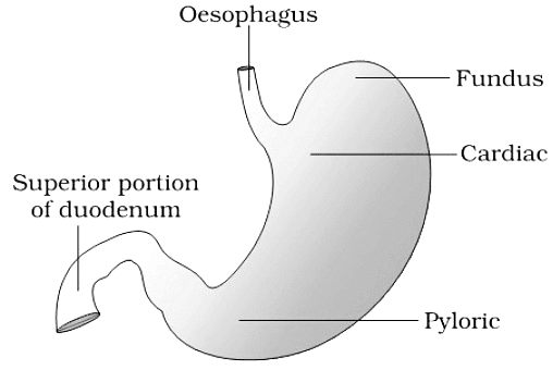
- Positioned in the upper left area of the abdominal cavity, the stomach can be divided into three main parts: the cardiac portion, where the esophagus connects; the fundic region; and the pyloric portion, which opens into the first section of the small intestine.
Small intestine
- The small intestine can be divided into three distinct regions: the duodenum, which has a 'C' shape; the jejunum, a long and coiled middle portion; and the ileum, which is highly coiled.
- The opening between the stomach and the duodenum is regulated by the pyloric sphincter, while the ileum connects to the large intestine.
- The small intestine, approximately 5 meters in length, possesses a high degree of coiling. It receives secretions from both the liver and the pancreas, in addition to secreting its own juices.
- Once food is digested, it is absorbed into the blood vessels within the intestinal wall, a process known as absorption.
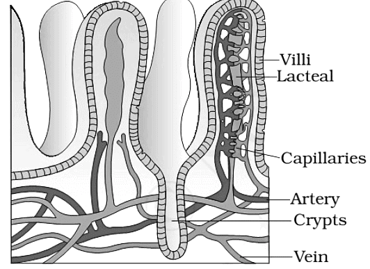
- The inner walls of the small intestine contain numerous finger-like projections called villi (singular: villus). These villi greatly increase the surface area available for the absorption of digested food.
- The villi are supplied with an extensive network of capillaries as well as a large lymph vessel called the lacteal.
- The absorbed substances are transported through the blood vessels to various organs within the body, where they are utilized to build complex substances like proteins, a process known as assimilation.
- Within the cells, glucose undergoes a breakdown with the aid of oxygen, resulting in the release of energy, carbon dioxide, and water.
- The remaining undigested and unabsorbed food then proceeds into the large intestine.
Large intestine
- The large intestine is wider and shorter compared to the small intestine, spanning a length of approximately 1.5 meters. Its primary function is to absorb water and certain salts from the undigested food material.
- The remaining waste material passes into the rectum, where it accumulates as semi-solid feces. Periodically, the fecal matter is expelled through the anus in a process known as egestion.
- The large intestine consists of three main parts: the caecum, the colon, and the rectum. The caecum is a small blind sac that houses symbiotic microorganisms. Extending from the caecum is a narrow, finger-like tube called the vermiform appendix, which is considered a vestigial organ—a remnant with reduced significance in modern humans. The appendix was once helpful in digesting roughage from plant-based food, but its role has diminished over time.
- The caecum opens into the colon, which is further divided into three sections: the ascending colon, the transverse colon, and the descending colon. The descending colon connects to the rectum, which ultimately leads to the anus for elimination of waste.
- The large intestine is not primarily involved in significant digestive activities. Its main functions include the absorption of water, minerals, and certain drugs, as well as the secretion of mucus, which aids in adhering waste particles together and lubricating them for easy passage.
- Undigested and unabsorbed substances, known as feces, enter the caecum of the large intestine through the ileocecal valve, which prevents the backflow of fecal matter. The feces are temporarily stored in the rectum until defecation occurs.
Layers of the Alimentary Canal
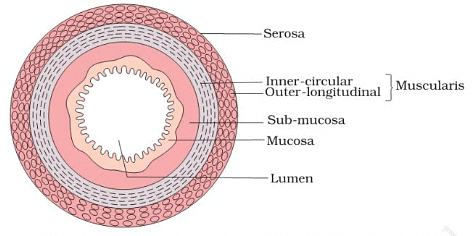
- The wall of the alimentary canal, from the esophagus to the rectum, consists of four layers: serosa, muscularis, submucosa, and mucosa.
- The serosa serves as the outermost layer and is composed of a thin mesothelium (epithelium of visceral organs) and connective tissues.
- The muscularis layer is formed by smooth muscles.
- The submucosal layer is composed of loose connective tissues that contain nerves, blood vessels, and lymph vessels. In the duodenum, glands are also present within the submucosa.
- The innermost layer lining the lumen of the alimentary canal is the mucosa. This layer exhibits irregular folds, known as rugae, in the stomach, and small finger-like projections called villi in the small intestine. The mucosal epithelium contains goblet cells, which secrete mucus to aid in lubrication. Additionally, the mucosa forms glands, such as gastric glands, within the stomach.
Digestive Glands
The alimentary canal is associated with various digestive glands, including the salivary glands, liver, and pancreas.
Salivary glands
- Saliva is primarily produced by three pairs of salivary glands: the parotids (located in the cheeks), the sub-maxillary glands (found beneath the lower jaw), and the sub-lingual glands (situated below the tongue).
- These glands, positioned just outside the buccal cavity, secrete salivary juice into the oral cavity.
- Saliva aids in the breakdown of starch into sugars.
Liver
- The liver is a reddish-brown gland located in the upper region of the abdomen on the right side.
- It holds the distinction of being the largest gland in the human body.
- The liver secretes bile juice, which is stored in a sac known as the gallbladder.
- Bile plays a vital role in the digestion of fats.
- The liver consists of two lobes, and within these lobes are structural and functional units called hepatic lobules, which contain hepatic cells.
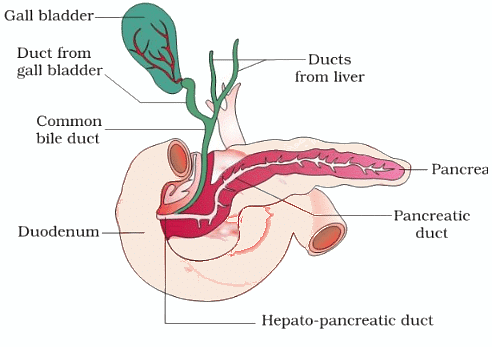
- The bile produced by the hepatic cells passes through hepatic ducts and is then stored and concentrated in a thin muscular sac called the gallbladder.
- The cystic duct, originating from the gallbladder, and the hepatic duct from the liver combine to form the common bile duct.
- The common bile duct and the pancreatic duct open together into the duodenum, forming the common hepato-pancreatic duct. This duct is regulated by a sphincter called the sphincter of Oddi.
Pancreas
- The pancreas is a large gland with a cream-colored appearance, located just below the stomach.
- Pancreatic juice, produced by the pancreas, acts on carbohydrates and proteins, breaking them down into simpler forms.
- As the partially digested food reaches the lower part of the small intestine, the process of digestion is completed with the assistance of intestinal juice, also known as succus entericus.
- The pancreas is a compound organ situated between the limbs of the 'U'-shaped duodenum, functioning as both an exocrine and endocrine gland.
- The exocrine portion secretes an alkaline pancreatic juice containing enzymes, while the endocrine portion secretes hormones such as insulin and glucagon.
Digestion - Enzyme Action in the Stomach
- The stomach serves as a storage site for food, holding it for approximately 4-5 hours. Through churning movements of its muscular wall, the food thoroughly mixes with the acidic gastric juice, forming a semi-fluid mixture called chyme.
- The proenzyme pepsinogen, when exposed to hydrochloric acid, is converted into its active form, the enzyme pepsin. Pepsin is a proteolytic enzyme responsible for breaking down proteins into proteoses and peptones (peptides).
- The gastric juice contains mucus and bicarbonates, which play important roles in lubricating and protecting the mucosal epithelium from the highly concentrated hydrochloric acid. The hydrochloric acid provides the optimal acidic pH (pH 1.8) for pepsin activity.
- Infants have an additional proteolytic enzyme called rennin in their gastric juice, which aids in the digestion of milk proteins.
Digestion - Enzyme Action in the Small Intestine
- The pancreatic juice contains inactive enzymes, including trypsinogen, chymotrypsinogen, procarboxypeptidases, amylases, lipases, and nucleases.
- Trypsinogen is activated into its active form, trypsin, by an enzyme called enterokinase, which is secreted by the intestinal mucosa. Once activated, trypsin further activates the other enzymes present in the pancreatic juice.
- Bile, released into the duodenum, consists of bile pigments (bilirubin and biliverdin), bile salts, cholesterol, and phospholipids, but it does not contain any enzymes.
- Bile aids in the emulsification of fats, breaking them down into very small micelles. Additionally, bile activates lipases. Although small amounts of lipases are secreted by gastric glands.
- The intestinal mucosal epithelium contains goblet cells, which secrete mucus. The secretions from the mucosa, combined with the mucus from goblet cells, constitute the intestinal juice.
- Intestinal juice contains various enzymes, such as disaccharidases (e.g., maltase), dipeptidases, lipases, nucleosidases, and more. The secretion of digestive juices is regulated by local hormones produced by the gastric and intestinal mucosa.
- The mucus, along with bicarbonates from the pancreas, protects the intestinal mucosa from acidity and provides an alkaline environment (pH 7.8) for enzymatic activities.
- The breakdown of biomacromolecules mentioned above occurs primarily in the duodenum region of the small intestine.
- The resulting simple substances are absorbed in the jejunum and ileum regions of the small intestine.
- Undigested and unabsorbed substances are passed on to the large intestine.
Absorption of Digested Products
- Absorption refers to the process by which the end products of digestion pass through the intestinal mucosa into the blood or lymph.
- Small amounts of monosaccharides like glucose, amino acids, and certain electrolytes like chloride ions are typically absorbed through simple diffusion. The passage of these substances into the blood relies on concentration gradients.
- In some cases, substances like glucose and amino acids are absorbed with the assistance of carrier proteins. This mechanism is known as facilitated transport.
- The transport of water depends on the osmotic gradient. Active transport occurs against the concentration gradient and requires energy. Various nutrients like amino acids, monosaccharides like glucose, and electrolytes like Na+ are absorbed into the blood through this mechanism.
- Due to their insolubility, fatty acids and glycerol cannot be absorbed directly into the blood. Instead, they are incorporated into small droplets called micelles, which move into the intestinal mucosa. There, they are re-formed into tiny fat globules coated with proteins called chylomicrons. These chylomicrons are transported into lymph vessels (lacteals) within the villi. Eventually, these lymph vessels release the absorbed substances into the bloodstream.
- The absorption of substances takes place in different parts of the alimentary canal, including the mouth, stomach, small intestine, and large intestine. However, the majority of absorption occurs in the small intestine.
Disorders of the Digestive System
- The most common ailment affecting the digestive system is inflammation of the intestinal tract, often caused by bacterial or viral infections.
- Infections can also be attributed to intestinal parasites such as tapeworms, roundworms, threadworms, hookworms, pinworms, and others.
- Jaundice: This condition occurs when the liver is affected, resulting in the yellowing of the skin and eyes due to the accumulation of bile pigments.
- Vomiting: Vomiting is the expulsion of stomach contents through the mouth. This reflex action is controlled by the vomit center in the medulla. Nausea is often experienced prior to vomiting.
- Diarrhea: Diarrhea refers to abnormally frequent bowel movements with increased fluidity of the fecal discharge. It can lead to reduced absorption of food.
- Constipation: Constipation occurs when feces are retained within the rectum due to irregular bowel movements.
- Indigestion: Indigestion occurs when food is not properly digested, resulting in a feeling of fullness. Inadequate enzyme secretion, anxiety, food poisoning, overeating, and consumption of spicy foods are common causes of indigestion.
|
745 videos|1444 docs|633 tests
|


