Locomotion and Movement Chapter Notes | Biology Class 11 - NEET PDF Download
| Table of contents |

|
| Locomotion and Movement |

|
| Types of Movement |

|
| Muscles |

|
| Mechanism of Muscle Contraction |

|
| Skeletal System |

|
Locomotion and Movement
Movement is a core feature exhibited by living organisms, encompassing a wide array of forms from basic protoplasmic flow in Amoeba to the coordinated motion of cilia, flagella, and tentacles in various species. Human beings, endowed with voluntary muscles, can manipulate their limbs, jaws, eyelids, tongue, and other organs. Among these movements, some involve changing position or location and are termed locomotion. Locomotion encompasses activities like walking, running, climbing, flying, and swimming.
The structures facilitating locomotion often serve dual purposes, contributing to other forms of movement as well. For example, in Paramecium, cilia aid in both food propulsion and locomotion, while Hydra utilizes its tentacles for prey capture and movement. In humans, limbs are instrumental in altering body posture as well as facilitating locomotion. These observations underscore the interconnectedness of movements and locomotion, leading to the assertion that "While all locomotions are movements, not all movements are locomotions."
Animals adapt various methods of locomotion according to their habitats and specific requirements. This diversity in locomotion serves purposes such as foraging for food, seeking shelter, finding suitable breeding grounds, responding to climatic conditions, or evading predators.
Types of Movement
The cells within the human body demonstrate three primary types of movement: amoeboid, ciliary, and muscular.
- Amoeboid movement is observed in specialized cells such as macrophages and leukocytes, as well as in organisms like Amoeba. It involves the formation of pseudopodia through protoplasmic streaming, facilitated by cytoskeletal components like microfilaments.
- Ciliary movement occurs in the internal tubular organs lined with ciliated epithelium. The synchronized beating of cilia, as seen in organs like the trachea, aids in clearing dust particles and foreign substances from inhaled air. Additionally, it facilitates the passage of ova through the female reproductive tract.
- Muscular movement is responsible for the motion of limbs, jaws, tongue, and other body parts. Muscles contract and relax to enable locomotion and various other movements in humans and most multicellular organisms. Achieving locomotion requires coordination among the muscular, skeletal, and neural systems.
Muscles
Muscle tissue, derived from the mesoderm, is a specialized component of the human body, constituting approximately 40-50% of the adult body weight. Examining muscle properties reveals their excitability, contractility, extensibility, and elasticity.
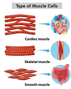
Muscles are categorized according to various criteria such as location, appearance, and regulatory nature of their activities. Primarily, muscles are classified into three types based on their location: skeletal muscles, visceral muscles, and cardiac muscles.

- Skeletal muscles, closely linked with the skeletal framework of the body, display a striped pattern under microscopic examination, earning them the designation of striated muscles. These muscles are voluntarily controlled by the nervous system and primarily facilitate movement and posture adjustments.
- Visceral muscles reside in the inner linings of hollow organs like the digestive and reproductive tracts. Their smooth appearance, lacking the striped pattern, labels them as smooth or non-striated muscles. Unlike skeletal muscles, visceral muscles operate involuntarily, assisting in functions such as food and gamete transportation, with regulation not solely governed by the nervous system.
- Cardiac muscles, found exclusively in the heart, derive their name from this association. Arranged in a branching pattern, cardiac muscle cells form the cardiac muscle tissue. Despite their striated appearance, their activities are not directly influenced by the nervous system.
Structure and Mechanism of Contraction
Skeletal muscles are structured with muscle bundles, or fascicles, bound together by fascia. Each bundle comprises muscle fibers encased by the sarcolemma and sarcoplasm. Within the sarcoplasm, the sarcoplasmic reticulum serves as a reservoir for calcium ions, while myofibrils, housing myofilaments, impart the striated appearance to the muscle fiber. Actin predominates in the I-band, whereas myosin is chiefly concentrated in the A-band. These filaments run parallel to each other along the longitudinal axis of the myofibril. The Z-line demarcates the I-band, and the M-line anchors the thick filaments within the A-band. Sarcomeres, positioned between Z-lines, act as the functional units governing muscle contraction. During rest, thin filaments partially overlap with thick filaments, leaving the H-zone unobstructed.
Structure of Contractile Proteins
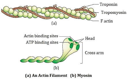
The structure of an actin (thin) filament comprises two helically wound "F" (filamentous) actins, each of which is a polymer formed by monomeric "G" (globular) actins. Alongside these "F" actins, two tropomyosin protein filaments run parallel to them. Tropomyosin, evenly distributed along the actin filaments' length, serves as a complex protein. During the resting state, troponin subunits cover or mask the active binding sites on the actin filaments, preventing myosin from binding to them.
- Conversely, the myosin (thick) filament is also a polymerized protein, composed of multiple monomeric proteins known as meromyosins. It consists of two primary components: heavy meromyosin (HMM) and light meromyosin (LMM).
- The HMM component of the myosin filament features globular heads housing an active ATPase enzyme. These heads also contain binding sites for ATP and active sites for actin. Projecting outward from the surface of the polymerized myosin filament at regular intervals and angles are globular heads with short arms, forming cross-arms.
- In essence, the actin filament comprises polymerized "F" actins along with tropomyosin and troponin, whereas the myosin filament consists of polymerized meromyosins, with the globular heads containing ATPase and actin-binding sites.

Mechanism of Muscle Contraction
- The sliding filament theory elucidates the process of muscle contraction, where thin filaments slide over thick filaments. Contraction begins with a signal from the central nervous system (CNS) transmitted via a motor neuron to the muscle fibers, forming a motor unit. At the neuromuscular junction, acetylcholine is released as a neurotransmitter, triggering an action potential in the sarcolemma.
- This action potential propagates through the muscle fiber, prompting the release of calcium ions into the sarcoplasm. Calcium ions bind to actin filaments, exposing active sites for myosin. Myosin heads, powered by ATP hydrolysis, bind to these active sites, forming cross-bridges. Cross-bridge formation pulls attached actin filaments toward the A-band center, shortening the sarcomere and inducing muscle contraction.
- During contraction, I-bands shrink while A-bands maintain length. Myosin releases ADP and P1, returning to its relaxed state, breaking the cross-bridge. ATP binds, initiating another cycle of cross-bridge formation and breakage, further filament sliding. This process persists until calcium ions are pumped back into the sarcoplasmic reticulum, concealing actin filaments and restoring Z-line position, leading to muscle relaxation.
- Muscle fiber reaction times vary. Repeated activation may lead to lactic acid accumulation via anaerobic glycogen breakdown, causing fatigue. Myoglobin, a red oxygen-storing pigment, is abundant in red fibers, which are rich in mitochondria for ATP synthesis. In contrast, white fibers have less myoglobin, fewer mitochondria, but ample sarcoplasmic reticulum, relying on anaerobic processes for energy.
Skeletal System
The skeletal system comprises bones and cartilage, serving as the body's framework and supporting movement. Bone, a specialized connective tissue, boasts a tough matrix rich in calcium salts, while cartilage possesses a somewhat flexible matrix thanks to chondroitin salts. Human skeletal makeup includes 206 bones and several cartilages, organized into two primary divisions: the axial skeleton and the appendicular skeleton.
Axial Skeleton
The axial skeleton comprises 80 bones running along the body's central axis, including the skull, vertebral column, sternum, and ribs. The skull consists of cranial and facial bones totaling 22, with 8 cranial bones protecting the brain and 14 facial bones shaping the front. The hyoid bone, positioned in the buccal cavity's base, is part of the skull, alongside three ear ossicles in each middle ear: malleus, incus, and stapes.
Vertebral Column
The vertebral column, situated along the body's dorsal side, encompasses 26 vertebrae extending from the skull base, forming the trunk's primary framework. Each vertebra contains a neural canal housing the spinal cord. Starting from the skull, the column divides into cervical (7), thoracic (12), lumbar (5), sacral (1-fused), and coccygeal (1-fused) regions. Its key functions include safeguarding the spinal cord, supporting the head, and anchoring ribs and back muscles.
Sternum
The sternum, or breastbone, is a flat bone in the thorax's ventral midline, connecting ribs and shielding internal organs. The 12 pairs of ribs attach dorsally to the vertebral column and ventrally to the sternum. True ribs (1-7) directly connect to the sternum via hyaline cartilage, while false ribs (8-10) attach indirectly. The last two pairs, floating ribs (11-12), lack ventral connections. Together with thoracic vertebrae, ribs, and sternum, they form the rib cage protecting thoracic organs.
Appendicular Skeleton
The appendicular skeleton encompasses limb bones and girdles. Each limb includes 30 bones. The forelimb contains humerus, radius, ulna, eight carpals, five metacarpals, and fourteen phalanges, while the hind limb features the femur, tibia, fibula, seven tarsals, five metatarsals, and fourteen phalanges, along with the patella guarding the knee joint.
Pectoral and Pelvic Girdles
The pectoral and pelvic girdles connect upper and lower limbs to the axial skeleton. Each half of the pectoral girdle comprises a clavicle and scapula, the latter articulating with the humerus via the glenoid cavity. The pelvic girdle, formed by the fusion of ilium, ischium, and pubis, supports the femur via the acetabulum and connects at the pubic symphysis.
Both girdles, along with associated bones, provide stability, support, and mobility, enabling limb articulation with the axial skeleton and various movements.
Bones in the Skeletal System
The skeletal system comprises various bones essential for supporting, protecting, and structuring the body. Here are the key bones in the human skeletal system:
- Cranium: The cranium is the bony structure surrounding and safeguarding the brain.
- Mandible: Also known as the lower jawbone, it forms the lower portion of the skull, crucial for biting and chewing.
- Clavicle: Commonly called the collarbone, it links the upper limbs to the axial skeleton, positioned between the sternum and scapula.
- Scapula: The scapula, or shoulder blade, is a triangular flat bone on the upper back, facilitating muscle attachment and shoulder movement.
- Sternum: Positioned centrally in the chest, the sternum serves as a point for rib attachment and protects vital thoracic organs.
- Ribs: These long, curved bones form the ribcage, encasing and safeguarding the heart, lungs, and other internal organs.
- Humerus: The humerus, the upper arm bone, extends from the shoulder to the elbow, crucial for arm movement and muscle attachment.
- Radius: Located on the thumb side of the forearm, the radius contributes to forearm and wrist movement and rotation.
- Ulna: Situated on the little finger side of the forearm, the ulna runs parallel to the radius, essential for forearm stability and movement.
- Carpals: These small bones in the wrist form the wrist joint, facilitating hand and wrist movements.
- Metacarpals: Found in the palm of the hand, metacarpals connect the carpals to the phalanges, providing hand support and flexibility.
- Phalanges: These bones compose the fingers and thumb, with each finger having three phalanges (proximal, middle, and distal), while the thumb has two.
Joints
- Joints are vital for facilitating body movements, serving as connections between bones. They come in three major structural forms:
- Fibrous joints, like those in the skull, fuse with dense fibrous connective tissue called sutures, immobilizing bones.
- Cartilaginous joints, such as those between adjacent vertebrae, allow limited movement by connecting bones with cartilage.
- Synovial joints contain a fluid-filled synovial cavity between articulating surfaces, enabling movement. Examples include ball and socket, hinge, pivot, gliding, and saddle joints.
Disorders of the Muscular and Skeletal System
Several disorders affect the muscular and skeletal systems:
- Myasthenia gravis: An autoimmune disorder causing weakness and paralysis of skeletal muscles due to immune attack on acetylcholine receptors.
- Muscular dystrophy: A genetic disorder leading to progressive muscle weakness and breakdown.
- Tetany: Characterized by sudden muscle spasms due to low calcium levels.
- Arthritis: Inflammation of joints causing pain, stiffness, and limited mobility.
- Osteoporosis: Age-related bone density loss increasing fracture risk.
- Gout: Arthritis resulting from uric acid crystal accumulation in joints, causing inflammation and pain.
|
150 videos|399 docs|136 tests
|


