NEET PG Exam > NEET PG Notes > Ear Nose Throat (ENT) > Mind Map: Embryology and Anatomy of Ear -1
Mind Map: Embryology and Anatomy of Ear -1

The document Mind Map: Embryology and Anatomy of Ear -1 is a part of the NEET PG Course Ear Nose Throat (ENT).
All you need of NEET PG at this link: NEET PG
FAQs on Mind Map: Embryology and Anatomy of Ear -1
| 1. What are the primary embryological derivatives of the ear? |  |
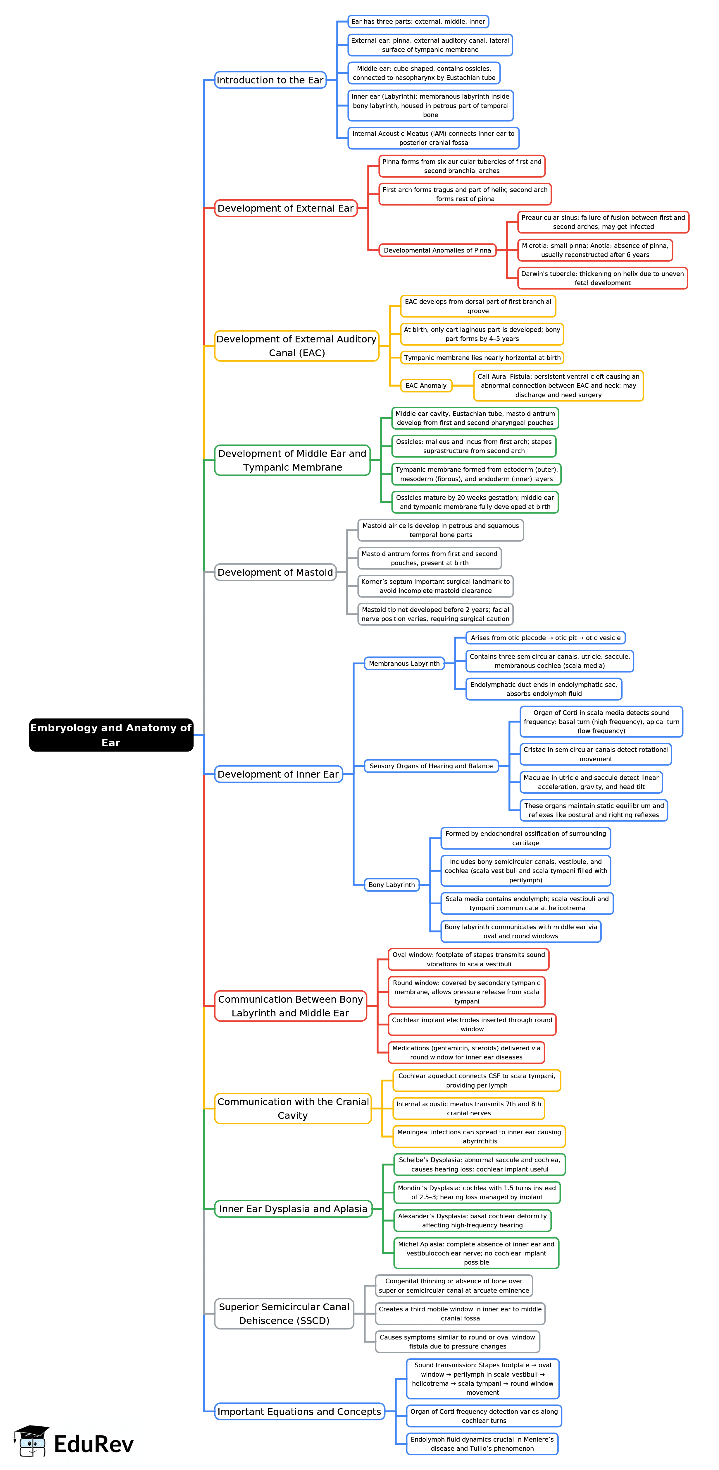
Ans. The ear develops from structures derived from the ectoderm, mesoderm, and endoderm. The outer ear (auricle) primarily originates from the first and second pharyngeal arches, while the middle ear structures arise from the first pharyngeal pouch. The inner ear, including the cochlea and vestibular system, develops from the otic placode, which is an ectodermal structure.
| 2. How does the anatomy of the ear contribute to its functions in hearing and balance? | |
Ans. The ear is divided into three main parts: the outer ear, middle ear, and inner ear. The outer ear collects sound waves and funnels them through the ear canal to the tympanic membrane (eardrum). The middle ear contains ossicles (malleus, incus, stapes) that amplify sound vibrations and transmit them to the inner ear. The inner ear contains the cochlea, which converts these vibrations into neural signals for hearing, and the vestibular system, which helps maintain balance by detecting changes in head position and motion.
| 3. What are common congenital malformations of the ear, and how do they affect hearing? | |
Ans. Common congenital malformations include microtia (small or absent external ear), atresia (absence of the ear canal), and cholesteatoma (abnormal skin growth in the middle ear). These conditions can lead to conductive hearing loss due to obstruction or malformation of the auditory pathway. Early diagnosis and surgical intervention can improve hearing outcomes for affected individuals.
| 4. What role does the tympanic membrane play in the auditory system? | |
Ans. The tympanic membrane, or eardrum, serves as a crucial boundary between the outer and middle ear. It vibrates in response to sound waves, converting these acoustic energy waves into mechanical vibrations. These vibrations are then transmitted through the ossicles to the oval window of the cochlea, where they are transformed into neural signals for interpretation by the brain.
| 5. How can understanding the anatomy of the ear assist in diagnosing ear-related diseases? | |
Ans. A thorough understanding of ear anatomy aids healthcare professionals in diagnosing various conditions, such as otitis media (middle ear infection), hearing loss, and tinnitus. Knowledge of anatomical structures allows for targeted examinations and imaging, leading to accurate diagnoses and effective treatment plans tailored to specific ear diseases.
About this Document

4.73/5 Rating

Apr 26, 2026 Last updated
Related Exams
Document Description: Mind Map: Embryology and Anatomy of Ear -1 for NEET PG 2026 is part of Ear Nose Throat (ENT) preparation. The notes and questions for Mind Map: Embryology and Anatomy of Ear -1 have been prepared according to the NEET PG exam syllabus. Information about Mind Map: Embryology and Anatomy of Ear -1 covers topics like and Mind Map: Embryology and Anatomy of Ear -1 Example, for NEET PG 2026 Exam. Find important definitions, questions, notes, meanings, examples, exercises and tests below for Mind Map: Embryology and Anatomy of Ear -1.
Introduction of Mind Map: Embryology and Anatomy of Ear -1 in English is available as part of our Ear Nose Throat (ENT) for NEET PG & Mind Map: Embryology and Anatomy of Ear -1 in Hindi for Ear Nose Throat (ENT) course. Download more important topics related with notes, lectures and mock test series for NEET PG Exam by signing up for free. NEET PG: Mind Map: Embryology and Anatomy of Ear -1
Description
Mind Map: Embryology & Anatomy of Ear of Ear Nose Throat provides you one-page visual summary of the chapter covering all the important topics. Download the PDF from EduRev.
Information about Mind Map: Embryology and Anatomy of Ear -1
In this doc you can find the meaning of Mind Map: Embryology and Anatomy of Ear -1 defined & explained in the simplest way possible. Besides explaining types of Mind Map: Embryology and Anatomy of Ear -1 theory, EduRev gives you an ample number of questions to practice Mind Map: Embryology and Anatomy of Ear -1 tests, examples and also practice NEET PG tests


Related Searches
MCQs, Free, Previous Year Questions with Solutions, Objective type Questions, Extra Questions, shortcuts and tricks, ppt, Sample Paper, past year papers, mock tests for examination, Exam, Semester Notes, Viva Questions, practice quizzes, pdf , study material, Mind Map: Embryology and Anatomy of Ear -1, video lectures, Summary, Mind Map: Embryology and Anatomy of Ear -1, Important questions, Mind Map: Embryology and Anatomy of Ear -1;