Practice Questions :Biological Bases of Behavior

SECTION I: MULTIPLE CHOICE
Directions
Each of the questions or incomplete statements below is followed by four suggested answers or completions. Select the one that is best in each case and record your choice on the answer sheet provided.
Question 1
Dr. Martinez is studying how different brain regions contribute to memory formation. She uses a brain imaging technique that measures blood flow to determine which areas are most active when participants encode new information.
Which neuroimaging technique is Dr. Martinez most likely using?
- Electroencephalogram (EEG)
- Functional magnetic resonance imaging (fMRI)
- Computed tomography (CT) scan
- Transcranial magnetic stimulation (TMS)
Question 2
A patient suffered damage to a specific brain region following a stroke. Afterward, the patient could understand spoken and written language perfectly but was unable to produce coherent speech. The patient's speech was limited to fragmented phrases and single words, though the patient clearly understood what they wanted to say.
Which brain area was most likely damaged?
- Wernicke's area
- Broca's area
- The primary motor cortex
- The angular gyrus
Question 3
Research Scenario: A neuroscientist administers a drug that increases the reuptake of serotonin in the synaptic cleft. She then measures participants' mood ratings over the following week.
Based on the known function of serotonin, what effect would this drug most likely have?
- Participants would experience elevated mood and increased energy
- Participants would likely experience decreased mood and potential depressive symptoms
- Participants would show improved motor coordination
- Participants would demonstrate enhanced memory consolidation
Question 4
The peripheral nervous system is divided into two main subdivisions. Which of the following correctly identifies these subdivisions and their primary functions?
- The sympathetic nervous system, which activates fight-or-flight responses, and the parasympathetic nervous system, which promotes rest-and-digest functions
- The somatic nervous system, which controls voluntary movements, and the autonomic nervous system, which regulates involuntary bodily functions
- The central nervous system, which processes information, and the peripheral nervous system, which transmits signals
- The afferent division, which sends motor commands, and the efferent division, which receives sensory information
Question 5
A researcher studying addiction examines the role of dopamine pathways in reward processing. She finds that repeated exposure to a particular substance leads to decreased dopamine receptor sensitivity in the nucleus accumbens.
This finding best explains which aspect of addiction?
- Why individuals develop tolerance and require increasing amounts of the substance to achieve the same effect
- Why withdrawal symptoms occur when the substance is discontinued
- Why genetic factors contribute to addiction vulnerability
- Why psychological dependence develops independently of physical dependence
Question 6
Case Study: Following a bicycle accident, Marcus experienced damage to his hippocampus. In the months following the injury, he can recall events from his childhood and teenage years clearly, but he has difficulty forming new long-term memories of daily events.
Marcus's memory pattern best illustrates the role of the hippocampus in which memory process?
- Storage of long-term memories
- Consolidation of new memories from short-term to long-term storage
- Retrieval of procedural memories
- Encoding of sensory information
Question 7
During an action potential, what causes the rapid depolarization phase of the neuron?
- Potassium ions rushing out of the neuron
- Sodium ions rushing into the neuron
- Chloride ions entering the neuron
- Calcium ions being released from internal stores
Question 8
Dr. Chen conducts a study on stress responses. She measures cortisol levels in participants before and after they give a public speech. She finds that cortisol levels increase significantly during the stressful task and gradually return to baseline over the following hour.
Which endocrine gland is primarily responsible for releasing cortisol in response to stress?
- The thyroid gland
- The pituitary gland
- The adrenal glands
- The pineal gland
Question 9
Experimental Results: Researchers use split-brain surgery to treat severe epilepsy in a patient. After recovery, they present the word "KEY" to the patient's left visual field only and ask what was seen.
What response would be expected from this split-brain patient?
- The patient would verbally report seeing the word "KEY"
- The patient would report seeing nothing but could draw a key with their left hand
- The patient would report seeing the word "KEY" and could draw it with either hand
- The patient would verbally report seeing nothing but could pick out a key from objects using their right hand
Question 10
A pharmaceutical company develops a new drug designed to treat anxiety disorders. The drug functions as an agonist for GABA receptors in the brain.
Based on GABA's function as the primary inhibitory neurotransmitter, what effect would this drug most likely produce?
- Increased neuronal firing and heightened arousal
- Decreased neuronal firing and reduced anxiety symptoms
- Enhanced memory consolidation during sleep
- Improved muscle coordination and motor control
Question 11
Twin Study Data: Researchers studying the heritability of intelligence tested 200 pairs of identical twins raised together and 200 pairs of identical twins raised apart. The correlation for IQ scores was \(r = 0.86\) for twins raised together and \(r = 0.72\) for twins raised apart.
What conclusion is best supported by these findings?
- Intelligence is determined entirely by genetic factors, with no environmental influence
- Both genetic and environmental factors contribute to intelligence, with genetics playing a substantial role
- Environmental factors account for most of the variation in intelligence scores
- The heritability of intelligence cannot be determined from twin studies
Question 12
The myelin sheath that covers many neuronal axons serves which primary function?
- Producing neurotransmitters for synaptic transmission
- Increasing the speed of neural impulse transmission
- Providing nutrients to the neuron cell body
- Receiving signals from other neurons
Question 13
A neuroscientist uses a lesioning technique to temporarily deactivate the amygdala in laboratory rats. She then exposes the rats to a stimulus that previously elicited a strong fear response.
What behavioral change would most likely be observed in these rats?
- The rats would show enhanced fear responses and increased avoidance behavior
- The rats would show diminished fear responses and reduced avoidance behavior
- The rats would lose their ability to form new procedural memories
- The rats would demonstrate impaired motor coordination
Question 14
Pharmacological Study: Participants are given a drug that blocks acetylcholine receptors at the neuromuscular junction. Researchers then ask participants to perform various physical tasks.
What effect would this drug most likely produce?
- Improved cognitive function and enhanced memory
- Muscle paralysis or weakness
- Elevated mood and increased energy
- Enhanced sensory perception
Question 15
During a laboratory exercise, students measure the resting membrane potential of a neuron and find it to be approximately -70 mV. They observe that the inside of the neuron is more negatively charged than the outside.
Which factor primarily maintains this resting potential?
- The equal distribution of sodium and potassium ions across the membrane
- The sodium-potassium pump actively transporting ions against their concentration gradients
- The continuous influx of calcium ions through voltage-gated channels
- The passive diffusion of chloride ions into the neuron
Question 16
Evolutionary Psychology Research: A research team examines brain structure differences between humans and other primates. They find that humans have a significantly larger prefrontal cortex relative to overall brain size compared to chimpanzees and other great apes.
This anatomical difference best supports which functional difference between humans and other primates?
- Humans have superior sensory processing abilities
- Humans have enhanced capacity for complex planning, decision-making, and impulse control
- Humans have better motor coordination and physical dexterity
- Humans have more acute emotional responses to environmental stimuli
Question 17
Neuroplasticity refers to the brain's ability to reorganize itself by forming new neural connections throughout life. Which scenario best illustrates neuroplasticity?
- A child's brain producing excess synapses during early development
- A stroke patient regaining speech ability as undamaged areas of the brain assume functions previously performed by damaged regions
- The thinning of the cerebral cortex that occurs with normal aging
- The genetic programming that determines brain structure before birth
Question 18
Hormone Study: Researchers investigate the relationship between testosterone levels and aggressive behavior. They measure baseline testosterone in 150 male participants and then observe their behavior during a competitive task. Results show a moderate positive correlation (\(r = 0.48\), \(p < 0.01\))="" between="" testosterone="" levels="" and="" aggressive="" behaviors="" during="">
What is the most appropriate conclusion based on these findings?
- High testosterone levels cause aggressive behavior in competitive situations
- Aggressive behavior causes increased testosterone production
- There is a significant positive relationship between testosterone levels and aggressive behavior, though causation cannot be determined
- Testosterone has no meaningful relationship with aggression since correlation does not equal causation
Question 19
A patient with damage to the cerebellum is asked to perform a series of tasks including walking in a straight line, touching their nose with their eyes closed, and maintaining balance on one foot.
Which deficit would this patient most likely demonstrate?
- Inability to understand spoken language
- Loss of emotional regulation and increased impulsivity
- Impaired coordination, balance, and fine motor control
- Difficulty forming new long-term memories
Question 20
Genetic Study: A behavioral geneticist uses adoption studies to examine the relative contributions of genetics and environment to the development of schizophrenia. She finds that adopted children whose biological parents had schizophrenia have a significantly higher risk of developing the disorder compared to adopted children whose biological parents did not have schizophrenia, even when raised in similar adoptive environments.
This finding provides the strongest support for which conclusion about schizophrenia?
- Schizophrenia is caused entirely by genetic factors with no environmental influence
- Genetic factors play a significant role in the vulnerability to developing schizophrenia
- Environmental factors are more important than genetic factors in causing schizophrenia
- Adoption itself increases the risk of developing schizophrenia regardless of biological heritage
SECTION II: FREE RESPONSE
Directions
Answer each of the following questions as completely as possible. Each question is designed to assess your understanding of psychological concepts, research methods, and your ability to apply psychological principles to real-world scenarios. Use complete sentences and appropriate psychological terminology in your responses.
FRQ 1: Article Analysis Question (AAQ)
Study Summary: Neural Mechanisms of Fear Extinction
Researchers at a university medical center conducted an experimental study to investigate the role of the prefrontal cortex in fear extinction learning. The study involved 60 healthy adult participants (32 female, 28 male) aged 18-35 years, recruited from the local community through online advertisements. All participants provided informed consent and were screened to exclude individuals with neurological disorders, psychiatric conditions, or current psychotropic medication use.
The study used a fear conditioning and extinction paradigm combined with functional magnetic resonance imaging (fMRI). During the acquisition phase, participants viewed a blue square (conditioned stimulus, CS+) paired with a mild electric shock to the wrist (unconditioned stimulus) on 75% of trials, and a yellow square (CS-) that was never paired with shock. Shock intensity was individually calibrated to each participant's tolerance level, defined as "uncomfortable but not painful." During the extinction phase conducted 24 hours later, both the CS+ and CS- were presented repeatedly without any shocks while brain activity was recorded using fMRI.
The dependent variables were: (1) skin conductance response (SCR) amplitude measured in microsiemens as an index of physiological fear response, and (2) blood-oxygen-level-dependent (BOLD) signal in the ventromedial prefrontal cortex (vmPFC) measured during CS+ presentation.
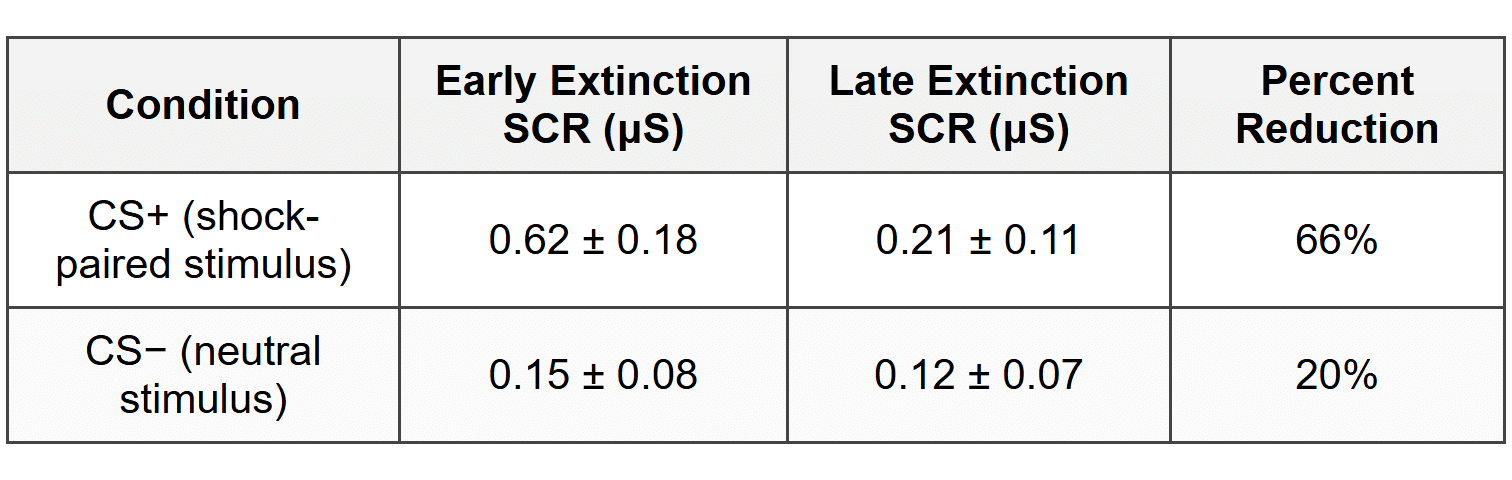
Results showed that during early extinction trials, SCR to the CS+ was significantly elevated (mean = 0.62 μS, SD = 0.18) compared to the CS- (mean = 0.15 μS, SD = 0.08). By late extinction, SCR to the CS+ decreased significantly (mean = 0.21 μS, SD = 0.11), indicating successful extinction learning. Neuroimaging data revealed that vmPFC activation increased progressively across extinction trials (\(r = 0.71\), \(p < 0.001\)).="" furthermore,="" participants="" showing="" greater="" vmpfc="" activation="" during="" late="" extinction="" demonstrated="" more="" complete="" fear="" reduction,="" with="" a="" significant="" negative="" correlation="" between="" vmpfc="" activity="" and="" scr="" amplitude="" (\(r="-0.58\)," \(p=""><>
Table 1: Mean Skin Conductance Response Across Experimental Phases
The researchers concluded that the ventromedial prefrontal cortex plays a critical role in inhibiting fear responses during extinction learning, supporting previous animal models. Participants were debriefed following the study and given contact information for psychological services if they experienced any distress. The study was approved by the institutional review board and followed APA ethical guidelines.
Based on the study summary above, answer the following questions:
- Identify the research method used in this study.
- State an operational definition of one key variable measured in this study.
- Describe what the data in Table 1 indicate about the success of the extinction procedure.
- Identify one ethical guideline that the researchers followed in conducting this study.
- Explain the extent to which the findings can be generalized to a broader population, using specific evidence from the study description.
- Explain how the finding regarding vmPFC activation supports the concept of top-down emotional regulation.
FRQ 2: Evidence-Based Question (EBQ)
Study A: Neurogenesis and Exercise
Van Praag and colleagues (1999) investigated the effects of voluntary exercise on adult neurogenesis in the hippocampus of mice. Mice were randomly assigned to either an enriched environment with running wheels or standard housing. After several weeks, researchers measured the number of newly generated neurons in the dentate gyrus of the hippocampus using cellular markers. Results showed that mice with access to running wheels produced significantly more new neurons (approximately 2-fold increase) compared to controls. Additionally, the exercising mice demonstrated superior performance on spatial learning tasks, including the Morris water maze. The researchers concluded that physical activity stimulates neurogenesis and may contribute to enhanced cognitive function.
Study B: Stress and Hippocampal Volume
Bremner and colleagues (1997) used magnetic resonance imaging (MRI) to measure hippocampal volume in military veterans with post-traumatic stress disorder (PTSD) compared to combat veterans without PTSD. Findings revealed that veterans with PTSD had significantly smaller right hippocampal volumes (8% reduction on average) compared to the control group. The degree of volume reduction correlated with the severity of PTSD symptoms and the duration of trauma exposure. The researchers proposed that chronic stress and elevated cortisol levels associated with PTSD may damage hippocampal neurons, leading to structural changes. This finding suggests that prolonged stress can have neurotoxic effects on brain structures critical for memory.
Study C: Enriched Environments and Brain Plasticity
Rosenzweig and Bennett (1996) conducted experiments examining the effects of environmental complexity on brain development in rats. Rats were assigned to either enriched environments (containing toys, tunnels, and opportunities for social interaction) or impoverished environments (isolated cages with minimal stimulation). After several weeks, neuroanatomical analysis revealed that rats in enriched environments developed thicker cerebral cortices, increased dendritic branching, and higher concentrations of neurotransmitters compared to rats in impoverished conditions. The enriched-environment rats also performed better on problem-solving tasks. These results demonstrated that environmental experiences can produce measurable structural and functional changes in the brain, supporting the principle of experience-dependent plasticity.
Based on the three studies summarized above, answer the following questions:
- Identify one commonality in the focus or methodology across all three studies.
- Evaluate the consistency of findings across the three studies regarding the relationship between environmental or experiential factors and brain structure or function.
- Apply the concept of neuroplasticity to explain the pattern of results observed across these three studies.
ANSWER KEY
Part A: Multiple Choice Answer Table
Part B: Free Response Answer Key
FRQ 1 - Answer Key
Part A: Identify the research method used in this study
The research method used in this study is an experiment (or controlled experiment). The researchers manipulated the independent variable (presence or absence of shock paired with the conditioned stimulus) and measured its effects on dependent variables (skin conductance response and brain activation) while using random assignment and controlling extraneous variables.
Part B: State an operational definition of one key variable measured in this study
One acceptable operational definition: Fear response was operationally defined as the skin conductance response (SCR) amplitude measured in microsiemens (μS) in response to presentation of the conditioned stimulus.
Alternative acceptable operational definition: Fear extinction was operationally defined as the decrease in skin conductance response amplitude from early extinction trials to late extinction trials when the CS+ was presented without shock.
Part C: Describe what the data in Table 1 indicate about the success of the extinction procedure
The data in Table 1 indicate that the extinction procedure was successful. The CS+ (shock-paired stimulus) showed a substantial reduction in skin conductance response from early extinction (0.62 μS) to late extinction (0.21 μS), representing a 66% decrease in physiological fear response. In contrast, the CS- (neutral stimulus) showed minimal change (only 20% reduction from an already low baseline). This differential pattern demonstrates that participants successfully learned that the CS+ no longer predicted shock, resulting in extinction of the conditioned fear response.
Part D: Identify one ethical guideline that the researchers followed in conducting this study
Acceptable answers include any of the following:
- The researchers obtained informed consent from all participants before the study began
- The researchers provided debriefing after the study and gave participants contact information for psychological services
- The researchers used screening procedures to exclude vulnerable populations (those with neurological or psychiatric conditions)
- The study received institutional review board (IRB) approval ensuring ethical oversight
- The researchers calibrated shock intensity to each participant's tolerance to minimize harm (protection from harm)
Part E: Explain the extent to which the findings can be generalized to a broader population, using specific evidence from the study description
The generalizability of these findings is moderately limited due to several sample characteristics. The sample consisted of only 60 participants recruited from a local community, which is a relatively small sample size that may not represent broader demographic diversity. Additionally, the sample was limited to healthy adults aged 18-35 years with no neurological disorders, psychiatric conditions, or medication use. This strict exclusion criteria means the findings may not generalize to older adults, individuals with mental health conditions, or those taking psychotropic medications. The gender balance (32 female, 28 male) was relatively even, which strengthens generalizability across sex. However, the use of community volunteers recruited through online advertisements may introduce self-selection bias, as participants may differ systematically from those who do not volunteer for research studies. Therefore, while the findings likely apply to healthy young adults, caution should be exercised in generalizing to clinical populations or other age groups.
Part F: Explain how the finding regarding vmPFC activation supports the concept of top-down emotional regulation
The finding that vmPFC activation increased during extinction and negatively correlated with fear responses supports the concept of top-down emotional regulation, which refers to the use of higher-order cognitive processes in the prefrontal cortex to modulate or inhibit emotional responses generated by subcortical structures like the amygdala. The ventromedial prefrontal cortex is a region involved in executive control and cognitive regulation of emotion. The data showed that as vmPFC activity increased across extinction trials, physiological fear responses (SCR) decreased, with a significant negative correlation (\(r = -0.58\)). This pattern suggests that the vmPFC actively inhibits or suppresses the fear response during extinction learning. Rather than fear simply fading passively, the prefrontal cortex exerts cognitive control to override the previously learned fear association. This demonstrates top-down processing because a higher-level cortical region (vmPFC) is regulating activity in lower-level emotional circuits, allowing participants to cognitively learn that the stimulus is now safe despite prior conditioning.
FRQ 2 - Answer Key
Part A: Identify one commonality in the focus or methodology across all three studies
Acceptable answers include:
Commonality in focus: All three studies investigated how environmental factors, experiences, or conditions influence brain structure and function. Each study examined the relationship between external or behavioral variables (exercise, stress, environmental enrichment) and measurable changes in neural anatomy or neurochemistry.
Alternative commonality: All three studies used animal models (mice or rats) or involved measurement of brain structures (hippocampus, cerebral cortex) to investigate neurobiological mechanisms underlying behavior and cognition.
Alternative commonality: All three studies provided evidence for brain plasticity - the capacity of the nervous system to change in response to experience or environmental conditions.
Part B: Evaluate the consistency of findings across the three studies regarding the relationship between environmental or experiential factors and brain structure or function
The findings across all three studies are highly consistent in demonstrating that environmental and experiential factors produce measurable changes in brain structure and function. Study A found that exercise (a positive environmental factor) increased neurogenesis and enhanced cognitive performance. Study C similarly found that enriched environments produced structural brain changes including thicker cortices and increased dendritic branching, along with improved problem-solving abilities. Both of these studies show that positive, stimulating environments enhance brain development and function. Study B, examining the opposite direction, found that negative environmental factors (chronic stress and trauma) were associated with reduced hippocampal volume and cognitive impairment. Taken together, these studies consistently support a bidirectional relationship: enriching, positive experiences promote neural growth and cognitive enhancement, while impoverished or stressful conditions are associated with neural damage or reduced development. The consistency across different populations (rodents and humans), different brain regions (hippocampus and cerebral cortex), and different environmental manipulations strengthens the conclusion that experience shapes brain structure.
Part C: Apply the concept of neuroplasticity to explain the pattern of results observed across these three studies
Neuroplasticity refers to the brain's capacity to change its structure and function in response to experience, learning, and environmental input throughout the lifespan. This concept directly explains the pattern of results across all three studies. In Study A, voluntary exercise stimulated the growth of new neurons in the hippocampus (neurogenesis), demonstrating that physical activity can trigger plastic changes at the cellular level that enhance cognitive function. In Study C, rats exposed to enriched environments showed increased cortical thickness and dendritic branching, illustrating experience-dependent plasticity - the brain physically reorganizes in response to environmental complexity and stimulation. In Study B, chronic stress was associated with reduced hippocampal volume, showing that neuroplasticity can also work in a negative direction when the environment is harmful; prolonged exposure to stress hormones can damage neural structures. All three studies demonstrate that the brain is not static but continuously adapts to environmental demands. Positive experiences (exercise, enrichment) promote neural growth and complexity, while negative experiences (chronic stress) can lead to neural deterioration. This plasticity represents an evolutionary adaptation allowing organisms to optimize brain function based on environmental challenges and opportunities, supporting survival and adaptive behavior.