NEET PG Exam > NEET PG Notes > Anatomy > CheatSheet: Thorax
CheatSheet: Thorax


1. Thoracic Wall
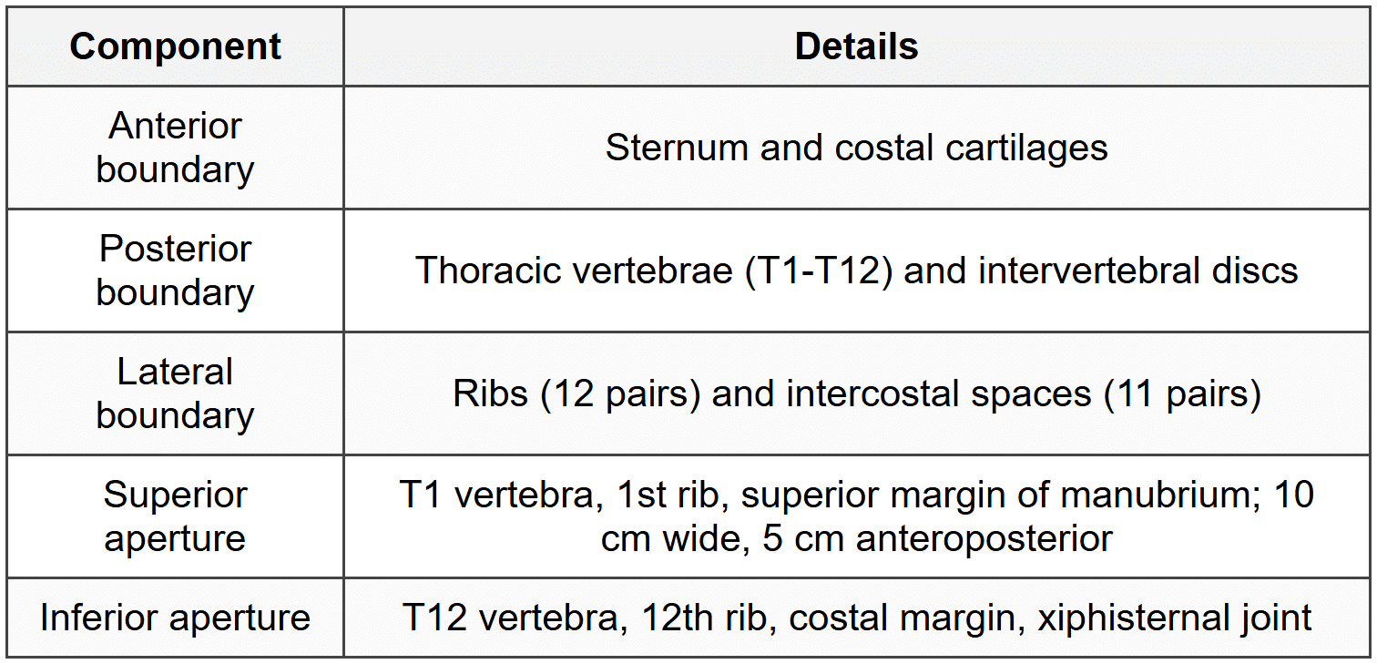
1.1 Boundaries and Components
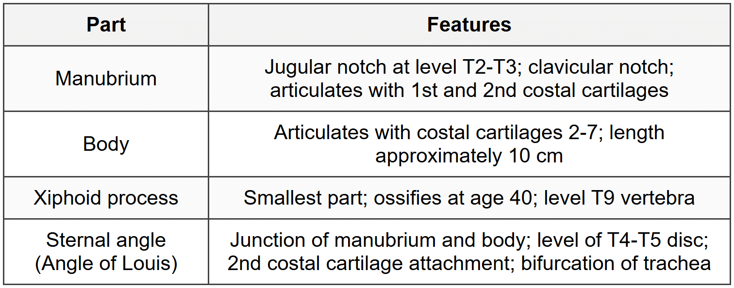
1.2 Sternum
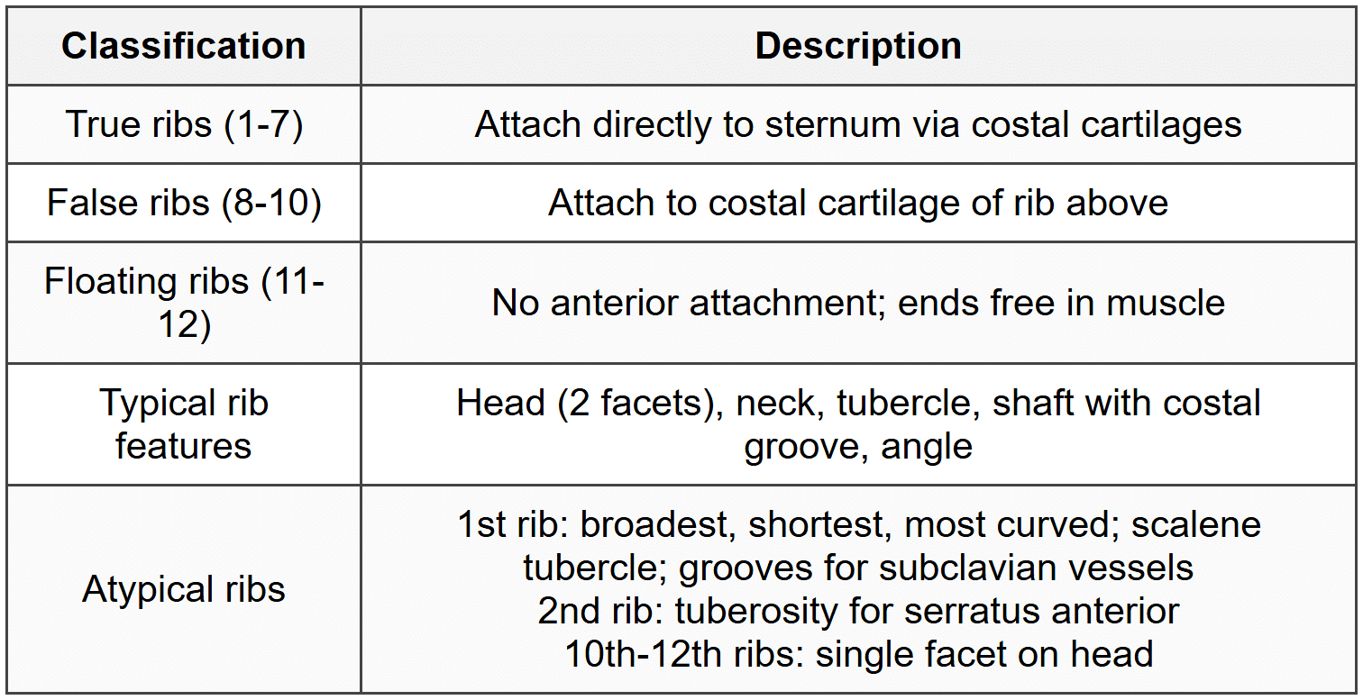
1.3 Ribs
1.4 Intercostal Spaces
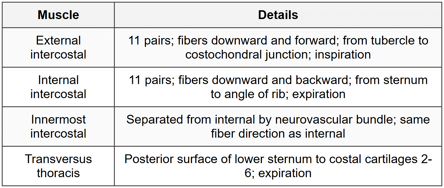
1.4.1 Intercostal Muscles
1.4.2 Neurovascular Bundle
- Position: Between internal intercostal and innermost intercostal
- Order (superior to inferior): Vein, Artery, Nerve (VAN)
- Location: Costal groove in upper part of intercostal space
- Safe zone for thoracentesis: Just above upper border of lower rib
1.4.3 Intercostal Arteries
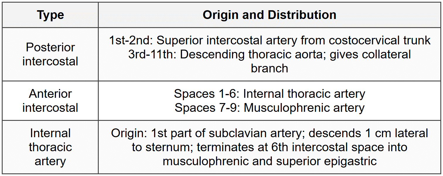
1.4.4 Intercostal Nerves
- Anterior rami of thoracic spinal nerves T1-T11
- T1: Contributes to brachial plexus; small 1st intercostal nerve
- T2-T6: True intercostal nerves
- T7-T11: Thoracoabdominal nerves (supply anterior abdominal wall)
- Branches: Collateral, lateral cutaneous (anterior and posterior divisions), anterior cutaneous
- Dermatomes: T4-nipple, T6-xiphoid, T10-umbilicus
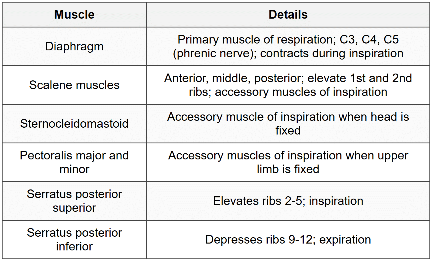
1.5 Muscles of Thoracic Wall
2. Mediastinum
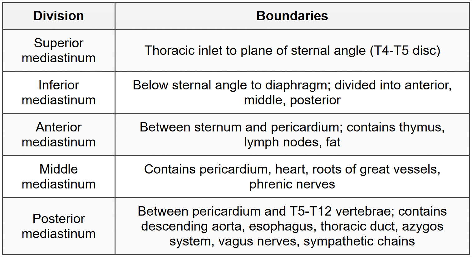
2.1 Divisions and Boundaries
2.2 Superior Mediastinum Contents
2.2.1 Thymus
- Location: Posterior to manubrium sterni in anterior superior mediastinum
- Maximum size at puberty (30-40 g); involutes after puberty
- Blood supply: Internal thoracic and inferior thyroid arteries
- Two lobes connected by connective tissue
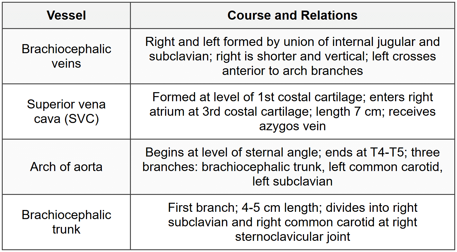
2.2.2 Great Vessels
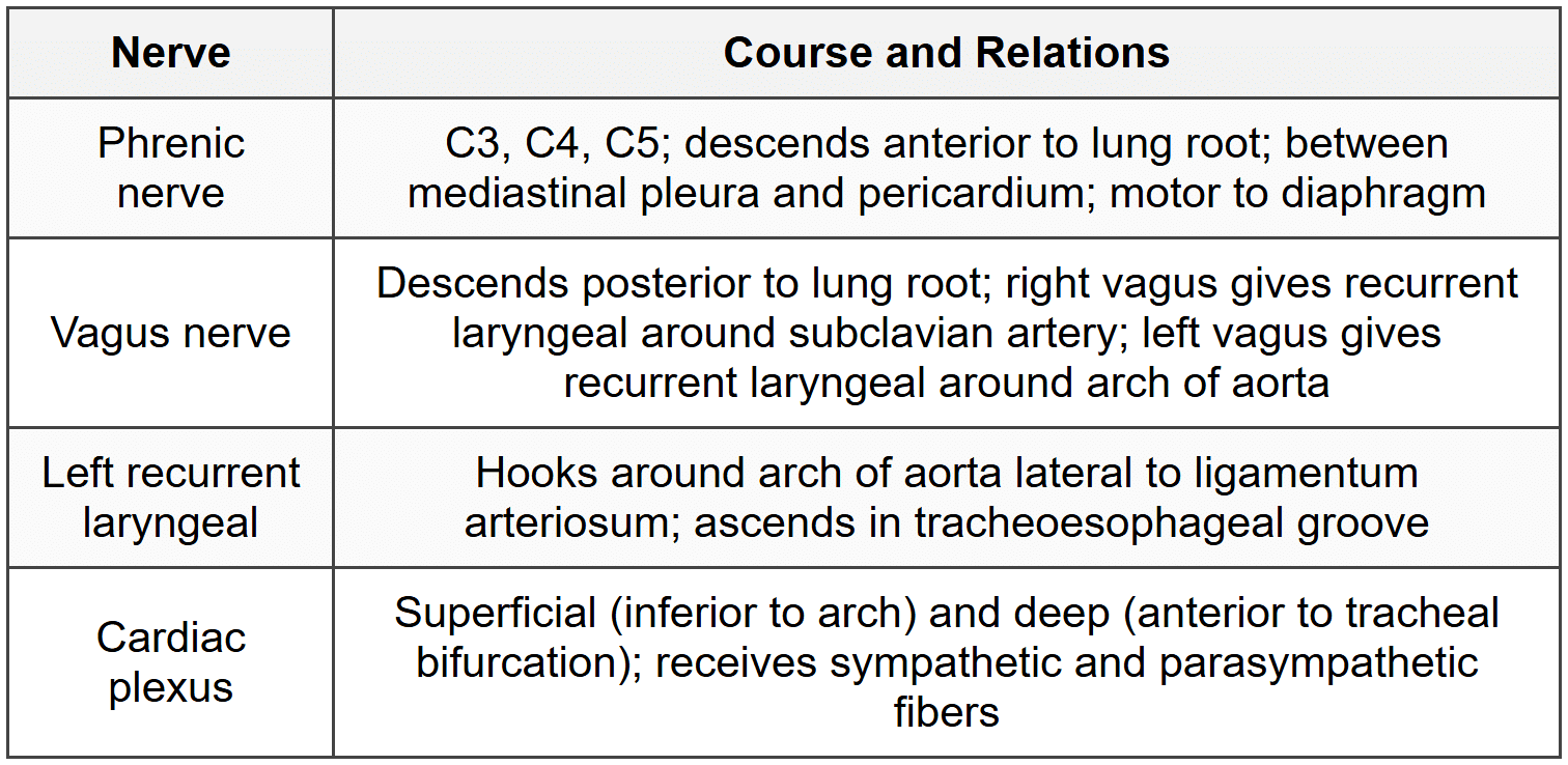
2.2.3 Nerves
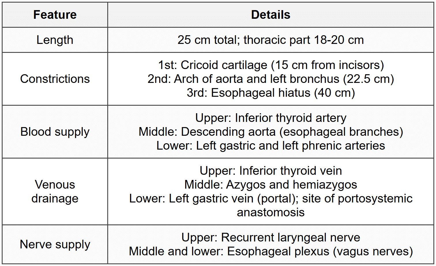
2.2.4 Trachea and Esophagus
- Trachea: 10-12 cm length; 16-20 C-shaped cartilages; bifurcates at T4-T5 (carina); right main bronchus wider, shorter, more vertical
- Esophagus in superior mediastinum: Posterior to trachea; related to left recurrent laryngeal nerve in tracheoesophageal groove
2.3 Posterior Mediastinum
2.3.1 Descending Thoracic Aorta
- Begins at T4-T5; ends at T12 (aortic hiatus); length 20 cm
- Branches: Posterior intercostal arteries (9 pairs), subcostal arteries, bronchial arteries (2 left, 1 right), esophageal arteries (4-5), superior phrenic arteries
- Relations: Esophagus (initially right, crosses anteriorly, then left); azygos vein on right; hemiazygos on left
2.3.2 Esophagus
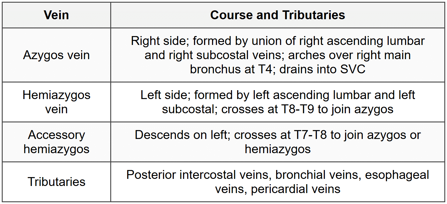
2.3.3 Azygos Venous System
2.3.4 Thoracic Duct
- Origin: Cisterna chyli at L1-L2
- Enters thorax through aortic hiatus at T12
- Ascends in posterior mediastinum between aorta (left) and azygos vein (right)
- Crosses to left at T5 level
- Terminates at junction of left internal jugular and left subclavian veins
- Length: 38-45 cm; largest lymphatic vessel
2.3.5 Sympathetic Trunk
- 12 pairs of thoracic ganglia
- Located on heads of ribs
- Branches: White and gray rami communicantes, splanchnic nerves
- Greater splanchnic nerve: T5-T9 ganglia; pierces diaphragm; ends in celiac ganglion
- Lesser splanchnic nerve: T10-T11 ganglia; ends in aorticorenal ganglion
- Least splanchnic nerve: T12 ganglion; ends in renal plexus
3. Heart and Pericardium
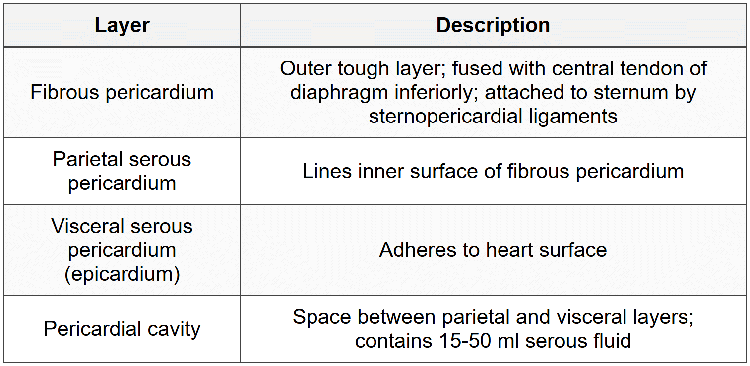
3.1 Pericardium
3.1.1 Layers
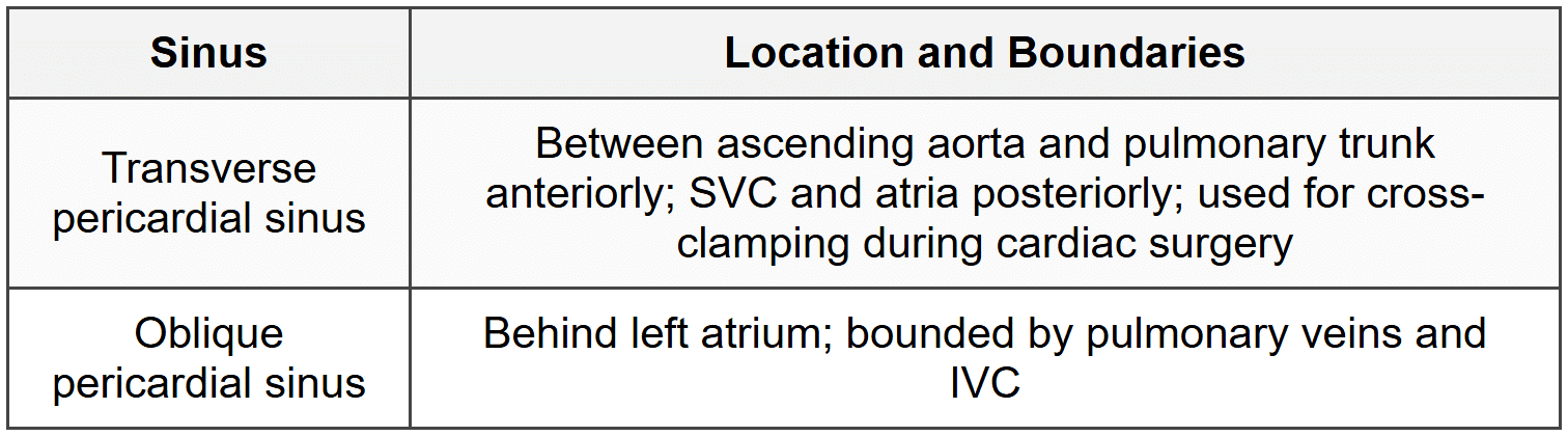
3.1.2 Pericardial Sinuses
3.1.3 Blood Supply and Nerve Supply
- Arterial: Pericardiophrenic artery (internal thoracic), descending thoracic aorta, superior phrenic artery
- Venous: Pericardiophrenic veins to brachiocephalic veins
- Nerve: Phrenic nerve (C3, C4, C5); fibrous pericardium highly sensitive to pain
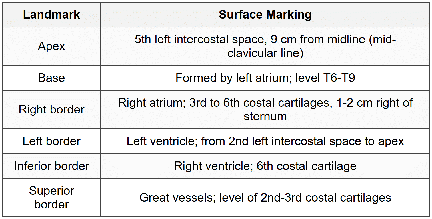
3.2 Heart Position and Surface Anatomy
3.3 Heart Chambers
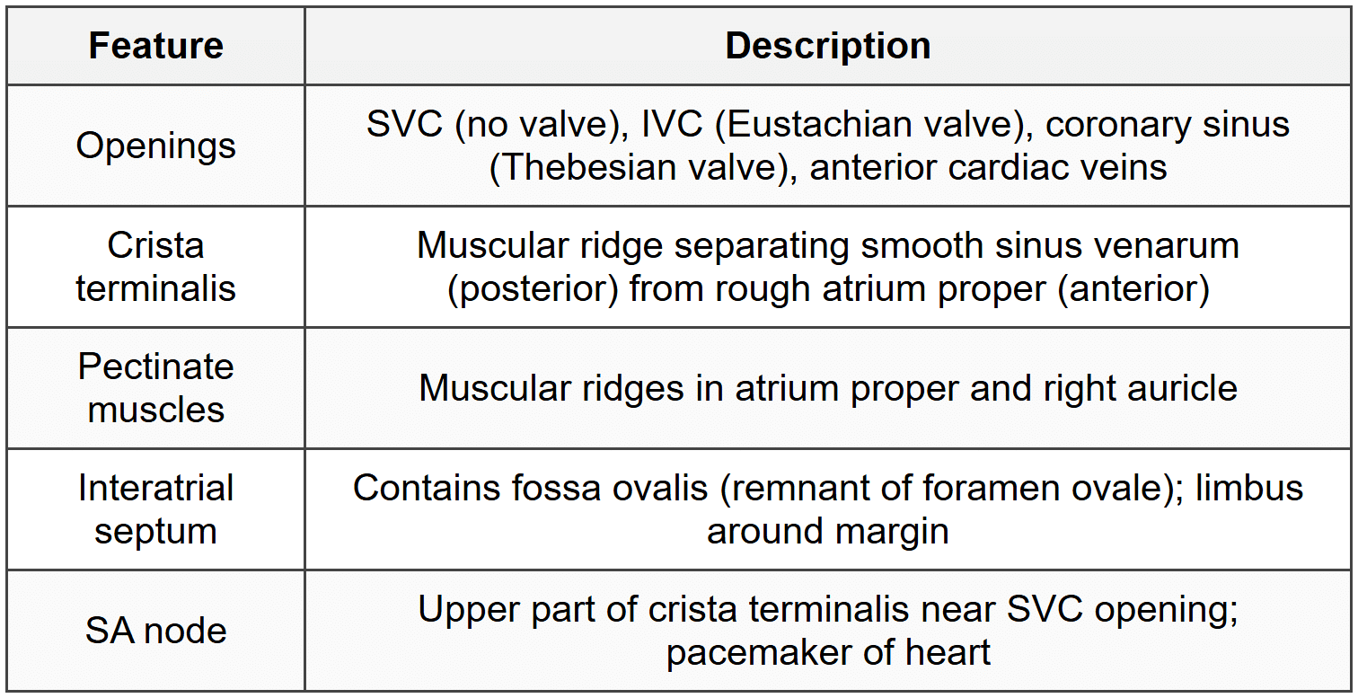
3.3.1 Right Atrium
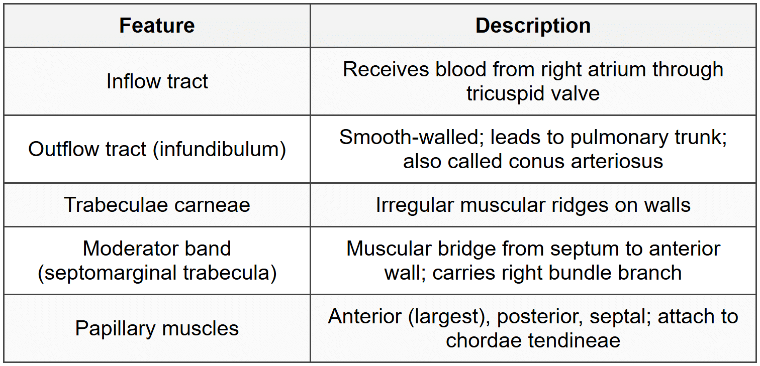
3.3.2 Right Ventricle
3.3.3 Left Atrium
- Forms base of heart
- Receives 4 pulmonary veins (2 right, 2 left); no valves
- Smooth-walled except left auricle (has pectinate muscles)
- Anterior wall related to transverse sinus
- Posterior wall related to esophagus
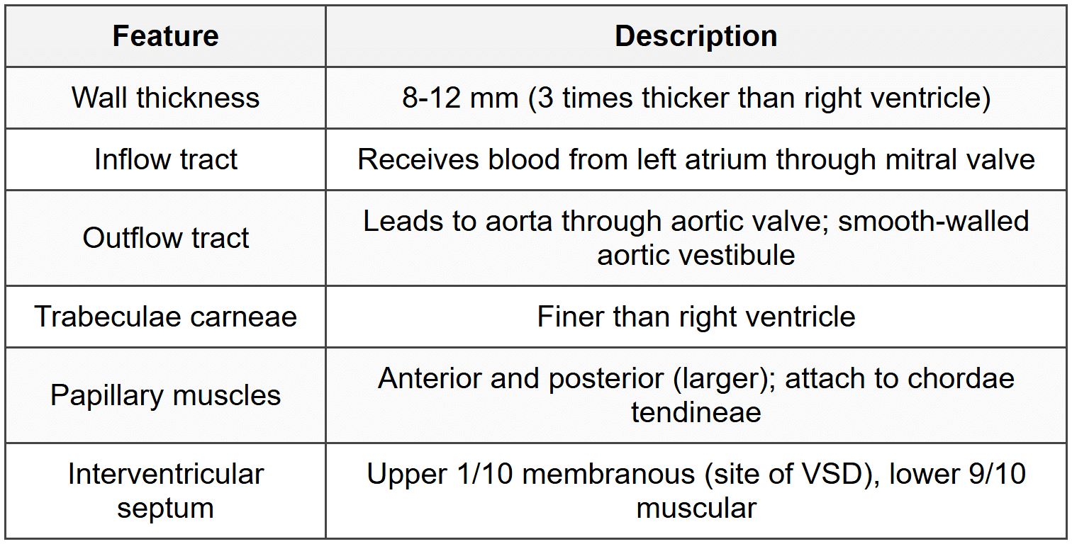
3.3.4 Left Ventricle
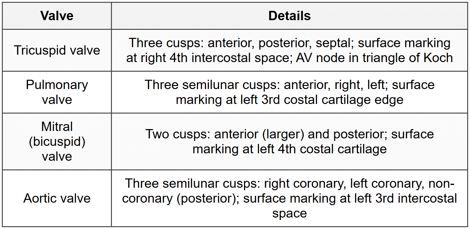
3.4 Heart Valves
3.5 Coronary Circulation
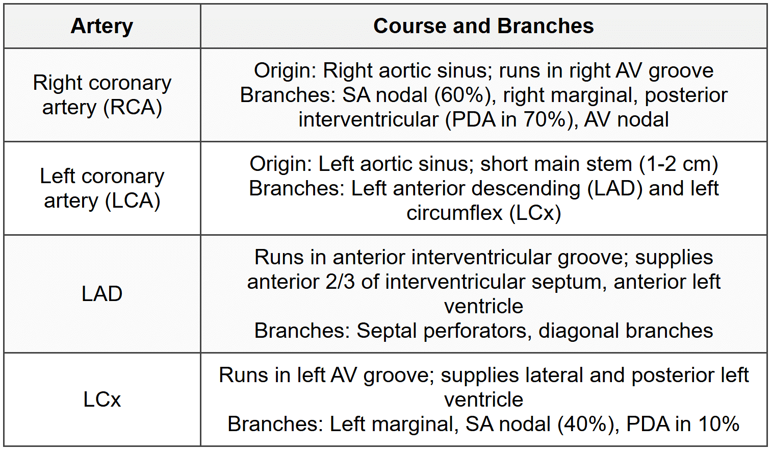
3.5.1 Coronary Arteries
3.5.2 Coronary Dominance
- Right dominant (70%): PDA from RCA
- Left dominant (10%): PDA from LCx
- Co-dominant (20%): Dual supply
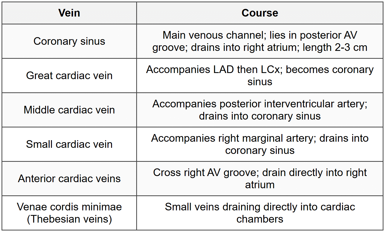
3.5.3 Venous Drainage
3.6 Conducting System
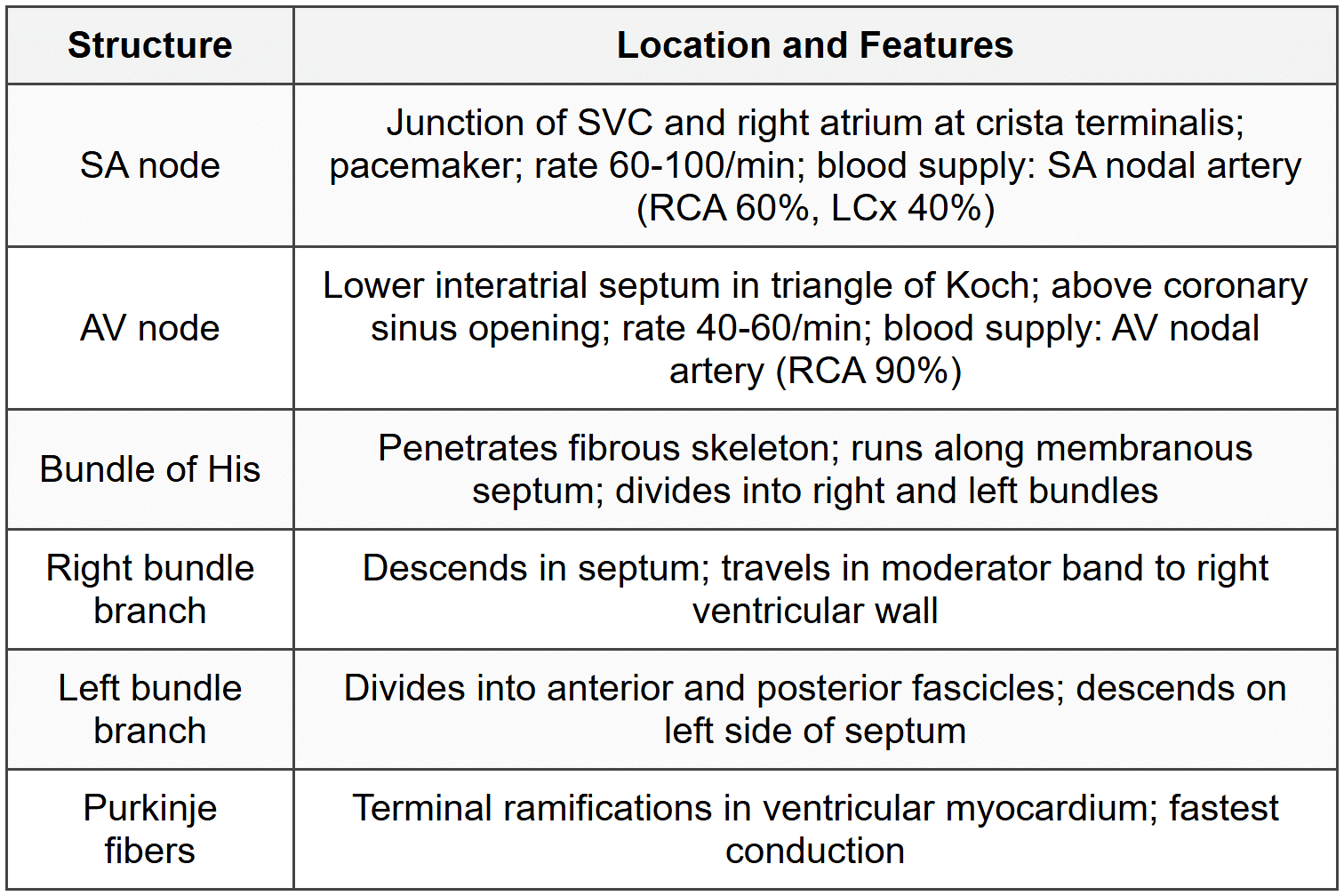
3.7 Nerve Supply of Heart
- Sympathetic: Cervical and upper thoracic sympathetic ganglia; increases heart rate and contractility
- Parasympathetic: Vagus nerve (CN X); decreases heart rate; right vagus to SA node; left vagus to AV node
- Cardiac plexus: Superficial (below arch of aorta) and deep (anterior to tracheal bifurcation)
- Pain fibers: Sympathetic pathway; referred pain to left shoulder and medial left arm (T1-T4 dermatomes)
4. Pleura and Lungs
4.1 Pleura
4.1.1 Types and Extent
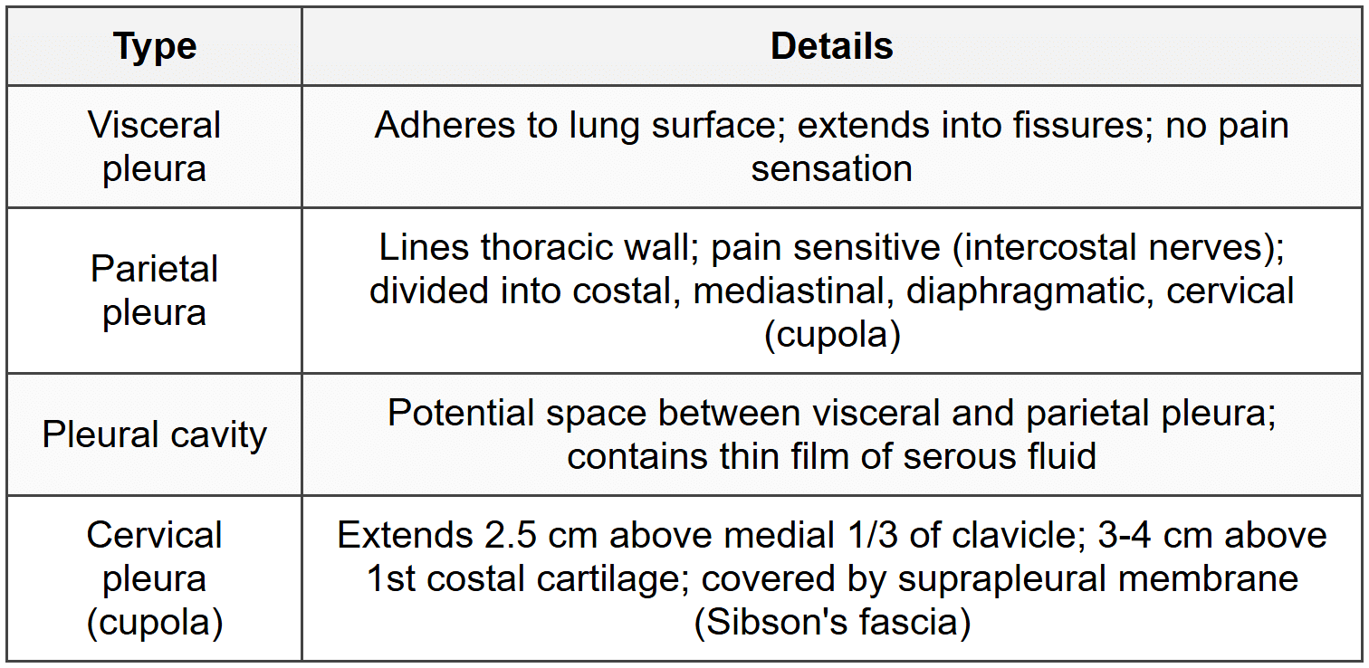
4.1.2 Pleural Recesses
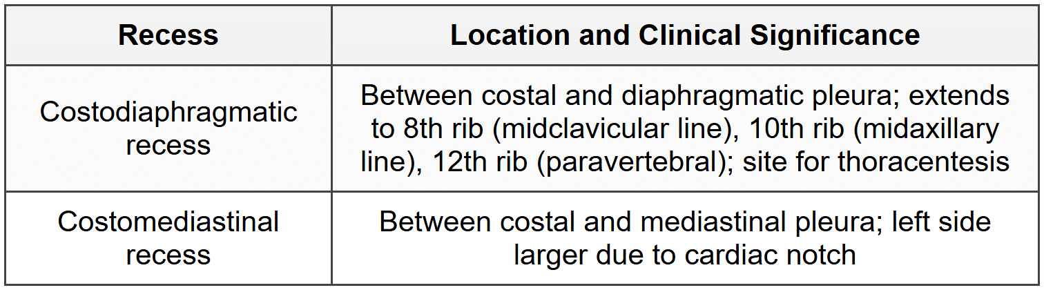
4.1.3 Surface Markings
- Apex: 2.5 cm above medial 1/3 of clavicle
- Anterior border: Both pleurae meet at sternal angle; right continues to 6th costal cartilage; left deviates laterally at 4th costal cartilage (cardiac notch)
- Lower border: 6th rib (midclavicular line), 8th rib (midaxillary line), 10th rib (paravertebral line)
- Lung lower border: 2 ribs higher than pleural reflection (6th, 8th, 10th ribs at respective lines)
4.2 Lungs
4.2.1 General Features
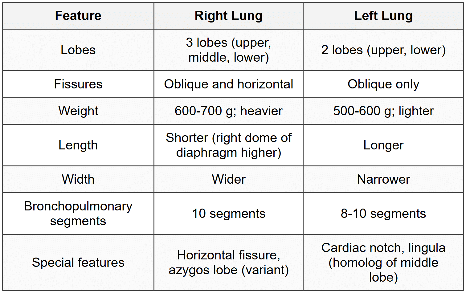
4.2.2 Surfaces and Borders
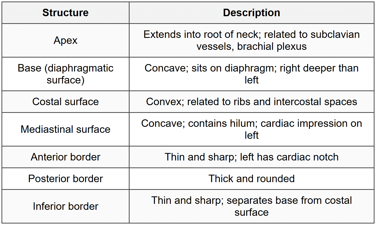
4.2.3 Fissures
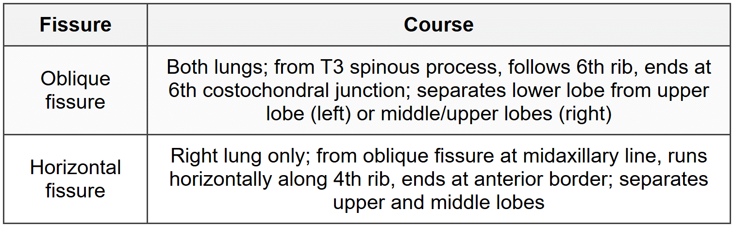
4.2.4 Lung Root and Hilum
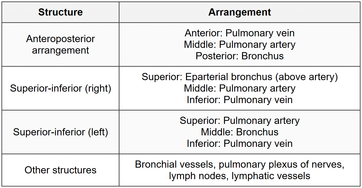
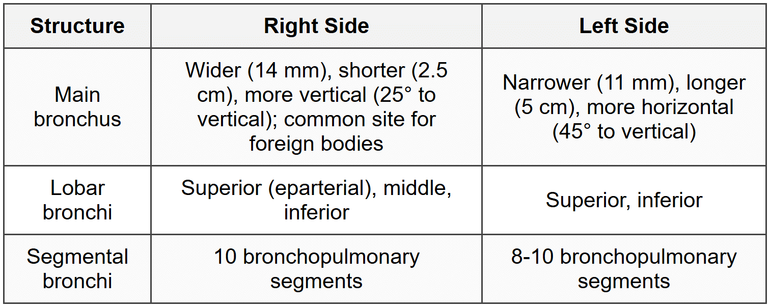
4.2.5 Bronchial Tree
4.2.6 Bronchopulmonary Segments - Right Lung
- Upper lobe: Apical (1), posterior (2), anterior (3)
- Middle lobe: Lateral (4), medial (5)
- Lower lobe: Apical/superior (6), medial basal (7), anterior basal (8), lateral basal (9), posterior basal (10)
4.2.7 Bronchopulmonary Segments - Left Lung
- Upper lobe: Apicoposterior (1+2), anterior (3), superior lingular (4), inferior lingular (5)
- Lower lobe: Apical/superior (6), anteromedial basal (7+8), lateral basal (9), posterior basal (10)
4.3 Pulmonary Vasculature
4.3.1 Pulmonary Arteries
- Pulmonary trunk: Origin from right ventricle; length 5 cm; bifurcates at T5-T6 level (below aortic arch)
- Right pulmonary artery: Longer; passes behind ascending aorta and SVC; anterior to right bronchus
- Left pulmonary artery: Shorter; connected to aortic arch by ligamentum arteriosum; superior to left bronchus
- Carry deoxygenated blood to lungs
4.3.2 Pulmonary Veins
- 4 veins (2 from each lung); no valves
- Superior and inferior pulmonary veins from each lung
- Drain into left atrium
- Carry oxygenated blood from lungs
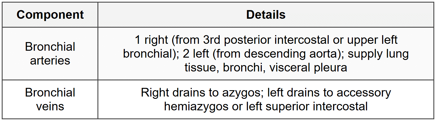
4.3.3 Bronchial Circulation
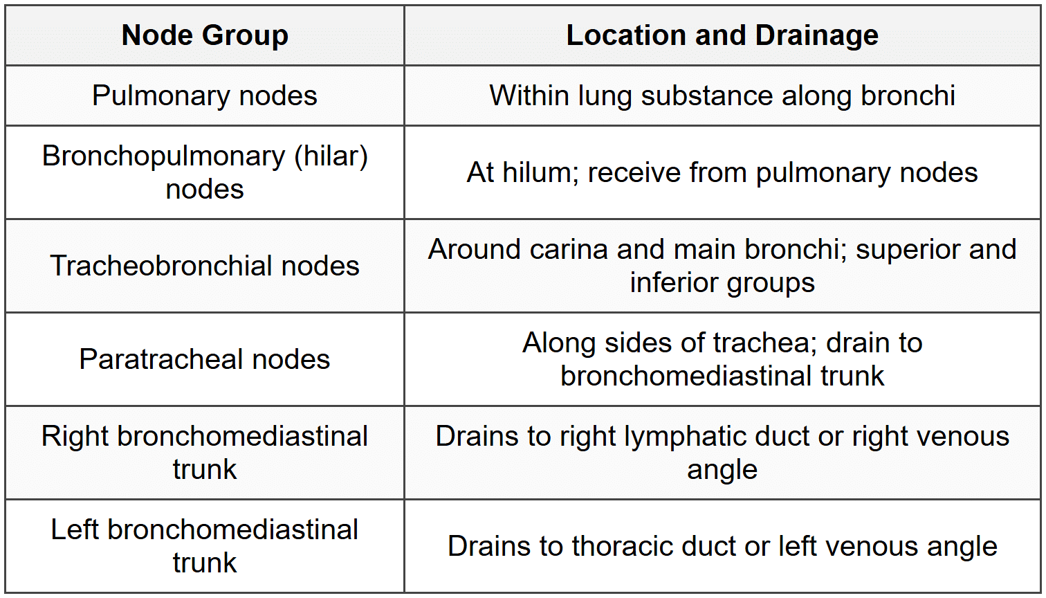
4.4 Lymphatic Drainage
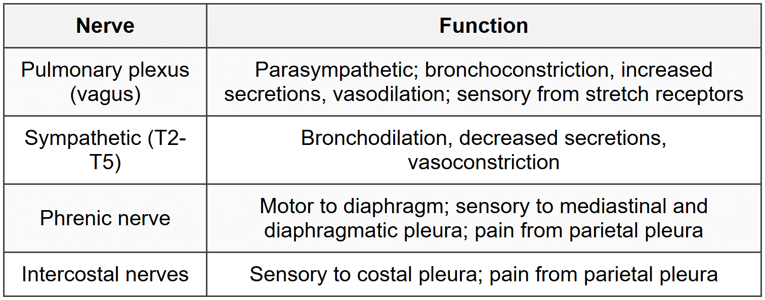
4.5 Nerve Supply
5. Diaphragm
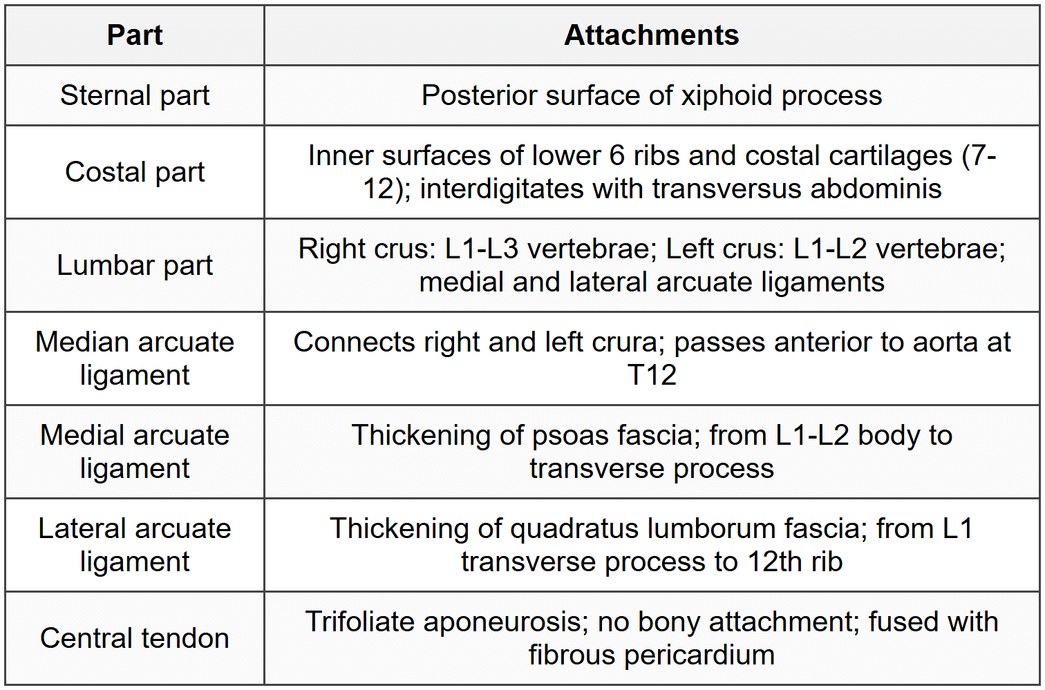
5.1 Attachments and Parts
5.2 Openings
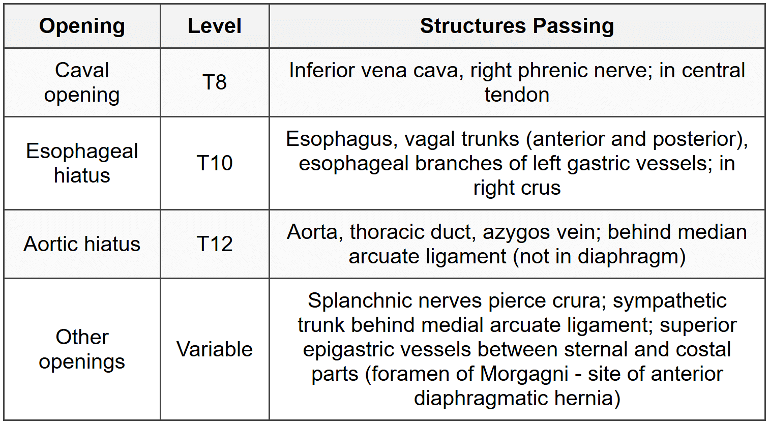
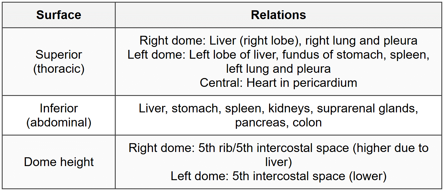
5.3 Relations
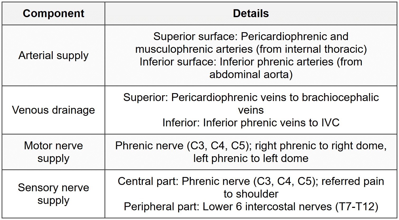
5.4 Blood Supply and Nerve Supply
5.5 Functions and Clinical Points
- Primary muscle of respiration; contraction increases vertical diameter of thorax
- Contributes 75% of inspiratory effort
- Assists in increasing intra-abdominal pressure (defecation, micturition, parturition, vomiting)
- Congenital diaphragmatic hernia: Posterolateral (Bochdalek) most common; anterolateral (Morgagni) rare
- Hiatus hernia: Sliding (95%) or paraesophageal (5%)
- Right dome higher than left by 2.5 cm due to liver
6. Clinical Correlations
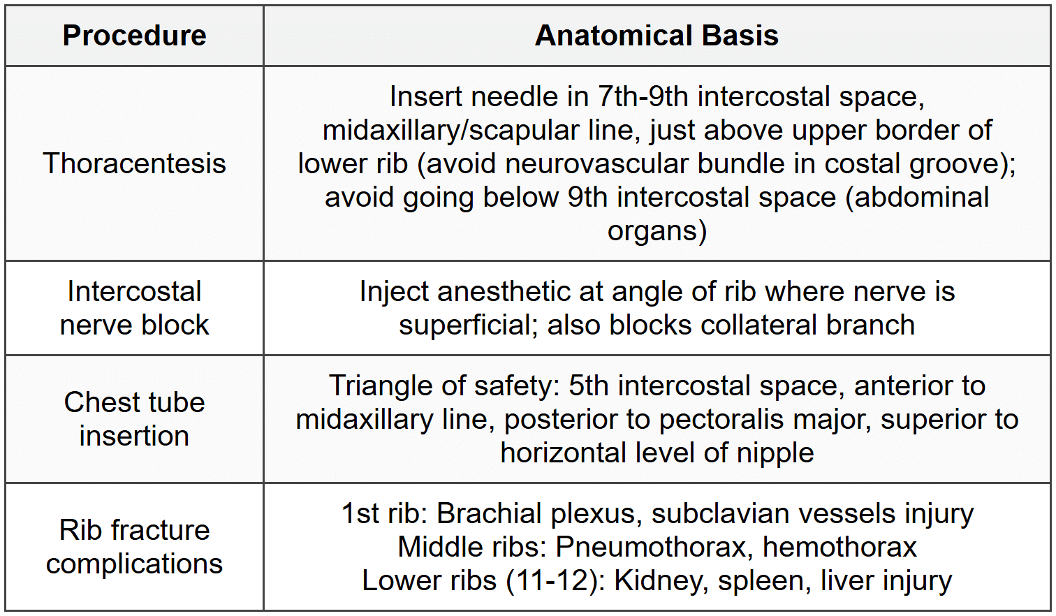
6.1 Thoracic Wall Procedures
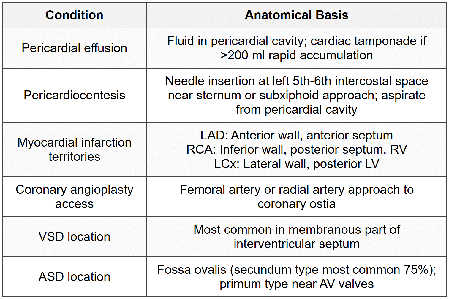
6.2 Cardiac Clinical Correlations
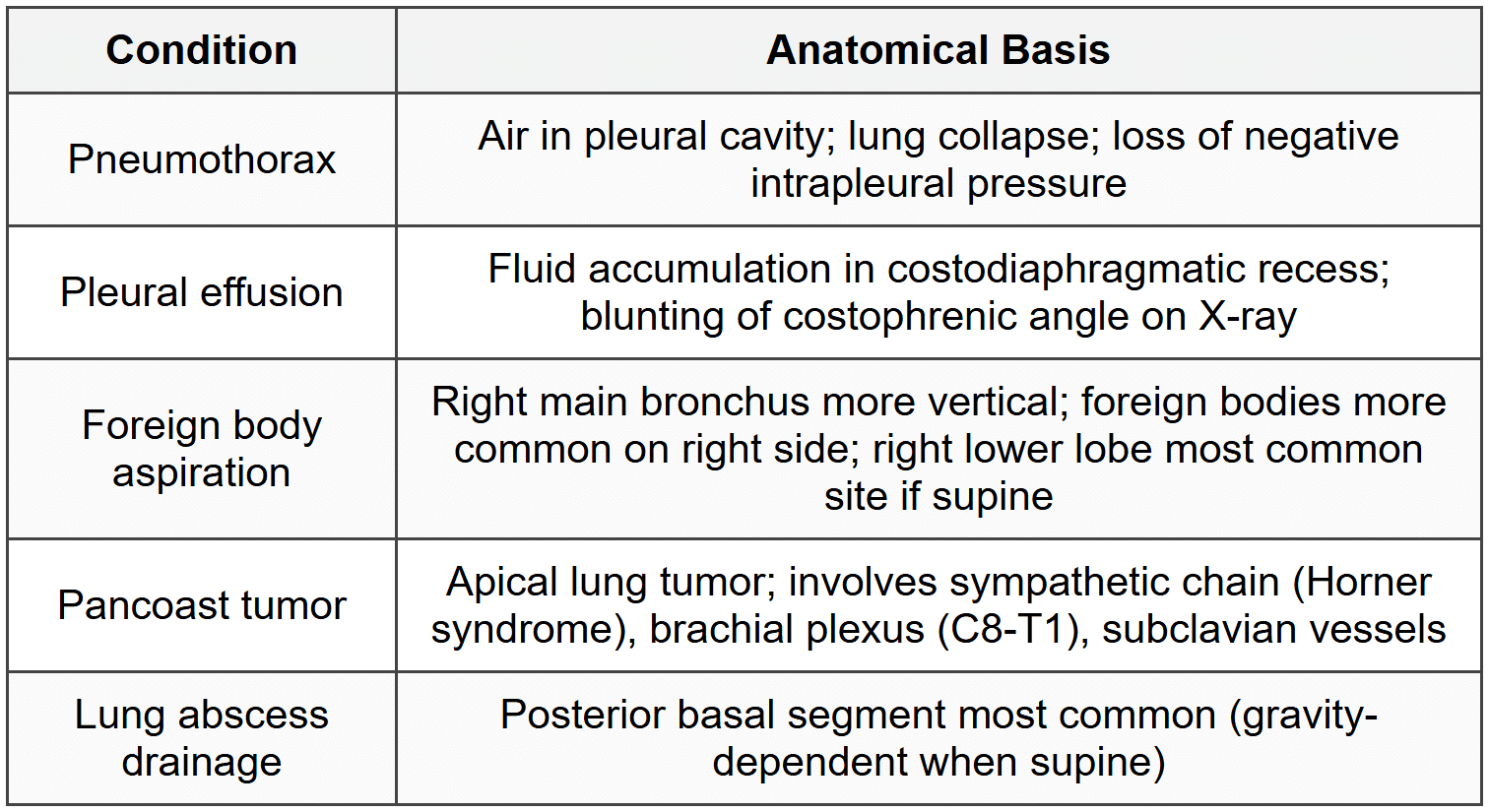
6.3 Respiratory Clinical Correlations
6.4 Surface Landmarks
- Sternal angle (Angle of Louis): T4-T5 disc; 2nd costal cartilage; tracheal bifurcation; aortic arch; azygos vein entry to SVC
- Jugular notch: T2-T3 vertebral level
- Xiphisternal joint: T9 vertebral level
- Cardiac apex beat: 5th left intercostal space, 9 cm from midline (mid-clavicular line)
- Nipple: 4th intercostal space in males
- Inferior angle of scapula: T7 vertebral level
About this Document

4.78/5 Rating

Apr 19, 2026 Last updated
Related Exams
Document Description: CheatSheet: Thorax for NEET PG 2026 is part of Anatomy preparation. The notes and questions for CheatSheet: Thorax have been prepared according to the NEET PG exam syllabus. Information about CheatSheet: Thorax covers topics like and CheatSheet: Thorax Example, for NEET PG 2026 Exam. Find important definitions, questions, notes, meanings, examples, exercises and tests below for CheatSheet: Thorax.
Description
CheatSheet: Thorax of Anatomy to help you remember important concepts with short tricks. Start learning for NEET PG exam & improve retention with EduRev.
Information about CheatSheet: Thorax
In this doc you can find the meaning of CheatSheet: Thorax defined & explained in the simplest way possible. Besides explaining types of CheatSheet: Thorax theory, EduRev gives you an ample number of questions to practice CheatSheet: Thorax tests, examples and also practice NEET PG tests


Related Searches
ppt, Free, CheatSheet: Thorax, Exam, MCQs, Objective type Questions, CheatSheet: Thorax, Sample Paper, video lectures, Extra Questions, pdf , Summary, Important questions, mock tests for examination, past year papers, Semester Notes, CheatSheet: Thorax, study material, shortcuts and tricks, practice quizzes, Previous Year Questions with Solutions, Viva Questions;