NCERT Based Activity: Cell : The Building Block of Life

Activity 2.1 : Let us estimate the size of a cell
1. Take a transparent ruler with millimetre (mm) markings.
2. Place the ruler on the stage of microscope, focus on it using the adjustment knob and observe the diameter of the circular field of view through the eyepiece and measure it in mm.
3. Convert the diameter from mm to micrometre (μm). Suppose the diameter of the visible field is 5 mm, meaning 5 × 1000 = 5000 μm.
4. Remove the ruler and place an onion peel slide on the stage of the microscope.
5. Focus on the slide and count the number of cells present along the diameter of the field of view in one straight line.
6. Estimate the real size of the cell using the formula:
Estimated size of the onion peel cell = Diameter of the visible field in micrometre ÷ Number of cells along the diameter
(Unit conversion: 1 millimetre (mm) = 1000 micrometre (μm))
Observation
Suppose 25 cells are seen along the diameter. In that case, the size of one onion cell would be 5000 μm/25 = 200 μm.
Explanation
A microscope allows us to see very small structures clearly and is an essential tool for studying cell structure. The total magnification of a microscope depends on the magnifying power of the eyepiece and the objective lens. If both have magnifying power of 10X, the total magnification is 100X - meaning a cell of estimated size 200 μm will appear 100 times larger.
Activity 2.2 : Let us experiment
1. With the help of a kitchen knife, carefully cut a potato into two pieces of roughly equal size(fig.).
2. Measure and record the initial weight of both the pieces using a weighing balance.
3. Put one piece of the potato in Beaker A with plain water.
4. Put the other piece of the potato in Beaker B with 20 per cent salt or sugar solution.
5. Leave them undisturbed for about an hour or until you notice a visible change in the size of the pieces.
6. Measure and record the final weight of each piece.
7. Calculate the difference between the initial and their final weights.
What do you observe? You may observe that -
- Beaker A - The potato piece swells.
- Beaker B - The potato piece shrinks.
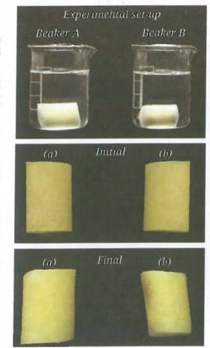 Experimental set-up, and initial and final states of potato pieces in (a) plain water, and (b) 20 per cent salt solution
Observation
- Beaker A - The potato piece swells.
- Beaker B - The potato piece shrinks.
The weight of the potato piece in Beaker A has increased, while the weight of the potato piece in Beaker B has decreased.
Explanation
The cell membrane allows water to move in and out of the cell but not the sugar or salt molecules. Water moves from an area with more water and less solute (dilute solution) to an area with less water and more solute (concentrated solution) until the concentrations in the two areas become equal. This movement of water through a selectively permeable membrane is called osmosis. Diffusion is the net movement of particles from a higher to a lower concentration. Osmosis is the diffusion of water across a selectively permeable membrane.
Activity 2.3 : Let us investigate
1. Prepare temporary slides of a thin peel of an onion leaf or a Rhoeo (Cradle lily) leaf and mount it with safranin using cover slip to observe plant cells under a microscope.
2. Similarly, prepare a temporary slide of cheek cells by gently scraping the inner side of your cheek with a cotton swab or the blunt end of a toothpick.
3. Spread the cheek cells on a clean glass slide.
4. Add a drop of water followed by a few drops of methylene blue stain and carefully place a coverslip.
5. Observe both the slides under a microscope.
What do you observe? Onion peel cells (Fig. a) or Rhoeo leaf peel cells are box-shaped and regularly arranged, whereas cheek cells are irregularly arranged (Fig. b). Why do you think this difference exists?
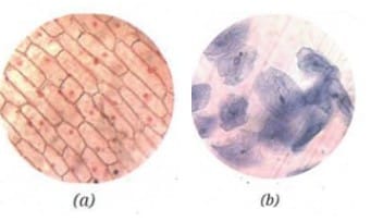 (a) The onion peel cells, and (b) human cheek cells
Prepare two slides of a Rhoeo leaf peel and human cheek cells again, and put 20 per cent sugar solution on them. Observe them under a microscope after half an hour. What do you observe? You must have observed that the boundaries of the plant cells remain the same but their inner content shrinks, and the space between the inner and outer boundaries increases (Fig. a and b). You may observe that the cheek cells, on the other hand, have shrunk considerably.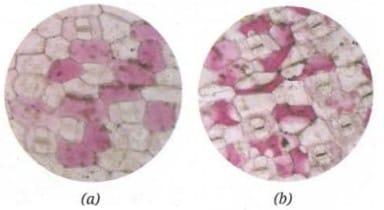 Cradle lily leaf peel cells (a) in water, and (b) in 20 per cent sugar solution
Observation
- Onion peel cells / Rhoeo leaf peel cells are box-shaped and regularly arranged.
- Cheek cells are irregularly arranged.
- Plant cells - boundaries remain the same but inner content shrinks; space between inner and outer boundaries increases (plasmolysis).
- Cheek cells - have shrunk considerably.
Explanation
Plant cells have a rigid cell wall outside the cell membrane. When placed in a concentrated sugar solution, plant cells lose water due to osmosis - the cell membrane pulls away from the cell wall, but the overall shape is maintained by the rigid wall. Animal cells like cheek cells do not have a cell wall, so they lose water and shrink completely when placed in a concentrated solution. This activity demonstrates the role of the cell wall in maintaining the shape and structure of plant cells.
Activity 2.4 : Let us study
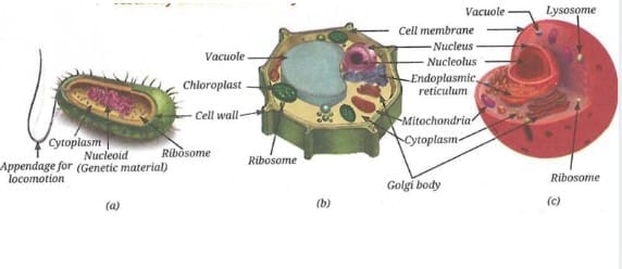 (a) A typical bacterial cell, (b) a typical plant cell, and (c) a typical animal cell
1. Study the given diagrams of a bacterial cell, a plant cell, and an animal cell (Fig. a, b and c).
2. Observe the different structures present in each of them.
3. Record your observations in Table .
| S. No. | Cell structures | Bacterial cell | Plant cell | Animal cell |
|---|---|---|---|---|
| 1. | Cell membrane | |||
| 2. | Cell wall | |||
| 3. | Cytoplasm | |||
| 4. | Well-defined nucleus (genetic material enclosed by a membrane) | |||
| 5. | Primitive nucleus / nucleoid (genetic material without membrane around it) | |||
| 6. | Membrane-bound organelles |
Observation
- Roots are observed growing from the base of the onion bulb after 5-6 days.
- Under the microscope, cells of the onion root tip are not all similar in structure.
- Some cells show different structural arrangements, corresponding to different stages of cell division.
- Cells at different stages of division are visible - some with clearly visible chromosomes, some mid-division, and some that appear normal.
Explanation
The cells of a growing tip of root of onion divide continuously. This process is called cell division. The cells exhibit different structures corresponding to different stages of cell division. Every day, an estimated hundreds of billions of cells in our body are replaced, which is almost 1 per cent of the total number of cells in our body. Both prokaryotic and eukaryotic cells divide, but eukaryotic cells divide in a more controlled and orderly manner by a process called the cell cycle. There are two major types of cell division - mitosis and meiosis. Mitosis is important for normal growth, repair, maintenance and asexual reproduction, while meiosis is important for sexual reproduction and creation of genetic diversity.
Activity 2.5 : Let us enhance our skills
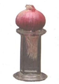 Growing roots of an onion in a jar containing water
1. Take a jar and fill it up with plain water.
2. Now, place an onion bulb over the jar in such a way that its base, bearing roots, is immersed in the water.
3. Leave the setup for 5-6 days and observe. Do you observe the roots growing? Cut 2-3 cm of the freshly grown roots and transfer them to freshly prepared aceto-alcohol (glacial acetic acid : ethanol :: 1:3). Keep the root tips in aceto-alcohol for 24 hours and then transfer them to 70 per cent ethanol (for preservation).
4. Take one or two preserved roots, wash them in water and then place them on a clean slide.
5. Put one drop of dilute Hydrochloric acid (HCl) on the root tips to soften the tissue. Rinse the roots after 10-15 minutes. Then add 2-3 drops of aceto-carmine stain on them.
6. Leave the slide for 5-10 minutes and then, gently warm it (with caution) over a spirit lamp.
7. Cut the tip portion of the root on the slide and put a coverslip. Gently squash the coverslip with your thumb to spread the cells on the slide.
8. Observe the slide under a microscope.
What do you observe? Do you observe the cells of the onion root tip? Are they similar in structure? Do you find any structural differences in these cells? If yes, why is it so?
Observation
The cells of a growing tip of the root of onion divide continuously. This process is called cell division. The cells exhibit different structures corresponding to different stages of cell division. Various stages of cell division are visible under the microscope.
Explanation
The cells of the onion root tip are at different stages of cell division because the root tip is a region of active growth. Both prokaryotic and eukaryotic cells divide, but eukaryotic cells divide in a more controlled and orderly manner by a process called the cell cycle. Every day, hundreds of billions of cells in our body are replaced - almost 1 per cent of the total number of cells in our body.
FAQs on NCERT Based Activity: Cell : The Building Block of Life
| 1. What is a cell? |  |
| 2. What are the key components of a cell? | |
| 3. How can the size of a cell be estimated? | |
| 4. What is the significance of studying cells in biology? | |
| 5. How do cells differ from one another? | |