NCERT Solutions: Body Fluids & Circulation

Q1: Name the components of the formed elements in the blood and mention one major function of each of them.
Ans: The formed elements of blood are:
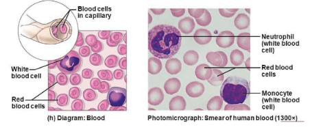
(i) Erythrocytes (red blood cells): These are the most abundant blood cells and contain the red pigment haemoglobin. Their primary function is to transport oxygen from the lungs to body tissues and to carry a small amount of carbon dioxide back to the lungs. In mammals, mature erythrocytes lack a nucleus and are produced in the bone marrow. Normal count is about 4-6 million cells per cubic millimetre of blood; their lifespan in humans is roughly 120 days.
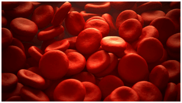 Erythrocytes
(ii) Leukocytes (white blood cells): Colourless cells that protect the body against infections and foreign substances. They are divided into:
1. Granulocytes: contain visible granules; include neutrophils (major phagocytic cells that destroy microbes), eosinophils (involved in allergic responses and defence against parasites) and basophils (release substances that promote inflammation).
2. Agranulocytes: lack visible cytoplasmic granules; include lymphocytes (mount specific immune responses and produce antibodies) and monocytes (phagocytic cells that remove debris and pathogens). Together, leukocytes provide immune defence and surveillance against infection.
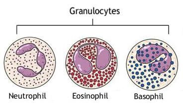 Granulocytes
(iii) Platelets (thrombocytes): Small, irregular cell fragments derived from megakaryocytes that contain chemicals essential for blood clotting. Their principal role is to plug injured blood vessels and initiate coagulation, thereby preventing excessive blood loss. A normal platelet count is about 150,000-400,000 per cubic millimetre of blood.
 Platelets
Q2: What is the importance of plasma proteins?
Ans: Plasma is the fluid component of blood (about 55% of blood volume). About 6.8% of plasma is made up of proteins. The major plasma proteins and their functions are:
- Albumin: the most abundant plasma protein; synthesised in the liver. It maintains the colloid osmotic (oncotic) pressure of blood and thereby helps retain fluid within the blood vessels, preventing excessive fluid loss into tissues.
- Globulins: a group that includes antibodies (immunoglobulins). They play a key role in immune defence by recognising and neutralising pathogens and by helping with transport of some molecules.
- Fibrinogen: a soluble plasma glycoprotein produced by the liver. On activation during injury, it is converted into fibrin, which forms the meshwork of a blood clot and thus is essential for blood coagulation. Fibrinogen also helps in wound healing by forming a temporary matrix for tissue repair.
 Plasma Protein
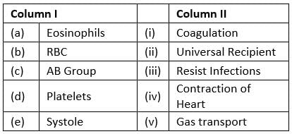
Ans:
Q4: Why do we consider blood as a connective tissue?
Ans: Connective tissues have cells embedded in an extracellular matrix and connect or support other body parts.
Blood is considered a connective tissue because:
- It is derived from the mesoderm during embryonic development, like other connective tissues.
- It has an extracellular matrix called plasma in which formed elements (RBCs, WBCs and platelets) are suspended.
- It links and supports body systems by transporting oxygen, nutrients, hormones and waste products between tissues, thereby connecting and integrating the functions of different organs.
 Connective Tissue
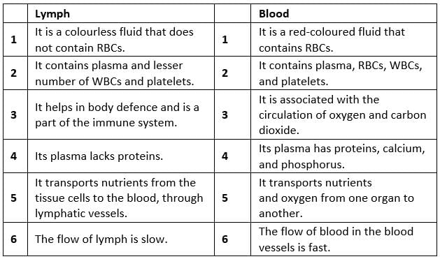
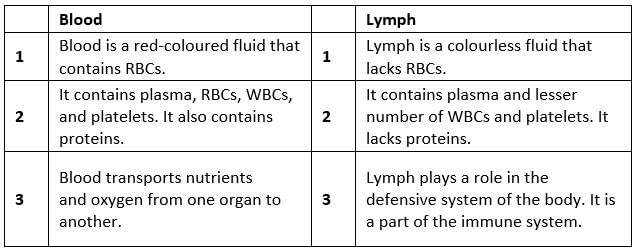
Q5: What is the difference between lymph and blood?
Ans:
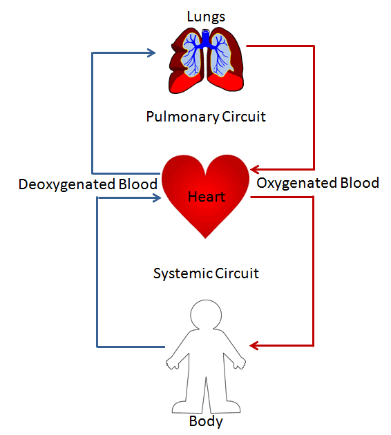
Q6: What is meant by double circulation? What is its significance?
Ans: Double circulation means that blood passes through the heart twice during one complete circuit of the body: once to be pumped to the lungs for oxygenation and once to be pumped to the body tissues.
 Double Circulation
- Pulmonary circulation: Deoxygenated blood is pumped from the right ventricle to the lungs via the pulmonary artery. In the lungs the blood receives oxygen and releases carbon dioxide. Oxygenated blood returns to the left atrium via the pulmonary veins.
- Systemic circulation: Oxygenated blood is pumped from the left ventricle into the aorta and delivered through arteries and capillaries to body tissues. Deoxygenated blood from tissues is collected by veins and returned to the right atrium via the superior and inferior venae cavae.
In birds and mammals, the heart is completely divided into four chambers (two atria and two ventricles), so oxygenated and deoxygenated blood remain separate. This enables an efficient supply of oxygen to tissues and supports higher metabolic activity.
Significance of double circulation: It ensures better separation of oxygenated and deoxygenated blood, allowing tissues to receive highly oxygenated blood. As a result, systemic circulation can operate at a higher pressure than pulmonary circulation, which improves the delivery of oxygen and nutrients and supports more efficient respiration and metabolism, especially in endothermic animals.
Q7: Write the differences between:
(a) Blood and Lymph
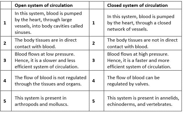
(b) Open and Closed system of circulation
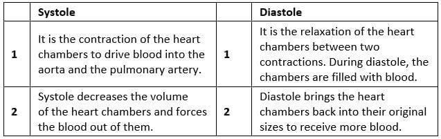
(c) Systole and Diastole
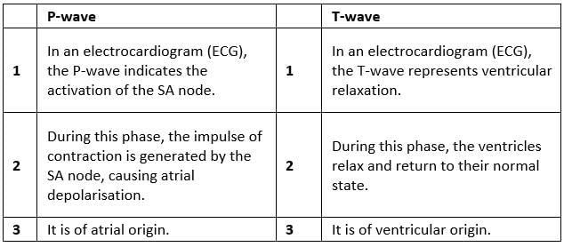
(d) P-wave and T-wave
Ans: (a) Blood and lymph
(b) Open and closed systems of circulation
(c) Systole and diastole
(d) P-wave and T-wave
Q8: Describe the evolutionary change in the pattern of heart among the vertebrates.
Ans: The heart shows increasing structural complexity during vertebrate evolution:
Fishes:
- 2-chambered heart (one atrium, one ventricle).
- Blood follows a single circulation route: heart → gills (oxygenation) → body → heart.
Amphibians and most reptiles:
- 3-chambered heart (two atria, one ventricle).
- There is incomplete separation of oxygenated and deoxygenated blood in the single ventricle, so some mixing occurs; this suits their metabolic needs and often supports cutaneous respiration in amphibians.
Crocodiles, birds and mammals:
- 4-chambered heart (two atria, two ventricles).
- Complete separation of oxygenated and deoxygenated blood enables efficient double circulation, supporting high metabolic demands and enabling sustained activity and high rates of oxygen delivery.
Q9: Why do we call our heart myogenic?
Ans: The heart is called myogenic because its rhythmical contraction is initiated by specialised heart muscle cells themselves rather than by nervous tissue. The sino-atrial (SA) node, located in the right atrium, generates spontaneous electrical impulses that set the heartbeat. These impulses spread through the conduction system of the heart and cause coordinated contractions. Thus, the initiating signal originates in the heart muscle, making the heart myogenic. Many vertebrates and some molluscs also have myogenic hearts.
Q10: Sino-atrial node is called the pacemaker of our heart. Why?
Ans: The sino-atrial (SA) node is called the pacemaker because:
- It automatically generates electrical impulses that start each heartbeat.
- These impulses spread across the atria and trigger their contraction in a coordinated manner, ensuring efficient atrial emptying into the ventricles.
- It sets the normal heart rhythm by producing impulses at a regular rate, typically about 70-75 per minute in a resting adult.
- Because it controls the timing of heartbeats, other parts of the conduction system follow the SA node's rhythm and coordinate ventricular contraction.
For these reasons the SA node is essential for initiating and regulating the heart's rhythmic activity.
Q11: What is the significance of atrio-ventricular node and atrio-ventricular bundle in the functioning of heart?
Ans: The atrioventricular (AV) node is located at the junction between the atria and the ventricles. Its functions include:
- Receiving the excitation wave from the atria and introducing a short delay before passing it on. This delay allows the atria to complete their contraction and empty blood into the ventricles before the ventricles contract.
- Giving rise to the bundle of His (atrioventricular bundle), which conducts impulses from the AV node down the interventricular septum.
- The bundle divides into right and left branches that terminate in a network of Purkinje fibres, which distribute the impulse rapidly through the ventricular myocardium and ensure a coordinated ventricular contraction from apex to base.
Together, the AV node and the bundle of His ensure orderly timing and efficient contraction of the ventricles following atrial excitation, which is essential for effective pumping of blood.
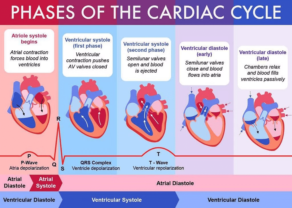
Q12: Define a cardiac cycle and cardiac output.
Ans: The cardiac cycle is the sequence of mechanical and electrical events that occur from the beginning of one heartbeat to the beginning of the next. It includes:
- Atrial systole - atrial contraction.
- Ventricular systole - ventricular contraction.
- Cardiac diastole - relaxation of the heart chambers.
A typical adult heart completes about 72 cycles per minute, each lasting roughly 0.8 seconds. The volume of blood ejected by each ventricle in one contraction is called the stroke volume (about 70 mL per beat in a healthy adult).
Cardiac output is the total volume of blood pumped by a ventricle in one minute. It is calculated as:
Cardiac output = Stroke volume × Heart rate
For an average adult at rest, cardiac output is about 5 litres per minute, though it increases during exercise to meet greater metabolic demand.
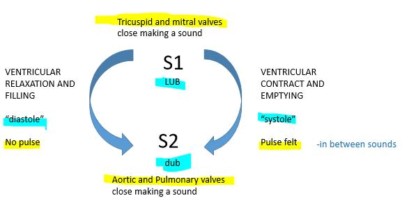
Q13: Explain heart sounds.
Ans: Heart sounds are the noises produced by valves closing during the cardiac cycle. The two main sounds in a healthy heart are:
- "Lub" (first heart sound, S1): produced when the atrioventricular valves (tricuspid and bicuspid/mitral) close at the beginning of ventricular systole.
- "Dub" (second heart sound, S2): produced when the semilunar valves (aortic and pulmonary) close at the start of ventricular diastole.
These sounds are useful clinically because changes in their timing, intensity or quality may indicate valve disorders or other cardiac abnormalities. Additional faint sounds or murmurs may suggest turbulent blood flow or valve defects.
 Heart Sounds
Q14: Draw a standard ECG and explain the different segments in it.
Ans: An electrocardiogram (ECG) is a recording of the heart's electrical activity. The main deflections and segments are:
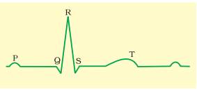 ECG
ECG- The P-wave signifies the electrical excitation (depolarisation) of the atria, causing both atria to contract.
- The QRS complex shows the depolarisation of the ventricles, triggering their contraction; it is normally the largest deflection and marks the onset of ventricular systole.
- The T-wave represents the repolarisation of the ventricles, as they return to their resting electrical state; its end signals the completion of ventricular systole.
- By counting the number of QRS complexes within a specified time on the ECG tracing, the heart rate of an individual can be determined.
FAQs on NCERT Solutions: Body Fluids & Circulation
| 1. What are the main components of blood and their functions? |  |
| 2. How does the circulatory system maintain homeostasis? | |
| 3. What is the difference between arteries, veins, and capillaries? | |
| 4. What role does the heart play in circulation? | |
| 5. How does the lymphatic system contribute to the circulatory system? | |


