Counter Current Mechanism | Biology A-Level - A Level PDF Download
| Table of contents |
|
| Introduction of Counter Current Mechanism |
|
| Regulation of Kidney Function |
|
| Functions of Kidney |
|
| Diseases related with Kidney |
|
| Autoregulation of GFR |
|
Introduction of Counter Current Mechanism
Countercurrent mechanism to maintain medullary hyperosmolality : The gradient of increasing hypersomolality of medullary interstitium is maintained by a peculiar countercurrent mechanism operated by the Henle's loops of juxtamedullary nephrons and vasa recta. About 15% to 20% of the nephrons in mammalian kidneys are situated at the level where cortex and medulla meet and, hence called juxtamedullary nephrons.
The Henle's loops of these nephrons are thin and long and extend almost upto the tips of medullary papillae.
The peritubular capillaries associated with these Henle's loops are also very thin and in the form of thin loops extending almost upto the tips of medullary papillae. These capillary loops are called vasa recta.
A countercurrent can be defined as the flow of a fluid in opposite directions in the two arms of a U-tube if the arms are rather very close together. Thus, the Henle's loops of juxtamedullary nephrons and vasa recta are anatomically ideal for the operation of countercurrent mechanism.
There are two aspects of this mechanism:
(1) countercurrent multiplication
(2) countercurrent exchange
Note: The Henle's loops play the role of countercurrent multipliers.
The vasa recta plays the role of countercurrent exchanger.
Since the concentration of tubular fluid in descending limb reflects the concentration of medullary interstitium, and since the concentration in the interstitium is raised by extrusion of salt from ascending limb, a positive feedback mechanism is created. The more salt the ascending limb extrudes, the more concentrated will be the fluid that enters into it from descending limb. Obviously, this feedback mechanism is the key point in the countercurrent multiplier system.
Countercurrent Exchange: In order for the countercurrent multiplier system to be effective in creating the gradient of medullary hyperosmolality, most of the salt extruded by the ascending limb of Henle's loop must remain in medullary interstitium, while most of the water coming out of the descending limb must be drained off into the blood. This is accomplished by the vasa recta by means of the mechanism known as countercurrent exchange. Salt is thus recirculated and trapped within the medullary interstitium, But contrarily, the water diffuses into the blood of ascending limb of vasa recta and is carried away into general blood circulation.
Significance of Urea : Permeability to urea is found only in the deeper parts of thin ascending limbs of Henle's loops and collecting ducts. Urea diffuses out of the collecting ducts and enters into the thin ascending limbs down its concentration gradients. A certain amount urea recycled in this way is trapped in medullary interstitium.
Role of Distal Convoluted Tubule, Collecting Duct and ADH : As described above, the role of countercurrent mechanism is to produce a small volume of highly hypotonic (osmolality only about 100 mL osmol/litre) tubular fluid which enters from the thick ascending limb of Henle's loop into the distal convoluted tubule and, thereafter into the collecting duct. To finally produce a very small volume of highly concentrated urine from this hypotonic tubular fluid is the role of distal convoluted tubule and collecting duct under the influence of the antidiuretic hormone (ADH). ADH triggers synthesis of a large number of molecules of a specific protein, named aquaporin, in the epithelial cells of distal convuled tubule and more particularly of collecting duct. Molecules of aquaporin become incorporated in the plasma membrane of these cells as integral proteins and act as water channels.
Consequently some water is lost from hypotonic tubular fluid by exosmosis while it flows through the distal convoluted tubule, but most of the water of tubular fluid is lost across the wall of collecting duct via aquaporin as this duct traverses through the medullary interstitium to empty into a calyx.
Due to this, the osmolality of the urine emptied into the calyx becomes 1200 to 1400 mL osmol/ litre.
Regulation of Kidney Function
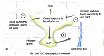
Two important hormonal controls of the kidney function by negative feedback circuits. These are as follows :-(i) Control by Antidiuretic Hormone (ADH) :- ADH is produced in the hypothalamus from the pituitary glands. ADH enhances fluid retention by making the kidneys reabsorb more water. The release of ADH is triggered when osmoreceptors in the hypothalamus detect an increase in the osmolarity of the blood above a set point of 300 mosm L–1. In this situation, the osmoreceptor cells also promote thirst. Drinking reduces the osmolarity of the blood, which inhibits the secretion of ADH, thereby completing the feedback circuit.
(ii) Control by Juxtaglomerular Apparatus (JGA) :- JGA operates a multi-hormonal ReninAngiotensin-Aldosterone System (RAAS). Whenever there is a fall in BP or blood volume, the JGA responds to this decrease in blood pressure or blood volume in the afferent arteriole of the glomerulus and releases an enzyme called renin, into the blood stream. In the blood, renin initiates chemical reactions that convert a plasma protein, called angiotensinogen to a peptide, called angiotension II, which works as a hormone. Angiotensin II increases blood pressure by causing arterioles to constrict. It also increases blood volume in two ways.
(1) Firstly, by signaling the proximal convoluted tubules to reabsorb more NaCl and water.

(2) Secondly, by stimulating the adrenal gland to release aldosterone, a hormone that induces the distal convoluted tubule to reabsorb more Na+ and water. This leads to an increase in blood volume and pressure, completing the feedback circuit by supporting the release of renin.
Still another hormone, a peptide called Atrial Natriuretic Factor(ANF), opposes the regulation by RAAS. Whenever there is rise in the BP or blood volume, the walls of the atria of the heart release ANF in response to this increase in blood volume and pressure. ANF inhibits the release of renin from the JGA, and thereby, inhibits NaCl reabosorption by the collecting duct and reduces aldosterone release from adrenal gland. Thus, ADH, the RAAS and ANF provide an elaborated system of checks and balance that regulate the kidney functioning, to control body fluid osmolarity, salt concentrations, blood pressure and blood volume.
Role of Lungs in Excretion
Human lungs eliminate around 18L of CO2 per hour and about 400ml of water per day in normal resting condition. Water loss via the lungs is small in hot humid climate and large in cold dry climates. The rate of ventilation and ventilation pattern (i.e. breathing through mouth or nose) also affect the water loss through the lungs. Different volatile materials are also readily eliminated through the lungs.
Role of Skin in Excretion
Human possess two types of glands:(1) Sweat glands : These excrete sweat, Sweat contain 99.5%, Water, NaCl, Lactic acid, Urea, Amino acid and glucose. Volume of sweat may vary from negligible to 14 litres a day.
(2) Sebaceous glands : These secrete sebum which contain waxes, sterols, other hydrocarbons and fatty acids. Integument in many aquatic animals excretes ammonia in surrounding medium by diffusion.

Role of Liver in Excretion
Liver is the main site for elimination of cholesterol, bile pigments (bilirubin & biliverdin), inactivated products of steroid hormones, some vitamins and many drugs. Bile carries these materials to the intestine from where they are excreted with the faeces.Functions of Kidney
- Regulation of water and electrolyte balance.
- Regulation of body fluid osmolarity and electrolyte concentration.
- Regulation of acid base balance.
- Regulation of arterial pressure.
- Excretion of metabolic waste and foreign chemicals.
- Secretion of hormones like erythropoeitin and renin.
- Gluconeogenesis from amino acids.
Diseases related with Kidney
1. Renal failure : It is a syndrome characterised by renal dysfunction, oliguria, anuria, sudden rise in metabolic waste products like urea & creatinine in blood (Uremia) . It is either of acute (sudden onset) or chronic (slow onset) nature.
2. Glomerulonephritis : It is a disease where due to infection or injury in the basement membrane, the inflammation of glomerulus progressively leads to renal failure and death.
3. Diabetic nephropathy : It is a complication due to diabetes mellitus where the kidney progressively gets damaged leading to death ultimately due to renal failure.
4. Urolithiasis : Formation of calculi (stone) in the urogenital tract at any point. These calculi are made of calcium phosphate, uric acid., cystine or calcium oxalate.
5. Abnormal urine : Various metabolic errors of kidney malfunctioning is reflected in the changes of the composition of urine.
Occurrence of ketone bodies, glucose, albumin, blood cells, excess pigments, pus cells, calculi or casts (kidney stones) are some of the major abnormal constituents of urine. Notable abnormal conditions are as follows : Proteinuria -excess protein level in urine.
6. Albuminuria -presence of albumin in urine, usually occurs in nephritis (inflammation of glomeruli), when the size of the filtering slits enlarges and basement membrane looses its negative charge.
7. Ketonuria - Presence of abnormally high ketone bodies in urine.
8. Haemoglobinuria - Presence of blood or blood cells in urine.
Autoregulation of GFR
Two important intrinsic mechanisms provide autoregulation of glomerular filtration rate :
(a) Myogenic Mechanism : An increase in blood pressure will tend to stretch the afferent arteriole, which would be expected to increase the blood flow to the glomerulus. The wall of the afferent arteriole, however, responds to stretch by contraction, this reduces the diameter of the arteriole, and therefore causes increase in the resistance to flow. This myogenic mechanism, thus, reduces variations in flow to the glomerulus in case of fluctuations in blood pressure.
(b) Juxtaglomerular Apparatus (JGA) : This specialised cellular apparatus is located where the distal convoluted tubule passes close to the Bowman's capsule between the afferent and efferent arterioles.
JGA cells secrete enzymes like renin that modulate blood pressure, and thus renal blood flow and GFR are regulated. This is discused earlier in the sheet.
Thus, myogenic and juxtaglomerular mechanisms work together to autoregulate the GFR over a wide range of blood pressure.
Composition of Urine
95% = Water
2% = Salts
2.7% = Urea Rest
0.3% = other materials like the drugs, Hippuric-acid, Uric acid, Vitamin-C, Dyes etc.
Pale yellow colour of urine is due to the Urochrome pigment. It is formed in the blood due to the reduction of Haemoglobin. So in the body of a healthy animal urochrome is found in a very less amount.
pH of urine = 6. The pH of urine is maintained by a Buffer-system. This is called the [ Na2HPO4– NaH2PO4] Buffer system
The specific-gravity of urine is 1.01 to 1.05
Osmoregulation by Kidneys
Kidneys regulate the amount of water and salts in the Extra-cellular fluid. Some hormones help in this process, which are:1. ADH or Pitressin or Vasopressin :- Main Controlling hormone of volume of urine. It mainly act on DCT & early CT. It promotes reabsorbtion of H2O. Due to deficiency. of ADH, diuresis occurs & water loss is increased.
This condition called as diabetes insipidus.
2. Aldosterone :- It is a hormone of the adrenal-cortex. The main salt found in the ECF is Na+ ions. They regulate the osmolality of the ECF. If Na+ number decreases in the blood the blood pressure also decreases and if Na+ number increases in the blood the blood pressure also increases. Aldosterone hormone, promotes the reabsorption of Na+ in the nephrons,ie. it checks the loss of Na+ ions through urine. When the ECF lacks Na+ ions, the secretion of Aldosterone increases.
3. Renin :- It is secreted by JG apparatus when there is decreased delivery of Na+ at macula densa, when fall of BP & Sympathatic stimulation occurs, then renin is secreted
4. ANF (Atrial Natriuretic Factor) :- It is released from atrial muscles. It decreases the effect of renin & aldosterone, It decrease BP & overload of heart & promotes vasodialation.
Old NCERT Syllabus
SOME TERMS
1. Oligouria :- Less production of urea/urine
2. Anuria :- No production of urine
3. Polyuria :- Excess production of urine. More urine formation takes place due to less secretion of ADH. Due to less secretion of ADH, the amount of water increases in the urine. So, the patient feels thirsty again and again.
This disease is calledDiabetes-insipidus.
4. Glycosuria :- Excretion of Glucose through Urine. This sign is present in Diabetes-mellitus. This disease is caused mainly due to less secretion of Insulin.
5. Uremia :- Excess of urea in blood is termed as Uremia.
6. Calculi and cast :- It is also termed as Kidney-stone. Due to deposition of Calcium-oxalate in the kidney, stone is formed. Sometimes, calcium - phosphate and calcium-sulphate are also found. These are insoluble-salts.
Normally, these are not excreted by the urine.
7. Haematuria :- Excretion of blood through urine. It is a symptom of many diseases like Black water fever, Bacterial-infection.
8. Diuresis :- The process of excess formation of urine in the kidney's is termed as diuresis.
9. Dysuria :- Condition of painful micturition
10. Urinode - Characteristic smell of the urine is due to urinode substances.
11. Cystitis :- Infection of urinary-bladder is termed as cystitis.
12. A person who is starving, will have more urea and ketone bodies in his blood and less urea in urine.
13. (a) The urine on standing gives a pungent smell. It is due to conversion of urea into ammonia by bacteria (b) The volume of urine produced per day will increase on a cold day, due to ADH seretion.
14. Highest concentration of urea is found in hepatic vein. (Because urea is synthesized in liver Least concentration of urea is found in renal vein. (Becasue urea is excreted through urine formed in kidney)
15. If one kidney is removed, the remaining one enlarges and performs function of both kidneys.
16. Camels can withstand water deprivation by reducing urinary water loss and water loss by sweat.
17. Earthworms excrete ammonia when sufficient water is available while they excrete urea instead of ammonia in drier surroundings.
18. When lung fishes and xenopus (African toad) live in water, they are normally ammonotelic but they become ureotelic when they live in moist air or mud during summer.
Crocodiles = normally ammonotelic
19. Bean shaped kidney are present only in mammals.
20. Number of pyramids is man = 8 – 12 Number of pyramids in Rabbit = only 1 “Renal column of Bertini” absent in Rabbit
21. Uric acid is the last product of purine metabolism in human 2,6,8–trioxy purine is uric acid
22. Renal blood flow = 1200 – 1300 ml./mt.
Renal plasma flow = 650 ml./mt.
23. Inulin clearance can be used to Estimate GFR
24. PAH (Para Amino Hippuric Acid) clearance can be used to Estimate RPF (Renal Plasma Flow)
25. Test of urea in urine is specific urease test.

Phenol Red is used as a indicator Optimum temp for reaction 60°C.
26. In brain, the major mechanism for removal of Ammonia is glutamine formation & in liver, the most important pathway is urea formation.
27. Urinary Bladder – Stimulation for voiding urine = 220 cc.
Generally micturation occur – 300 – 400 ml.
Discomfort condition after 500 ml.
Capacity of bladder 1000 cc.

29. Basement membrane is a meshwork of collagen and proteoglycan fibrills. It prevents filteration of plasma proteins because of stronge negative charge present on it due to proteoglycans.
30. Urinary excretion rate = filteration rate – reabsorption rate + secretion rate.
31. High threshold substances are completely reabsorbed after filtration while athreshold substances like creatinine are freely filtered but not at all reabsorbed.
32. No. of functioning nephrons decrease 10% for every 10 years after the age of 40 years.
33. In each kidney there are about 250 collecting duct each of which collects urine from 4000 nephrons.
Development of Kidney
During embryonic development, nephrotome plate develops from mesoderm which is made up of fine tubules called nephrons.
Nephrotome develops into kidney while nephros develops into Nephrons or uriniferous tubules. On the basis of development, kidney are of 3 types;
(1) Pronephric Kidney :- Develop from anterior part (Pronephros) of Nephrotome plate, Its nephrons are in simple tubular shape. Nephrons are not differentiated Eg. Cyclostomates & Tadpole of frog.
(2) Mesonephric Kidney or opisthonephros :- develop from middle part (Mesonephros) of Nephrotome plate & remaining part of nephtotome is destroyed. Only Bowman's capsule is found in nephrons while remaining part is simple tubular.
Eg. Most of the fishes & adult Amphibians.
(3) Metanephric Kidney :- Develops from posterior part (Metanephros) of nephrotome while remaining part is destroyed. Nephrons are well differentiated in to Bowman's capsule PCT, DCT & loop of Henle's Eg. Reptile, Aves, Mammals Henle's Loop :-
Less developed in Reptiles
Incompletely Developed in Aves
Mammals have most developed Henles loops.

|
280 videos|166 docs|147 tests
|
FAQs on Counter Current Mechanism - Biology A-Level - A Level
| 1. What is the counter current mechanism? |  |
| 2. How does the counter current mechanism regulate kidney function? | |
| 3. What are the functions of the kidney? | |
| 4. What are some diseases related to the kidneys? | |
| 5. How does autoregulation of GFR (glomerular filtration rate) work? | |

|
4K Views |

|
4.92/5 Rating |

|
Jan 03, 2025 Last updated |

|
Explore Courses for A Level exam
|
|
MCQs
,Extra Questions
,Important questions
,Semester Notes
,Viva Questions
,ppt
,study material
,Summary
,Counter Current Mechanism | Biology A-Level - A Level
,Free
,shortcuts and tricks
,Previous Year Questions with Solutions
,Exam
,Counter Current Mechanism | Biology A-Level - A Level
,past year papers
,practice quizzes
,Counter Current Mechanism | Biology A-Level - A Level
,video lectures
,Objective type Questions
,Sample Paper
,mock tests for examination
;


Counter Current Mechanism Free PDF Download
Importance of Counter Current Mechanism
Counter Current Mechanism Notes
Counter Current Mechanism A Level Questions
Study Counter Current Mechanism on the App
|
© EduRev
|
Education Revolution
|


|


within 7 days!