Cell Wall, Cell Membrane and Endomembrane System | Biology Class 11 - NEET PDF Download
| Table of contents |

|
| Cell Wall |

|
| Cell Membrane |

|
| Endomembrane System |

|
| 1. Endoplasmic Reticulum |

|
| 2. Golgi Complex |

|
| 3. Lysosome |

|
| 4. Vacuoles |

|
Cell Wall
- The cell wall was discovered by Robert–Hooke.
- The outermost but dead boundary of plant cells is the cell wall.
- Bacteria are included in plants because they have a cell wall.

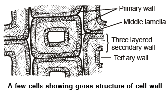
- Middle lamella consists of Ca & Mg pectates (Plant cement). The Amount of Ca is more.
- Fruits become soft and juicy due to dissolved middle lamella.
- Cellulose is the main constituent of the cell wall but in addition to cellulose – Hemicellulose, Cutin, Pectin, Lignin, Suberin are also present in the cell wall.
- Cell walls worked as a frame or protective layer of the cell.
- Cellulose microfibrils and microfibrils are arranged in layers to form the skeleton of the cell wall. In between these layers, other substances like pectin, hemicellulose may be present. These form matrix of the cell walls.
- A network of cellulose fibre forms the skeleton of the cell wall. 35-100 cellulose chain = 1 micelle. 20 micelle = 1 Microfibril 250 micro fibril = 1 Macrofibril in cell wall.
- Middle lamella is cement material between two adjacent cells in multi-cellular plants or the outermost layer of the cell wall. (the primary wall is considered as an outermost layer in a cell)
- Martinez and Palamo (1970) discovered cell-coat in animal cells, which is known as Glycocalyx. [Made by sialic acid, mucin and hyaluronic acid (animal cement)].
- Cell wall materials (Cellulose, Hemicellulose, Pectin, lignin) are synthesized in plant Golgi bodies or dictyosomes. Material of lipid nature (cutin and suberin) are synthesized in sphaerosome.
Formation of Cell Wall occurs by Two Methods
- Intussusception: This is a deposition of wall material in the form of fine grains.
- Apposition: Deposition of layers.
- The primary wall is formed mainly by intussusception, while the secondary wall is formed by both methods. Growth of already formed cell wall takes place only by intussusception.
- A Special protein called expansin helps in the growth of the cell walls by loosing the cellulose microfibril and addition of new cell wall material takes place in the space. Thus expansin is called a "cell wall loosening factor".
Specialization of Cell Wall
- Lignification: Lignin (coniferyl alcohol) is a cellulose derivative carbohydrate that deposits on walls of sclerenchyma, vessels and tracheids.
 Excess of lignin decreases the economic importance of pulp.
Pits
- Pits are formed in the lignified cell walls.
- Deposition of lignin occurs throughout the cell wall leaving some small thin-walled areas called pits.
- Pits are generally formed in pairs on the wall of adjacent cells.
- Two pits of a pair are separated by a thin membrane called pit membrane (completely permeable) (earlier composed of the middle lamella and primary wall).
- But, after some time primary wall may be dissolved.
- There are two types of pit pairs
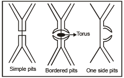
(i) Simple pits - When the diameter of a pit cavity is uniform throughout its length then such types of pits are called simple pits.
(ii) Bordered pits - When the diameter of the pit cavity increases from inside to outside then such pits are called Bordered pits. In such pits, the pit membrane has a thickening, composed of suberin called Torus. Torus functions like a valve to regulate the flow of materials. - Pits occur in sclerenchyma, vessels and tracheids.
- Tracheids in gymnosperms have a maximum number of bordered pits.
- Suberisation: Suberisation occurs on cork and Casparian strips of endodermal cells. Suberin is a highly impermeable material. It is water-tight and air-tight material. So suberisation also leads to the death of the cell. Maximum suberisation occurs in middle lamella. It reduces the transpiration rate in plants.
- Cutinization: Cutin - is also hydrophobic, waxy substance. Cutinization is the deposition of cutin on the cell walls of the leaf epidermis. It reduces the transpiration rate in plants.
- Cuticularisation: Deposition of cutin on the surface of the leaf. It leads to the formation of cuticles.
- Mucilage deposition: Mucilage deposits on the surface of hydrophytes.
- Deposition of silica: Occurs on the Leaves of grasses, Equisetum, Atropa, Diatoms, rice.

Cell Membrane
- The cell membrane is also known as the plasma membrane.
- An outermost envelope-like membrane or a structure, which surrounds the cell and its organelles are called the plasma membrane.
- It is a double membraned cell organelle, which is also called the phospholipid bilayer and is present both in prokaryotic and eukaryotic cells.
- In all living cells, the plasma membrane functions as the boundary and is selectively permeable, by allowing the entry and exit of certain selective substances.
- Along with these, the plasma membrane also acts as a connecting system by providing a connection between the cell and its environment.
- The main functions of the cell membrane include:
Protecting the integrity of the interior cell.
Providing support and maintaining the shape of the cell.
Helps in regulating cell growth through the balance of endocytosis and exocytosis.
The cell membrane also plays an important role in cell signalling and communication.
It acts as a selectively permeable membrane by allowing the entry of only selected substances into the cell.
Structure of Plasma Membrane
- A plasma membrane is mainly composed of carbohydrates, phospholipids, proteins, conjugated molecules, which are about 5 to 8 nm in thickness.
- The plasma membrane is a flexible and lipid bilayer that surrounds and contains the cytoplasm of the cell.
- Based on their arrangement of molecules and the presence of certain specialized components, it is also described as the fluid mosaic model.
Fluid Mosaic Model
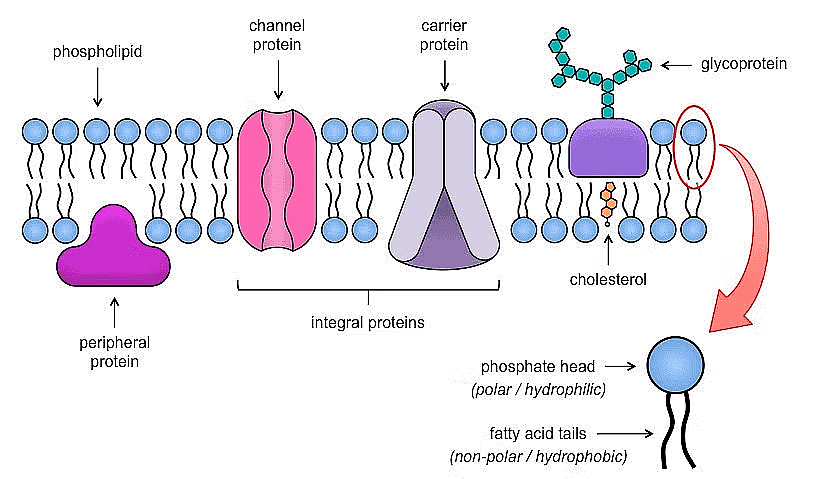
- The description of the structure of the plasma membrane can be carried out through the fluid mosaic model as mosaic cholesterol, carbohydrates, proteins and phospholipids.
- First proposed in 1972 by Garth L. Nicolson and S.J. Singer, the model explained the structure of plasma membranes.
- The model evolved with time however, it still accounts for the functions and structure of plasma membranes the best way.
- The model describes plasma membrane structure as a mosaic of components that includes proteins, cholesterol, phospholipids, and carbohydrates; it imparts a fluid character on the membrane.
- The thickness of the membrane is in the range of 5-10nm.
- The proportion of constituency of plasma membrane i.e., the carbohydrates, lipids and proteins vary from cell to cell.
- For instance, the inner membrane of the mitochondria comprises 24% lipid and 76% protein, in myelin, 76% lipid is found and 18% protein.
- The chief fabric of this membrane comprises phospholipid molecules that are amphiphilic.
- The hydrophilic regions of such molecules are in touch with the aqueous fluid outside and inside the cell.
- The hydrophobic or the water-hating molecules on the other hand are non-polar in nature.
- One phospholipid molecule comprises a three-carbon glycerol backbone along with 2 fatty acid molecules associated with carbons 1 and 2 and one phosphate-containing group connected to the third carbon.
- This organization provides a region known as the head to the molecule on the whole. The head, which is a phosphate-containing group possesses a polar character or a negative charge while the tail, another region containing fatty acids, does not have any charge.
- They tend to interact with the non-polar molecules in a chemical reaction, however, do not typically interact with the polar molecules.
- The hydrophobic molecules when introduced to water, have the tendency to form a cluster.
- On the other hand, hydrophilic areas of the phospholipids have the tendency to form hydrogen bonds with water along with other polar molecules within and outside the cell.
- Therefore, the membrane surface interacting with the exterior and interior of cells is said to be hydrophilic.
- On the contrary, the middle of the cell membrane is hydrophobic and does not have any interaction with water.
- Hence, phospholipids go on to form a great lipid bilayer cell membrane separating fluid inside the cell from the fluid to the exterior of the cell.
- The second major component is formed by the proteins of the plasma membrane. Integrins or integral proteins integrate fully into the structure of the membrane, along with their hydrophobic membrane, ranging from regions interacting with hydrophobic regions of the phospholipid bilayer.
- Typically, single-pass integral membrane proteins possess a hydrophobic transmembrane segment consisting of 20-25 amino acids.
- Few of these traverse only a portion of the membrane linking with one layer whereas others span from one to side of the membrane, thereby exposing to the flip side.
- Few complex proteins consist of 12 segments of one protein, highly convoluted to be implanted in the membrane.
- Such a type of protein has a hydrophilic region/s along with one or more mildly hydrophobic areas.
- This organization of areas of the proteins has the tendency to align the protein along with phospholipids where the hydrophobic area of the protein next to the tails of the phospholipids and hydrophilic areas of protein protrudes through the membrane is in touch with the extracellular fluid or cytosol.
- The third most important component of the plasma membrane is carbohydrates.
- They are generally found on the outside of the cells and linked either to lipids to form glycolipids or proteins to form glycoproteins.
- The chain of this carbohydrate can comprise two to sixty monosaccharide units which could be branched or straight.
- Carbohydrates alongside peripheral proteins lead to the formation of concentrated sites on the surface of the cell which identify each other.
- This identification is crucial to cells as they permit the immune system to distinguish between the cells of the body and the foreign cells/tissues.
- Such glycoproteins and glycoproteins are also observed on the surface of viruses, which can vary thereby preventing the immune cells to identify them and attract them.
- On the exterior surface of cells, these carbohydrates, their components of both glycolipids and glycoproteins are together known as glycocalyx, which is extremely hydrophilic in nature attracting huge quantities of water on the cell surface.
- This helps the cell to interact with its fluid-like environment and also in the ability of the cell to acquire substances dissolved in water.
 Fluid Mossaic Model
Functions of Plasma Membrane
- The plasma membrane functions as a physical barrier between the external environment and the inner cell organelles.
- The plasma membrane is a selectively permeable membrane, which permits the movement of only certain molecules both in and out of the cell.
- The plasma membranes play an important role in both the endocytosis and exocytosis processes.
- The plasma membrane also functions by facilitating the communication and signalling between the cells.
- The plasma membrane plays a vital role in anchoring the cytoskeleton to provide shape to the cell and also maintain the cell potential.
Facts about Plasma Membrane
- Both cell membrane and plasma membrane are often confused because of the similarity in words.
- But these two are the protective organelles of the cell and are very much different in their structure, composition and functions.
- The cell membrane is a type of plasma membrane and is not always the outermost layer of the cell.
Plasma Membrane Structure
- Also referred to as the cell membrane, the plasma membrane is the membrane found in all cells, which separates the inner part of the cell from the exterior.
- A cell wall is found to be attached to the plasma membrane to its exterior in plant and bacterial cells.
- A plasma membrane is composed of a lipid layer that is semipermeable.
- It is responsible to regulate the transportation of materials and the movement of substances in and out of the cell.
- In addition to containing a lipid layer sitting between the phospholipids maintaining fluidity at a range of temperatures, the plasma membrane also has membrane proteins.
- This also includes integral proteins passing through the membrane which act as membrane transporters and peripheral proteins attaching to the sides of the cell membrane.
- It loosely serves as enzymes that shape the cell.
- The plasma membrane is selectively permeable to organic molecules and ions, it regulates the movement of particles in and out of organelles and cells.
Plasma Membrane Function
- This membrane is composed of a phospholipid bilayer implanted with proteins.
- It forms a stable barrier between two aqueous compartments, which are towards the outside and inside of a cell in a plasma membrane.
- The embedded proteins perform specialized functions which include cell-cell recognition and selective transport of molecules.
- Plasma membrane renders protection to the cell along with providing a fixed environment within the cell.
- It is responsible for performing different functions. In order for it to allow the movement of substances such as white and red blood cells, it must be flexible such that they could alter the shape and pass through blood capillaries.
- In addition, it also anchors the cytoskeleton to render shape to a cell and in associating with extracellular matrix and other cells to assist the cells in forming a tissue.
- It also maintains the cell potential.
- The plasma membrane is responsible for interacting with other, adjacent cells which can be glycoprotein or lipid proteins.
- The membrane also assists the proteins to monitor and maintain the chemical climate of the cell, along with the assistance in the shifting of molecules across the membrane.
Plasma Membrane – Components
It is composed of the following constituents:
- Phospholipids – forms the ultimate fabric of the membrane
- Peripheral proteins – present on the outer or inner surface of phospholipid bilayer but are not implanted in the hydrophobic core
- Cholesterol – folded between the hydrophobic tails of phospholipid membrane
- Carbohydrates – found to be attached to the lipids or proteins on the extracellular side of the membrane, leading to the formation of glycolipids and glycoproteins
- Integral proteins – found to be implanted in the phospholipid bilayer
Difference between Cell Membrane and Plasma Membrane
- Plasma membrane and cell membrane are often confused to be similar terms. However, they are quite different from each other.
- The plasma membrane encloses the organelles of the cell, whereas, the cell membrane encloses the entire cell components.
Difference Between Cell Membrane and Plasma Membrane
Following are the important difference between the cell membrane and plasma membrane:
Cell Membrane | Plasma Membrane |
It surrounds the entire components of the cell. | It surrounds only the cell organelles. |
It regulates the tonicity of the cell. | It does not regulate the tonicity of the cell. |
The cell membrane can be transformed to stimulate movement and feeding in organisms such as Paramaecium. | The plasma membrane cannot be modified. |
It contains initial receptors for signal transduction and is the first step in cell signalling. | It is not the first step in cell signalling. However, it is involved in the process. |
Always protects the cell from bacteria and viruses. | Does not always protect the cell from outside invaders. |
Plays an important role in cytokinesis during cell division. | Do not play a key role in cytokinesis during cell division. |
Cilia are present and are involved in feeding and movement. | Cilia are absent. |
Is a target for antimicrobials | Is not a target for antimicrobials |
Key Points on Cell Membrane
- The cell membrane is a type of plasma membrane that encloses the cell and all its components.
- Both the membranes are selectively permeable and regulate the entry and exit of components.
- The cell membrane is the only membrane involved in cytokinesis.
Endomembrane System
While each of the membranous organelles is distinct in terms of its structure and function, many of these are considered together as an endomembrane system because their functions are coordinated. The endomembrane system include endoplasmic reticulum (ER), golgi complex, lysosomes and vacuoles. Since the functions of the mitochondria, chloroplast and peroxisomes are not coordinated with the above components, these are not considered as part of the endomembrane system.
1. Endoplasmic Reticulum
"Garnier" (1897) first observed them and called Ergastoplasm. E. R. name proposed by "Porter" (1961). (Credit for discovery of ER goes toPorter) Components of E.R.
- Cisternae - These are long flattened and unbranched units arranged in stacks.
- Vesicles - These are oval membrane bound structures.
- Tubules - These are irregular, often branched tubes bounded by membrane. Tubules may free or associated with cisternae.
(i) Structure of E.R. is like the golgi body but in E.R. cisternae, vesicles and tubules are isolated in cytoplasm and these do not form complex.
(ii) Golgi body is localised cell organelle while E.R. is widespread in cytoplasm. E.R. is often termed as “System of Membranes”.
| Rough E.R. (Granular) | Smooth E.R. (Agranular) |
(1) 80s ribosomes binds by their larger subunit, with the help of two glycoproteins (Ribophorin I and II) on the surface of Rough E.R. | (1) Ribosomes and Ribophorins absent |
| 2) More Stable structure | 2) Less Stable structure |
| 3) Mainly Composed of cisternae and vesicles | (3) Mainly composed of tubules. |
| 4) Abundantly occurs in cells which are actively engaged in protein synthesis e.g. liver, pancreas, Goblet cells. | (4) Abundantly occurs in cells concerned with glycogen and lipid metabolism. e.g. Adipose tissue, Interstitial cells, Muscles,Glycogen storing liver cells, and adrenal cortex. |
Enzymes of E.R.
Sucrases, NADH diphosphatase, Gulcose-6-phosphatase, NADH-cytochrome-C-reductase, Mg+2 activated ATPase, Nucleotide diphosphatase, Ascorbic acid synthase are enzymes of E.R.
Functions of E.R.
- Mechanical support: Microfilaments, Microtubules and E.R. forms endoskeleton of cell.
- Intracellular exchange: E.R. forms intracellular conducting system. Transport of materials in cytoplasm from one place to another may occurs through the E.R.
(i) At some places E.R. is also connected to P.M. So E.R. can secrete the materials outside the cell. - Rough E.R.: Provides site for the protein synthesis, because rough E.R., has ribosomes on its surface.
- Lipid Synthesis: Lipids (cholesterol & phospholipids) synthesized by the agranular portion of E.R. (Smooth E.R.). The major lipids synthesized by S. E. R. are phospholipids and Cholesterol.
- Release of Glucose from Glycogen: Endoplasmic reticulum seems to play a role in breakdown of glycogen (glycogenolysis).(The polymerisation of glucose to form glycogen probably occur in the cytosol not in the wall of S.E.R.)
- Cellular metabolism:The membranes of the reticulum provides an increased surface for metabolic activities within the cytoplasm.
- Formation of nuclear membrane: Fragmented vesicles of disintegrated nuclear membrane and ER elements arranged around the chromosomes to form a new nuclear membrane during cell division.
- Formation of lysosomes, Golgi–body & Micro–bodies. All the organelles are form by E.R. which have membrane except chloroplast and mitochondria (semi autonomous organelles)
- Detoxification: Smooth ER concerned with detoxification of drugs, pollutants and steroids.
- Cytochrome P450 in E.R. act as enzyme which function in detoxification of drugs and other toxins
- E.R. provides the precursor of secretory material to golgi body.
2. Golgi Complex
2. Golgi Complex
Discovered by C. Golgi (1898) - In nerve cells of owl and named "internal reticular apparatus" (Golgi body first observed by L.S. George) (Impregnated with silver nitrate).
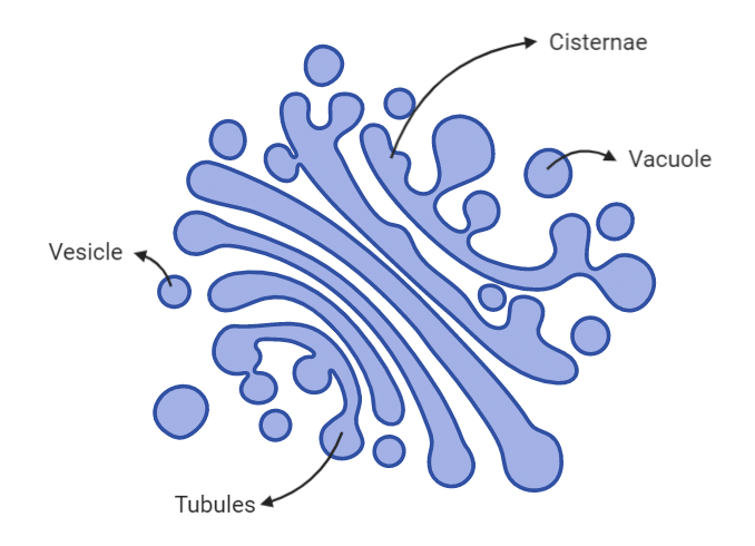 Golgi Body
The cytoplasm surrounding Golgi body have fewer or no other organelles. It is called Golgi ground substance or zone of exclusion. Golgi bodies are pleomorphic structures because component of golgi body are differ in structure & shape in different cells.
Structure
Golgi complex is made up of four parts:
- Cisternae: These are unbranched saccules likes smooth E.R., many cistenae are arranged in a stack. Dense opaque material inside cisternae is called nodes.
(i) Convex surface of cisternae which is towards the nucleus is called cis face or forming face.
(ii) Concave surface of cisternae which is towards the membrane is called trans face or maturing face. - Tubules: These are branched and irregular tube like structures associated with cisternae.
- Vacuoles: Large spherical structures associated to tubules.
- Vesicles: Spherical structures arise by budding from tubules. Vesicles are filled with secretory materials.
- Golgibody is single membrane bound cell organelle.
- About 60% proteins and 40% phospholipid occur in golgi body.
Functions
- Cell Secretion: Chief function of golgi body is secretion (export) of macromolecules.
- Secretion involve three steps:
(i) Golgi body recieves the materials from E.R. through it's cis - face.
(ii) These materials are chemically modified by golgi body. (For e.g. glycosidation (glycosylation) of proteins and lipids takes place in golgi body and it yields glycoprotiens and glycolipids).
(iii) After chemical modifications materials are packed in vesicles. These vesicles are pinched off from trans face of golgi body and discharged out side the cell (Reverse pinocytosis) - Golgi complex involves secretion of zymogen granules from pancreas, secretion of lactoprotein from mammary Glands.
- The secretion of hormone by endocrine glands is mediated through golgibodies.
- All the macromolecules which are to be sent out side the cell, move through the golgi body. So golgi body is termed as “Director of macromolecular traffic in cell” or middle men of cell.
- Synthesis of cell wall Material (Polysaccharide synthesis)
- Cell plate formation (Phragmoplast) during cell formation.
- Formation of acrosome during spermiogenesis. (formation of male gametes)
- Vitelline membrane of egg is secreted by golgi body.
- Formation of Lysosome = It is collective function of golgi body and E.R.
- Mucilage secretion by root cells for lubrication to soil.
- Secretion of hormones by cells of glands.
3. Lysosome
- Christian De Duve (1955) discovered lysosome as cell organelle and also named Lysosomes. (Lysosome were first observed by Novikoff)
- With the exception of mammalian RBC they were reported from all cells.
- In plant cells large central vacuole functions as Lysosome. So in higher plants lysosomes are less frequent. But number of lysosomes is high in fungi.
- Periplasmic Space :– space between cell wall and cell membrane in bacteria,may play similar role.
- Lysosomes are spherical bag like structures (0.1-0.8 mm) which is covered by single unit membrane. They are larger in Phagocytes (WBC) (0.8 to 2mm).
- Lysosomes are filled with 50 different type of digestive enzymes termed as Acid hydrolases. These acid hydrolases function in acidic medium ( pH=5).Membrane of lysosome has an active H+ pump mechanism which produce acidic pH in lumen of lysosome.
- Lysosomes are highly polymorphic cell organelle. Because, during functioning, lysosomes have different morphological and physiological states.
Types of Lysosomes
- Primary Lysosomes or Storage Granules: These lysosomes store enzyme Acid Hydrolases in the inactive form. (Enzymes synthesized on ribosomes in cytoplasm) these are newly formed lysosome.
- Digestive Vacuoles or Heterophagosomes: These lysosome forms by the fusion of primary lysosomes and phagosomes. These are secondary Lysosomes.
- Residual Bodies: Lysosomes containing undigested material are called residual bodies. These may be eliminated by exocytosis. These are also called as Telolysosomes. (Tertiary lysosomes)
- Autophagic Lysosomes or Cytolysosomes or Autophagosomes: Lysosomes containing cell organelles to be digested are known as Autophagosomes.
Functions
- Heterophagy :– This is digestion of foreign materials received in cell by phagocytosis and pinocytosis.
- Autophagy :– Digestion of old or dead cell organelles. Autophagy also takes place during starvation of cell. [Ambilysosomes :– Lysosomes which perform both heterophagy and autophagy.]
- Extracellular digestion :– Lysosomes of osteoclast (bone eating cells) dissolve unwanted part of bones. (Extracellular digestion also occurs by fungal lysosomes.)
- Crinophagy :– Excessive secretory granules of hormone in endocrine gland may be digested by lysosomes. This event is called crinophagy. Thyroglobulin stores in thyroid gland with its follicles and after crinophagy by proteases itproduces thyroxine.
- Cellular digestion (Autolysis) :– Sometimes all lysosomes of a cell burst to dissolve the cell completely. Old cells are removed by autolysis. unwanted organs of embryo are destroyed by autolysis Cathepsin of lysosome digests the tail of tadpole of frog during metamorphosis.
- Lysosomes are helpful in digestion of egg membrane to assist fertilisation.
- Lysosome also trigger the cell division or mitosis.
(i) Membrane stabilizers are substances, which stabilize the lysosome membrane and stop its rupture, thus prevents autolysis. e.g. cholesterol, chloroquine, cortisone etc.
(ii) Membrane labilizers are substances which make the lysosome membrane fragile and increase the chance of autolysis e.g. Progesterone, testosterone, Vitamin A, D, E, K, U.V. radiations, bile salts etc.
Sometime Lysosomes burst it's whole cell so Lysosome called as suicidal bags of cell.
(iii) Biogenesis of Lysosome Lyosomes originates from G E R L - (Golgi associated Endoplasmic Reticulum from which Lysosomes arise).
E.R. → Golgi body → Lysosome
4. Vacuoles
“Vacuoles are membrane-bound cell organelles present in the cytoplasm and filled with a watery fluid containing various substances.”
What are Vacuoles?
The term “vacuole” means “empty space”. They help in the storage and disposal of various substances. They can store food or other nutrients required by a cell to survive. They also store waste products and prevent the entire cell from contamination. The vacuoles in plant cells are larger than those in the animal cells. The plant vacuoles occupy more than 80% of the volume of the cell. The vacuoles may be one or more in number.
Structure of Vacuole
A vacuole is a membrane bound structure found in the cytoplasmic matrix of a cell. The membrane surrounding the vacuole is known as tonoplast. The components of the vacuole, known as the cell sap, differ from that of the surrounding cytoplasm. The membranes are composed of phospholipids. The membranes are embedded with proteins that help in transporting molecules across the membrane. Different combinations of these proteins help the vacuoles to hold different materials.
Functions of Vacuole
The important functions of vacuole include:
Storage
A vacuole stores salts, minerals, pigments and proteins within the cell. The solution that fills a vacuole is known as the cell sap. The vacuole is also filled with protons from the cytosol that helps in maintaining an acidic environment within the cell. A large number of lipids are also stored within the vacuoles.
Turgor Pressure
The vacuoles are completely filled with water and exert force on the cell wall. This is known as turgor pressure. It provides shape to the cell and helps it to withstand extreme conditions.
Endocytosis and Exocytosis
The substances are taken in by a vacuole through endocytosis and excreted through exocytosis. These substances are stored in the cells, separated from the cytosol. Lysosomes are vesicles that intake food and digest it. This is endocytosis and it varies in different cells.
Frequently Asked Questions
Why do plant cells have larger vacuoles?
The plant cells have larger vacuoles because they require more water, organic and inorganic components for the proper functioning of the cell.
Why are vacuoles an important cell organelle?
Vacuoles store nutrients and water on which a cell can rely for its survival. They also store the waste from the cell and prevents the cell from contamination. Hence, it is an important organelle.
Old NCERT Syllabus
Golgi body is known by several other names:
(1/16) Golgi body
(1/16) Dalton complex
(1/16) Golgi complex
(1/16) Lipochondria ( rich in lipids)
(1/16) Baker's body
(1/16) Idiosome
(1/16) Dictyosome (plant golgi body)
(1/16) Trophospongium
Modifications of E.R.
- Sarcoplasmic Reticulum (S.R.): These smooth E.R. occurs in skeletal and cardiac muscles. S.R. Stores Ca+2 and energy rich compounds required for muscle contraction.
- T-tubules: These are transversely arranged tubules in skeletal and cardiac muscle cells. These transmits stimulus for contraction of muscles.
- Ergastoplasm: When the ribosomes are accumulated on the small parallel cisternae of E.R., then called Ergastoplasm. Ergastoplasm of nerve cells is called as Nissl's bodies.
- Myeloid Bodies: Myeloid bodies are the specialised smooth E.R. which found in pigmented epithelial cells of the retina. Myeloid body is light sensitive structure and may be involved in pigment migration.
- Microsomes: These are pieces of E.R. with associated ribosomal particles (Claude 1951). These can be obtained by Fragementation and high speed centrifugation of cell. They do not exist as such in the living cell.
|
150 videos|399 docs|136 tests
|
FAQs on Cell Wall, Cell Membrane and Endomembrane System - Biology Class 11 - NEET
| 1. What is the function of the cell wall? |  |
| 2. How is the cell membrane different from the cell wall? |  |
| 3. What is the endomembrane system? |  |
| 4. What is the function of the endoplasmic reticulum? |  |
| 5. What is the role of the Golgi complex in the endomembrane system? |  |


