Cell Membrane, Cell Wall and Endomembrane System

The structural organization of the cell depends on membrane-bound structures that define cellular compartments and regulate molecular transport. This section covers three critical components: the plasma membrane (cell boundary), the cell wall (external protective layer in plants/fungi), and the endomembrane system (coordinated internal membrane network). Understanding their composition, structure, and functions is essential for NEET preparation, particularly for questions on transport mechanisms, membrane fluidity, and organelle functions.
1. Cell Membrane (Plasma Membrane)
The plasma membrane is the outermost boundary of all living cells. It regulates entry and exit of molecules, providing selective permeability.
1.1 Historical Background and Chemical Composition
- Detailed structure studied: After the advent of the electron microscope in the 1950s.
- Early chemical studies: Conducted on human red blood cells (RBCs) to deduce membrane structure.
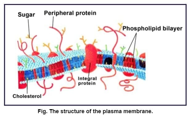
- Main components: Lipids (phospholipids and cholesterol) and Proteins.
- Carbohydrates: Also present, forming glycoproteins and glycolipids.
- Composition ratio (human erythrocyte): Approximately 52% protein and 40% lipids.
1.2 Lipid Bilayer Structure
- Major lipids: Phospholipids arranged in a bilayer.
- Orientation: Polar hydrophilic heads face outward (towards aqueous environment on both sides). Nonpolar hydrophobic tails (saturated hydrocarbons) face inward (towards each other).
- Function of arrangement: Protects hydrophobic tails from the aqueous environment.
- Cholesterol: Present in addition to phospholipids, contributes to membrane stability and fluidity.
1.3 Membrane Proteins
Membrane proteins are classified based on their association with the lipid bilayer:
- Peripheral proteins: Lie on the surface of the membrane. Loosely attached, easily extractable.
- Integral proteins: Partially or totally buried in the membrane. Difficult to extract. Many function as transport channels or carriers.
1.4 Fluid Mosaic Model
- Proposed by: Singer and Nicolson (1972).
- Key concept: The membrane is a quasi-fluid structure where lipids and proteins can move laterally within the bilayer.
- Fluidity: The ability of lipids and proteins to move within the membrane. Measured as membrane fluidity.
- Mosaic arrangement: Proteins are embedded in or attached to the lipid bilayer in a mosaic pattern.
1.5 Importance of Membrane Fluidity
Fluid nature of the membrane is crucial for several cellular processes:
- Cell growth: Allows membrane expansion.
- Formation of intercellular junctions: Enables cell-cell communication.
- Secretion: Facilitates vesicle fusion with plasma membrane.
- Endocytosis: Membrane invagination requires fluidity.
- Cell division: Membrane division during cytokinesis.
1.6 Transport Across Plasma Membrane
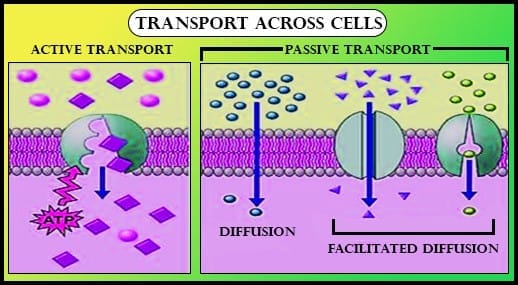
The plasma membrane is selectively permeable. It allows selective passage of molecules. Transport mechanisms include:
1.6.1 Passive Transport
- Definition: Movement of molecules across the membrane without energy requirement.
- Simple diffusion: Movement of neutral solutes along the concentration gradient (from higher to lower concentration). Does not require carrier proteins.
- Osmosis: Movement of water across the membrane by diffusion, from higher to lower concentration.
- Facilitated diffusion: Polar molecules cannot pass through the nonpolar lipid bilayer directly. They require a carrier protein to facilitate their transport across the membrane.
1.6.2 Active Transport
- Definition: Transport of molecules against the concentration gradient (from lower to higher concentration).
- Energy requirement: ATP is utilized.
- Example: Na⁺/K⁺ Pump (Sodium-Potassium pump). Pumps sodium ions out and potassium ions into the cell.
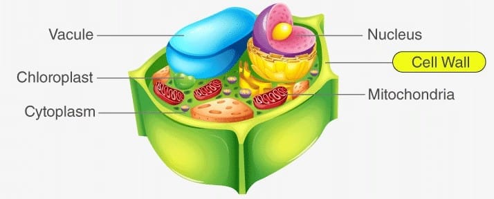
2. Cell Wall
The cell wall is a non-living, rigid structure that forms an outer covering for the plasma membrane in fungi, plants, and algae. It is absent in animal cells.
2.1 Functions of Cell Wall
- Provides shape: Gives definite shape to the cell.
- Mechanical protection: Protects the cell from mechanical damage.
- Protection from infection: Acts as a barrier against pathogens.
- Cell-to-cell interaction: Facilitates communication between adjacent cells.
- Barrier to macromolecules: Prevents entry of undesirable large molecules.
2.2 Chemical Composition
Cell wall composition varies across different organisms:
- Algae: Made of cellulose, galactans, mannans and minerals like calcium carbonate.
- Plants: Composed of cellulose, hemicellulose, pectins, and proteins.
- Fungi: Contains chitin (not mentioned in given content, but cell wall is present in fungi as stated).
2.3 Layers of Plant Cell Wall
- Primary wall: Present in young plant cells. Capable of growth. Thin and flexible.
- Secondary wall: Formed as the cell matures. Located on the inner side (towards the plasma membrane) of the primary wall. Thicker and rigid. Growth diminishes after secondary wall formation.
- Middle lamella: A layer mainly composed of calcium pectate. Located between adjacent plant cells. Holds or glues neighbouring cells together.
2.4 Plasmodesmata
- Definition: Cytoplasmic channels that traverse the cell wall and middle lamella.
- Function: Connect the cytoplasm of neighbouring cells, facilitating cell-to-cell communication and transport.
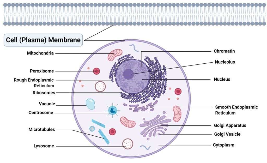
3. Endomembrane System
The endomembrane system is a group of membrane-bound organelles whose functions are coordinated. Each organelle has distinct structure and function, but they work together.
3.1 Components of Endomembrane System
- Endoplasmic Reticulum (ER)
- Golgi Complex (Golgi Apparatus)
- Lysosomes
- Vacuoles
Not part of endomembrane system: Mitochondria, Chloroplast, and Peroxisomes are not included because their functions are not coordinated with the endomembrane system components.
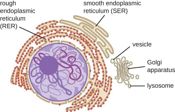 Endomembrane System
3.2 Endoplasmic Reticulum (ER)
The ER is a network or reticulum of tiny tubular structures scattered in the cytoplasm, revealed by electron microscopy in eukaryotic cells.
3.2.1 Structure and Compartmentalization
- Structure: Network of membrane-bound tubules and flattened sacs.
- Compartments:ER divides the intracellular space into two distinct compartments:
- Luminal compartment: Inside the ER.
- Extra-luminal compartment: Cytoplasm outside the ER.
3.2.2 Types of ER
Rough Endoplasmic Reticulum (RER):
- Appearance: Bears ribosomes attached to its outer surface, giving it a rough appearance.
- Location: Frequently observed in cells actively involved in protein synthesis and secretion.
- Structure: Extensive and continuous with the outer membrane of the nucleus.
- Function: Site of protein synthesis (by attached ribosomes) and protein modification.
Smooth Endoplasmic Reticulum (SER):
- Appearance: No ribosomes on its surface, appears smooth.
- Main function: Major site for synthesis of lipids.
- In animal cells: Lipid-like steroidal hormones are synthesized in SER.
3.3 Golgi Apparatus (Golgi Complex)
3.3.1 Discovery and Structure
- Discovered by: Camillo Golgi (1898). He observed densely stained reticular structures near the nucleus.
- Named after: Camillo Golgi.
- Structure: Consists of many flat, disc-shaped sacs or cisternae.
- Size of cisternae: 0.5 μm to 1.0 μm diameter.
- Arrangement: Cisternae are stacked parallel to each other. The Golgi cisternae are arranged concentrically near the nucleus.
- Number of cisternae: Varied number present in a Golgi complex.
3.3.2 Polarity of Golgi Apparatus
- Cis face (forming face): Convex side. Receives vesicles from ER.
- Trans face (maturing face): Concave side. Releases vesicles to target destinations.
- Faces are: Entirely different but interconnected.
3.3.3 Functions of Golgi Apparatus
- Packaging materials: Principal function is packaging of materials into vesicles.
- Delivery targets:Materials are delivered to:
- Intra-cellular targets (within the cell).
- Secreted outside the cell (exocytosis).
- Vesicle transport: Vesicles from ER fuse with the cis face and move towards the trans face (maturing face).
- Association with ER: Golgi apparatus remains in close association with ER due to this vesicle transport.
- Protein modification: Proteins synthesized by ribosomes on ER are modified in the cisternae of Golgi before release from trans face.
- Formation of glycoproteins and glycolipids: Important site for their synthesis.
3.4 Lysosomes
3.4.1 Structure and Formation
- Structure: Membrane-bound vesicular structures.
- Formation: Formed by the process of packaging in the Golgi apparatus.
3.4.2 Enzymatic Composition
- Enzyme content: Very rich in almost all types of hydrolytic enzymes (hydrolases).
- Types of hydrolases: Lipases, proteases, carbohydrases.
- Optimal pH: These enzymes are optimally active at acidic pH.
- Substrates digested: Capable of digesting carbohydrates, proteins, lipids, and nucleic acids.
3.5 Vacuoles
3.5.1 Structure and Contents
- Definition: Membrane-bound space found in the cytoplasm.
- Membrane: Bound by a single membrane called tonoplast.
- Contents: Contains water, sap, excretory products, and other materials not useful for the cell.
- Size in plant cells: Can occupy up to 90% of the cell volume.
3.5.2 Functions of Vacuoles
In Plant Cells:
- Tonoplast function: Facilitates the transport of ions and other materials against concentration gradients into the vacuole.
- Concentration difference: Concentration of substances is significantly higher in the vacuole than in the cytoplasm.
In Amoeba:
- Contractile vacuole: Important for osmoregulation and excretion.
In Protists:
- Food vacuoles: Formed by engulfing food particles.
Understanding the structural and functional relationships between the cell membrane, cell wall, and endomembrane system is fundamental to cell biology. The membrane systems provide compartmentalization, regulate transport, and coordinate cellular activities. The Fluid Mosaic Model, transport mechanisms (passive vs. active), and the coordinated functions of ER-Golgi-Lysosomes are high-yield topics for competitive exams. Remember the specific percentages, names of scientists, years of discovery, and unique features of each organelle for accurate recall during examinations.
FAQs on Cell Membrane, Cell Wall and Endomembrane System
| 1. What is the function of the cell wall? |  |
| 2. How is the cell membrane different from the cell wall? | |
| 3. What is the endomembrane system? | |
| 4. What is the function of the endoplasmic reticulum? | |
| 5. What is the role of the Golgi complex in the endomembrane system? | |


