NEET Exam  >  NEET Notes  >  Biology Class 11  >  Mitochondria, Plastid, Ribosome & Cytoskeleton

Mitochondria, Plastid, Ribosome & Cytoskeleton | Biology Class 11 - NEET PDF Download

Term "Cytoplasm", was given by Strasburger for the part of cell present between the nucleus and cell membrane. Cytoplasm can be divided into two parts.
Mitochondria, Plastid, Ribosome & Cytoskeleton | Biology Class 11 - NEET

  • Ground plasm / Hyaloplasm / Cytosol → Liquid matrix of cytoplasm except organelles.
  • Trophoplasm → Part of cytoplasm containing organelles & non living Inclusions. 

Cell Organelles

Permanent Metabolically active and living structures of cytoplasm are called organelles.

Mitochondria


  • Kolliker (1880) first observed mitochondria as cytoplasmic granules in striped muscles of insects.
    Mitochondria, Plastid, Ribosome & Cytoskeleton | Biology Class 11 - NEET
  • Altman (1894) established them as cell organelles and called Bioblast. Flemming and Altman was credited for the discovery of mitochondria. Term 'Mitochondria, was given by C.Benda.
  • F.Meves (1904) first observed them in plants ( Nymphea). 
  • Kinsburg and Hogeboom (1948) related them with cell respiration.
  • Seikevitz called them power house of cell.
  • Number: 1000–1600 per cell.
  • One in Microasterias, Chlorella fusca (alga). 
  • 50,000 mitochondria in an Amoeba Chaos Chaos. 
  • All the mitochondria present in a cell are collectively called chondriome.
  • Usually plant cells have fewer mitochondria as compared to animal cell.
  • In higher animals maximum mitochondria are found in flight muscles of birds.
  • Mitochondria can make its shape as ellipsoidal, oval, spherical or spiral. 

Question for Mitochondria, Plastid, Ribosome & Cytoskeleton
Try yourself:
What is the function of mitochondria in a cell?
View Solution

Mitochondria 

Power house of cell or ATP-mill in cell.
Cell within cell second largest organelle.
Cell furnaces or storage batteries.
Most busy and active organelle in cell.
Semi autonomous cell organelle.

Structure

  • Mitochondria is covered by double unit membrane. Outer membrane has more phospholipids (Phosphatidyl choline) and cholesterol as compared to inner membrane. Phospholipid in inner membrane is mainly diphosphatidyl glycerol and Inner membrane have more protein.
  • Each membrane is 60-75 Å thick and separated by a space (80-100 Å ) called perimitochondrial space.
  • This space have enzymes required for oxidation of fats and pyruvic acid.
  • If outer membrane of mitochondria is removed then structure is called as mitoplast. 
  • Outer surface of inner membrane is called C- face while inner surface is called M- face.
  • Some sessile particles attached to outer membrane are known as "subunit of parsons".
  • Inner membrane is folded into a number of finger like cristae. Cristae are tubular finger like but in fungi cristae are plate like while in Euglena cristae are vesicle shaped. Cristae word was coined by Palade.
  • In metabolically active mitochondria number of cristae is higher.
  • Many electron carrier cytochromes are arranged in a definite sequence in Inner membrane of mitochondria, which forms Electron  transport system (ETS) for oxidative phosphorylation.
  • Inner membrane is studded with pin head particles called oxysomes or elementary particles or F1 – Fparticles ( 104 to 106 in number).These particles first described by Fernandez Moran (1962).  
  • Head of Oxysomes composed of ATPase (ATP synthase) enzymes and are concerned with Oxidative phosphorylation (Recker1967). 
  • Mitochondrial matrix have enzyme for Kreb's cycle. Beside these enzymes matrix have a complete protein synthesis apparatus (Ribosome- 70-s, DNA, few RNA's & enzymes) so mitochondria called as semi autonomous cell organelles.
  • One to many (6 kb to 36 kb long) double stranded mainly circular naked DNA present in mitochondrial matrix.
  • Mitochondrial DNA is 1% of total DNA in a cell. It is rich in G-C content.
  • Mitochondrial DNA can code the synthesis of 10 to 37 different types of proteins. Rest of the proteins and enzymes of mitochondria are synthesized under the control of nuclear genes.
  • Mitochondrial DNA was discovered by Nass and Margit .
  • Enzymes for replication and transcription of DNA like DNA- polymerase and RNA- polymerase are found in mitochondrial matrix.
  • Mitochondria of mammals have 55s ribosomes (35s,25s units) 

Functions of Mitochondria

  • Most of the oxidative metabolism and ATP production occurs in mitochondria, thus mitochondria are the power house of cell, where organic compounds are broken down to release & store metabolic energy in the the form of ATP molecules. (Resp. metabolism).
  • Mitochondria help in vitellogenesis in oocytes, Mitochondrial Kinase makes the yolk viscous and insoluble for a longer duration storage. Mitochondria of oocytes called Yolk nuclei.
  • In cytoplasmic inheritance.

Biogenesis of Mitochondria

  • By Division
  • Endosymbiotic origin from Purple Sulphur bacteria or prokaryotic cells, because mitochondria are similar to prokaryotic cell (rickettsial bacteria) in Structure of DNA and DNA sequences, Type of ribosome (70s), Sensitivity of Antibiotic chloremphenicol with protein synthesis, Divided by amitosis or fission.

Plastids


  • The term "Plastid" first used by Haeckel . 
  • Schimper coined the term chloroplastid for green plastids. Meyer called them Autoplast 
  • Chloroplast name proposed by Erera.


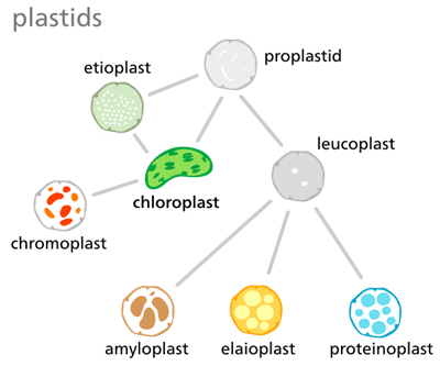
Types of Plastids

  • Chromoplasts: These are plastids, which contain different types of pigments (carotenes, Xanthophylls etc.). Chlorophylls either absent or occur in very less amount. Chromoplasts occurs mainly in pericarp and petals. Red colour of chillies and red tomatoes is due to the red pigment "Lycopene" of chromoplasts,. Lycopene is a type of carotene. Yellowish - orange colours of fruits are due to a-carotene, b-carotene and g–carotene.b– carotene is precursor of vitamin-A. Richest source ofb-carotene are carrot roots.
    (i) Chromoplasts also occurs in petals but colour of petals are mainly due to water soluble pigments  occur in cell sap. e.g. Anthocyanin - (Blue or violet or red pigment) Anthochlor (yellow pigment).
  • Chloroplasts: Green plastids with chlorophylls and other photosynthetic pigments.

Question for Mitochondria, Plastid, Ribosome & Cytoskeleton
Try yourself:
What is the main difference between chromoplasts and chloroplasts?
View Solution

  • Leucoplasts: These store food in different forms like starch (Amyloplasts), Fat and oil (Elaioplasts) and protein (Aleuroplasts). Pigments and lamellar structure absents in Leucoplasts. Generally occurs in non green and underground plant cells.
  • All types of plastids have common origin from proplastids, sac like non-lamellar structures.
  • Different types of plastids may transform from one form to another. Because genetic meteral is similar. But chromoplasts never transform to chloroplasts.
  • Etioplasts: These are plastids without pigments, stored food and lamellar structures. These plastids occur in etiolated plants due to the absence of light.

Mitochondria, Plastid, Ribosome & Cytoskeleton | Biology Class 11 - NEET

  • Chromatophores: These are clusters of pigment granules in cytoplasm of  photosynthetic bacteria.
  • In Blue-green algae pigments are located on membranous lamellar structures, scattered in Cytoplasm. These structures are known as Chlorosomes or Lamellisomes or Carboxysomes.


Structure of Chloroplast

Shape of chloroplasts

Plant types

Discoidal or oval 

higher plants

Girdle shaped 

Ulothrix

Cup shaped 

Chlamydomonas

Reticulate 

Spirogyra

Stellate 

Zygnema


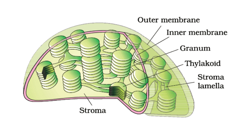
  • Chloroplast is a double membrane bound cell organelle, and is the largest organelle of cell. (4-6mm x 1-3mm). (largest component is nucleus) 
  • Internally chloroplast contains stroma (Matrix) and thylakoids or lamellae. Matrix part of chloroplast contains circular or rarely linear DNA, RNA, 70-s Ribosomes, starch grains, enzymes of calvin cycle or dark reaction. Rubisco is the most abundant enzyme on the earth. It made 16 % protein of the chloroplast

Mitochondria, Plastid, Ribosome & Cytoskeleton | Biology Class 11 - NEET

  • The number of chloroplast in cell of higher plants 20-40.(One in chlamydomonas)
  • Thylakoids (Term by Menke 1962) are membrane lined flattend sacs, which forms stacks called granum (Plu. grana). Each chloroplast contains about 20-100 granum. Fret channel or stromal thylakoids is connection between two granum. Photosynthetic pigments (chlorophylls) are located in the thylakoid membranes.
  • A photosynthetic functional unit, which consists of about 230 to 400 molecules of various pigments (Chl-a, Chl-b, carotenes, xanthophylls etc.) is called as Quantasomes (By Park & Biggins 1964).
  • Chloroplasts has their own genetic system & complete protein synthesis machinary (ds-DNA, RNA, Ribosomes, enzymes, Amino Acids), thus chloroplasts are called as semiautonomous organelle of the cell.
  • DNA of chloroplast was discovered by Ris & Plaut. (Upto 14% DNA of cell) * Chloroplast have more genes as compare to mitochondria. (100 or more genes) 

Functions

  • Photosynthesis : The chloroplasts trap the light energy of sun and transform it into the chemical energy in the form glucose.
  • Balancing of O2 & CO2 in nature.
  • Chloroplasts can be changed into chromoplasts during ripening of fruits.
  • Chloroplasts impart in cytoplasmic inheritance. 
  • Chloroplasts impart the pleasing greenary to the earth.
  • Chloroplasts store vitamin K, E, Rubisco protein and Fe etc.

Biogenesis

  • From Proplastid
  • From Division of pre-existing plastids.
  • Origin from Endosymbiotic origin by a cyanobacterium

Difference Between Mitochondria and Plastids

Mitochondria and plastids are the cell organelles present in eukaryotes. Mitochondria is found in all the eukaryotic cells including plants and animals, whereas, plastids are found only in plant cells.

Mitochondria vs Plastids

The important difference between mitochondria and plastids are:
Mitochondria, Plastid, Ribosome & Cytoskeleton | Biology Class 11 - NEET

Difference Between Mitochondria and Plastids

Mitochondria

Plastids

Found in all eukaryotic cells

Found in only plant cells

Produces ATP

Produces glucose and stores it as starch

The main function is cell respiration

Main organelle for photosynthesis

Smaller in size

Comparatively larger in size

Pigments are absent

Pigments are present

Question for Mitochondria, Plastid, Ribosome & Cytoskeleton
Try yourself:
What is the main function of chloroplasts?
View Solution

Ribosomes (Engine of Cell)

  •  Claude first observed them. Palade (1955) coined the term Ribosome.

  •  In plants, Robinson and Brown (1953) first observed them in bean roots.

  •  Except mammalian RBC all living cells have ribosomes (Both prokaryotes & Eukaryotes).

  •  Ribosomes are smallest cell organelles (150x250 A0 ) Ribosomes are organelle without membranes.

  •  Ribosomes are also called as ‘‘Organelle with in an organelle’’ & "Protein factory of cell".

RibosomeRibosome

[Question: 906869]

Types of Ribosomes

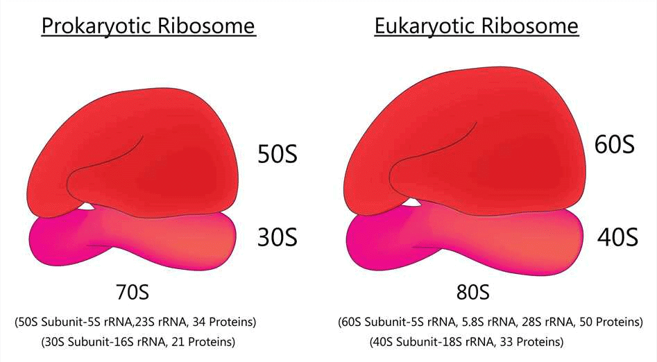
(1) Eukaryotic ribosomes :– 80 s - Occur in cytoplasm of eukaryotic cells.

(2) Prokaryotic ribosomes :– 70 s - Occur in cytoplasm of prokaryotes, and also in mitochondria, and Chloroplast of eukaryotes. (55 S ribosome present in mitochondria of mammals)

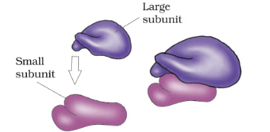
  • Each ribosome composed of two subunits i.e. larger and smaller subunits. Larger subunit is dome shaped and smaller unit is ovoid. 

  • Smaller subunit has a platform, a cleft, a head and a base. The larger subunit has a protuberance, a ridge and a stalk. Larger subunit also contains a tunnel which opens in cavity of E.R. 80s = 60s + 40s 70s = 50s + 30s

  • Magnesium ion is essential for the binding the ribosome sub units. Mg+2 form ionic bond with phosphate groups of r- RNA of two subunits. Minimum 0.001 M Mg+2 concentration is required for structural formation of ribosomes. If Mg+2 concentration  increased 10 times then ribosome dimer are formed.

  • 80s +80s = 120s (Dimer)
  • 70s + 70s = 100s (Dimer)

Chemical Composition of Ribosomes 

70s– 60% r- RNA  + 40% proteins

80s– 40% r-RNA + 60% proteins

60s– r-RNA 28s, 5.8s, 5s

40s– r-RNA 18s

50s– r-RNA 23s,5s

30s– r-RNA 16s

At the time of protein synthesis, several ribosomes become attached to m-RNA with the help of smaller subunits. This structure is called polyribosome or polysome or Ergosome. Ribosomes move along the mRNA like beads on a string, during protein synthesis. Larger subunit contains peptidyl transferase enzyme which helps in the formation of peptide during protein synthesis. This is an example of Ribozyme.

Two sites are found on larger sub units :

(i) A- site → Acceptor site for t-RNA

(ii) P-site → site for growing polypeptide chain

  •   S= Svedberg unit or Sedimentation rate

Mitochondria, Plastid, Ribosome & Cytoskeleton | Biology Class 11 - NEET

After synthesis on ribosomes, protein are transported in cytoplasm and organelles.
The proper foldin g and transp ort o f p roteins is assisted by specific proteins called Chaperons .


Cytoskeleton

“Cytoskeleton is the structure that maintains the shape and internal organization of the cell, and provides it mechanical support. “

What is Cytoskeleton?

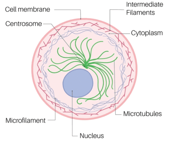
The cytoskeleton is the network of fibres forming the eukaryotic cells, prokaryotic cells and archaeans. These fibres in the eukaryotic cells contain a complex mesh of protein filaments and motor proteins that help in cell movement.

It provides shape and support to the cell, organizes the organelles and facilitates transport of molecules, cell division and cell signalling.


Cytoskeleton Structure

A cytoskeleton structure comprises the following types of fibres:

  1. Microfilaments
  2. Microtubules
  3. Intermediate Filaments

1. Microtubules

Microtubules appear like small, hollow, round tubes with a diameter of about 24 nanometers. They are made up of a protein, tubulin. Thirteen tubulins link to form a single tube. Microtubules are very dynamic structures, which reveal that they can change quickly. They keep growing or shrinking steadily. These help in transporting cellular materials and dividing chromosomes during cell division.


2. Microfilaments

Microfilaments are thread-like protein fibres, 3-6 nm in diameter. They are particularly found in muscle cells. They consist of the protein actin, responsible for muscle contraction. These are also responsible for cellular movements including cytokinesis, contraction, and gliding.


3. Intermediate Filaments

The intermediate filaments are about 10 nm in diameter and provide tensile strength to the cell. They facilitate the formation of keratins and neurofilaments.

The cytoskeleton is also made up of certain motor proteins. These include:

  1. Kinesin
    These proteins move along the microtubules carrying the cellular components. They pull the organelles along the cell membrane.
  2. Dynein
    These pull the cell organelles towards the nucleus.
  3. Myosin
    These interact with actin protein and are responsible for muscle contractions. They also perform cytokinesis, exocytosis, and endocytosis.

Cytoskeleton Functions

 The important cytoskeleton functions are mentioned below:

  1. It provides shape and support to the cell.
  2. It helps in the formation of vacuoles.
  3. It holds different cell organelles in place.
  4. It assists in cell signalling.
  5. It supports intracellular movements like the migration of cell organelles, transportation of vesicles in and out of the cell, etc.
The document Mitochondria, Plastid, Ribosome & Cytoskeleton | Biology Class 11 - NEET is a part of the NEET Course Biology Class 11.
All you need of NEET at this link: NEET
182 videos|367 docs|152 tests

Top Courses for NEET

FAQs on Mitochondria, Plastid, Ribosome & Cytoskeleton - Biology Class 11 - NEET

1. What is the function of mitochondria in a cell?
Ans. Mitochondria are responsible for producing energy in the form of ATP through a process called cellular respiration. They are known as the powerhouse of the cell.
2. What are plastids and what are their roles in a cell?
Ans. Plastids are double-membraned organelles found in plant cells. They have various functions including storing and synthesizing important molecules such as pigments, starch, and lipids. Chloroplasts, a type of plastid, are responsible for photosynthesis.
3. How do ribosomes contribute to cell function?
Ans. Ribosomes are the cellular structures responsible for protein synthesis. They read the genetic information from mRNA and assemble amino acids in the correct order to form proteins. They play a crucial role in the growth and maintenance of the cell.
4. What is the cytoskeleton and what are its components?
Ans. The cytoskeleton is a network of protein filaments that provides structural support and maintains cell shape. It is composed of three main components: microtubules, microfilaments, and intermediate filaments. These filaments also play a role in cell movement, intracellular transport, and cell division.
5. How are mitochondria, plastids, ribosomes, and the cytoskeleton interconnected in a cell?
Ans. Mitochondria and plastids are both membrane-bound organelles involved in energy production and storage. Ribosomes, located in the cytoplasm or attached to the endoplasmic reticulum, synthesize proteins that are targeted to various organelles including mitochondria and plastids. The cytoskeleton provides the structural framework for all cellular components, including organelles, and also facilitates their movement within the cell. So, all these components work together to ensure the proper functioning of a cell.
182 videos|367 docs|152 tests
Download as PDF
Explore Courses for NEET exam

Top Courses for NEET

Signup for Free!
Signup to see your scores go up within 7 days! Learn & Practice with 1000+ FREE Notes, Videos & Tests.
10M+ students study on EduRev
Related Searches

Ribosome & Cytoskeleton | Biology Class 11 - NEET

,

practice quizzes

,

shortcuts and tricks

,

ppt

,

Semester Notes

,

Objective type Questions

,

Important questions

,

Exam

,

mock tests for examination

,

Free

,

study material

,

Plastid

,

Sample Paper

,

Previous Year Questions with Solutions

,

past year papers

,

Ribosome & Cytoskeleton | Biology Class 11 - NEET

,

Viva Questions

,

video lectures

,

Summary

,

Mitochondria

,

Mitochondria

,

Plastid

,

Plastid

,

Ribosome & Cytoskeleton | Biology Class 11 - NEET

,

Mitochondria

,

Extra Questions

,

MCQs

,

pdf

;