Nucleus & Chromosomes | Biology for JAMB PDF Download
| Table of contents |
|
| Nucleus |
|
| Structure of Nucleus |
|
| Functions of Nucleus |
|
| Chromosomes |
|

Nucleus
- First of all Leeuwenhoek observed, nucleus in RBCs, of fish.
- Detail studied in orchid root cells and named by Robert Brown in 1831. Credit of discovery goes to Robert Brown.
- "Nucleus is double membrane bound dense protoplasmic body, which controls all cellular metabolism and encloses the genetic information of cell".
 Nucleus
- Nucleus is consider as controller or director of cell. Importance of nucleus in control of heredity, growth and metabolism was experimentally proved by Hammerling. (Experiment was on Acetabularia a single cell largest alga).
- If the nucleus of a cell is, experimentally removed, then unicellular organism will die after some time. Thus nucleus is very important and largest component of cell.
- Strasburgar stated that :- "Nucleus arises from divison of pre-existing nucleus only. The study of nucleus is known as Karyology.
- Generally eukaryotic cell contain at least one nucleus but nucleus is absents in mature phloem sieve tube elements and mature RBCs of mammals. (exceptionaly nucleus of RBCs of camel & lamma remains for longer time and degenerates later on).
- Dikaryotic (Paramecium) and multikaryotic cells are also known. Multinucleated cells may be following type :
(a) Coenocytic Cells: This type of cells, are formed by free nuclear divisions.
Example :- Phycomycetes fungi, Endosperm, rhizopus, vaucheria, etc.
(b) Syncytium: Syncytial condition is formed by the fusion of cells.
Example :- Epidermis of nematods, striped muscles.
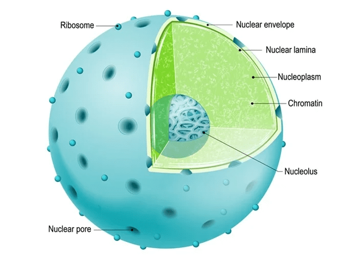
Structure of Nucleus
(i) Nuclear membrane or nuclear envelope or karyotheca
(ii) Nucleoplasm/Karyoplasm/Karyolymph
(iii) Chromatin net
(iv) Nucleolus/little nucleus/Ribosome factory
(i) Nuclear Membrane
Nucleus is surrounded by two unit membranes, thus nucleus is double membranous component of cell. Space (150 to 300 Å) between two membranes is known as perinuclear space. Outer membrane, of nucleus may connected with E.R. at several places and ribosome also may found on it.
- Nuclear membrane is perforated by minute nuclear pores of size, 300 to 1000Å diameter. Each nuclear pore is guarded by a octagonal discoid structure of nucleoplasmin protein this structure is called as annulus or Bleb. (Annulus + Pore = Nuclear Pore complex).
- The inner side of inner nuclear membrane is lined by nuclear lamina. This structure is formed by filaments of lamin protein.
- Pore complex provides the main channel, between nucleoplsm and cytoplam, while nucleoplasmin regulates nucleocytoplasmic traffic.
(ii) Nucleoplasm or Karyolymph
Term was given by Strasburger in 1882. Nucleoplasm or Nuclear sap is a ground substance of nucleus which is a complex colloidal formed of a number of chemicals like nucleotides, nucleosides, ATPs, proteins & enzymes of RNA & DNA polymerases, endonucleases, minerals, (Ca++, Mg++) etc.
Nucleoplasm contain high concentration of Nucleotides in the form of triphosphate.
- Nucleoplasm also have enzymes for glycolysis, thus nucleus may obtain energy by glycolysis.
- Chromatin net and nucleolus are embeded in nucleoplasm. Nucleoplasm provides site for process of transcription.
(iii) Chromatin Net
- Term was given by Flemming.
- It is an intranuclear, (stained with basic dyes) long, thread like fine fibres, which embeded in nucleoplasm. Chromatin net is mainly formed of DNA and histone protein complexes. Chromatin fibres contain genetic information and condensed to form constant number of chromosomes during cell division.
- Chemically chromatin consists of DNA (31%), RNA (2-5%), Histone protein (36%) and non histone(28%). 20 to 30% part of histone is made up of arginine and lysine amino acids.
On the basis of relative amount of arginine and lysin there are five type of Histone protein.
(H2A, H2B, H3, H4, H1)
Amino acid | Type of histone |
Lysin rich | H1 |
Slightly lysin rich | H2A, h2b |
Arginine rich | H3 , H4 |
Chromatin net has two type of chromatins (by Emil Heitz):
(a) Euchromatin:- This is lightly stained and diffused part of chromatin. Which is transcriptionally or genetically more active. Generally euchromatin lies at central part of nucleus.
(b) Heterochromatin:- This is dark stained, thick and condensed part of chromatin this part have more histone and less acidic protein. Heterochromatin is genetically less active chromatin and forms stop point in transcription. Heterochromatin occurs near nuclear membrane.
(i) Constitutive heterochromatin:- Occurs in all cells in all stages e.g. centromeric region.
(ii) Facultative heterochromatin:- Occurs in some cells in some stages e.g. barr body.
- Barr body in female cells is a facultative heterochromatic structure. (By M.Barr)
- Number of Barr body in nucleus of an individual is number of X-chromosome minus one.
Difference between Euchromatin and Heterochromatin. | |
Euchromatin | Heterochromatin |
1)Consist of thin, extended, light stained part of chromatin. | 1)Consist of thick aoild, condensed part of Chromatin and dark stained. |
2) Genetically more active chromatin | 2) Less active or inert chromatin. |
3) Lies at centre of nucleus. | 3) Lies near the nuclear membrane. |
4) less histone protein | 4) More histone protein |
5) Replicate in early s phase | 5) Replicate in late s phase |
6) DNA → mRNA | 6) DNA → rRNA, tRNA |
- Heterochromatin takes light stain during cell division stages (M-phase) & takes dark stain during Interphase.
(iv) Nucleolus
- Discovered by Fontana and term was given by Bowman.
- Nucleolus is naked or membraneless, rounded or slightly irregular structure present in nucleus and usually attached to chromatin (or chromosomes) of specific site called Nucleolar organiser region/ NOR.
- Number of nucleolus in a nucleus is one. Onion cell has 4, and in oocytes of amphibian has 2000 nucleoli. Nucleoli absent in sperm cell, muscle cells etc. Human cell has 5 nucleoli.
- Calcium is essential for maintenance of nucleolus. Nucleolus disappears during Prophase and reappears in telophase.
- Chemistry of nucleolus:
- Proteins 8 5%
- RNA 10%
- DNA 5%
Functions of Nucleolus
Ribosome formation is the chief role of nucleolus, thus its called as Ribosme factory of cell, the proteins of ribosomes are synthesised in cytoplasm but it diffused in to nucleus and reach at nucleolus. Here r-RNA and ribosomal proteins are assembled to form ribosomes which move to cytoplasm through nuclear pores.
Note: At the some places heterochromatin forms thickned dense granules which are known as karyosomes or chromocentre or false nucleoli.
Functions of Nucleus
(i) Genetic information :- Nucleus contains genetic information in its chromatin. (store house of genetic material)
(ii) Transmission of genetic information :- Nucleus takes part in transmission of genetical information from parent cell to daughter cell or the one generation to next.
(iii) In cell-division :- Division of nucleus is pre-requisite to cell division.
(iv) Control of metabolism :- Nucleus controls metabolism of cell by sending m-RNA in cytosol (Basically biomolecule DNA controls cellular activities through directing synthsis of enzyme).
(v) Variations :- Variation develops due to change in genetic material of nucleus. (Evolutionary role).
Chromosomes
- At the time of cell division the chromatin material get condensed to form chromosomes, thus chromosome is highly condensed form of the chromatin. Chromosomes are not visible during interphase stage.
- First of all, chromosomes was observed by Hofmeister (1818) and Karl Nageli in pollen mother cells (PMC) of Tradescantia.
- Strasburger (1875) described chromosome structure appeared in nucleus during cell division. (Credit of discovery of chromosomes goes to Strasburger).
- Term "Chromosome" was proposed by Waldeyer in 1889. (Term 'Chromatin, was suggested by Flemming).
- Generally chromosomes are rod-shaped, elongated or dot like in shape with size of 0.5 to 32m (Trillium plant has longest chromosome)
- Chromosomes can be best studied at metaphase stage because size of chromosomes is the shortest during metaphase due to highly condensation of chromatin threads by gelation, dehydration and coiling. (Shape of chromosome (V.L.J.I.) is studied at Anaphase stage)
- Generally chromosomes in plants are larger than chromosomes of animals, but number of chromosome is high in animals as compared to plants.
- The number of chromosomes has no relation with any specific feature like size, complexity of organism.
Structure of Chromosome
(Parts which appears in metaphase chromosome)
1. Pellicle – This is outermost, thin proteinaceous covering or sheath of chromosome.
2. Matrix – This is a liquid nongenetic achromatic ground substance of chromosome, which has different type of enzymes, minerals, water, proteins.
3. Chromonema (singular Chromonemata) →Term by Vejdovsky. This is an important, genetical, highly coiled thread, throughout the length of a chromosome or chromatid. It was called chromonema.
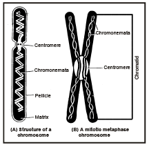
- Chromonema lie embeded in matrix. Each chromonemata is consist of a single long thread of DNA associated with histone.
- Sometimes bead like structure are seen on chromonema fibres, which are called as chromomeres.
4. Centromere/Kinetochore :- (Primary constriction)
- Each chromosome (at metaphase) is consist of two half chromosome or two chromatids. Both the chromatids of a chromosome are joined or connected by a structure called Centromere. At this point or centromere two protein discs are present which is called Kinetochore.
- Kinetochores constitute the actual site of attachement of spindles to chromosomes during cell division.
Centromeric DNA is called as alphoid DNA.
- At the region of centromere the chromosome is comparatively narrower than remaining part of chromosome, thus it is termed as Primary constriction.
5. Chromatid – At metaphase stage each chromosome is consist of two cylindrical structures - called chromatids.
Both sister chromatids or longitudinal half chromosome are joined together by a common centromere. A chromosome, may have single chromatid (in Anaphase or Telophase) or two chromatid. (as in metaphase)
6. Secondary Constriction : Besides primary constrictions one or two, other constriction may also occurs on some chromosome, which are known as secondary constriction.
Secondary constriction is also known as NOR (Nucleolar organizer region)(13,14,15,21,22 chromosomes in human)
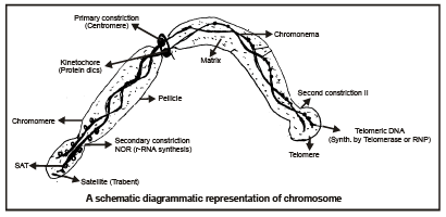
7. Satellite : part of chromosome remains after the NOR is known as chromosomes satellite/ trabent.
Chromosomes with satellite part are called as SAT chromosome (SAT = Sine Acid Thymonucleinico)
8. Telomere : Chromosomes have polarity and polar ends of chromosomes is known as Telomere.
Telomere prevents fusion of one chromosomes to other chromosome. Telomere rich in Guanine base.
Enzyme Telomerase presents in telomere part of chromosome, which is a Ribonucleoprotein.
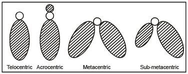
Types of Chromosomes on the basis of Position of Centromere
(i) Telocentric :- When centromere is terminal or located at the tip of chromosome.
ii) Acrocentric :- When the centromere is sub-terminal or located near the tip.
(iii) Metacentric :- When the centromere is located at mid of the chromosome.
(iv) Sub metacentric :- When the centromere located near centre or mid point of chromosome.
The ratio of length of the long arm to the short arm of a chromosome is called arm ratio. Arm ratio is maximum in acrocenteric chromosome.
Ultra Structure or Fine Structure of the Chromosome
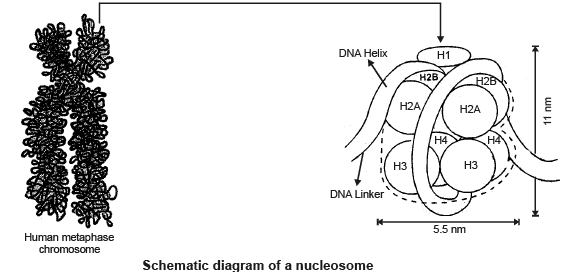
Nucleosome Model
Bead like structure in chromatin was first observed by Olin's etal. This model was proposed by Kornberg & Thomas in 1974, which is most important and universally accepted model for the structure of chromosome. This model explain that how giant DNA molecule & histone (Chromatin) packaged in to a chromosome. Term nucleosome was given by P. Oudet in 1975. "Nucleosome is a unit of chromatin (chromosome) which is composed of about 200 base pairs of the
DNA and an Octamer (Core particle) of four types ( H2A, H2B, H3& H4) of histone proteins". Nucleosome is also known as Nu-body or g-particle.
Nucleosome= Binding DNA (146 bp)+Octamer Core (H2A, H2B, H3, H4 × 2)+Linker DNA +H1 Histone
- 6 Nuclesome units united (or super coiling) to forms Solenoid structure. (by Klug - 1982)
- H1 histone protein (sealing histone) joined the turns of binding DNA in nucleosome.
- Nucleosome unit have 1.75 or (1*3/4) turns of binding DNA.
Special Types of Chromosomes
1. Salivary Gland Chromosome
This type of chromosome was discovered by E.G. Balbiani, in Chironomous larva of Drosophila . Size of this chromosome may upto 2000 micron (2mm) and number of chromatids may be 512 to many thousands. Thus, this type of chromosome also known as Giant chromosome.
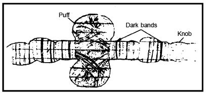
- Koller named it as Polytene chromosome, because number of chromatids is very high.
- Swollen areas present at some places in polytene chromosome, which are called as Balbiani rings or puffs. These puffs helps in synthesis of RNA & proteins.
- Salivary gland chromosome concerns with metamorphosis and moulting process of insect larva. (This chromosome is related to a moulting hormone - Ecdyson)
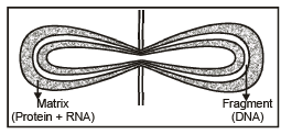
2. Lampbrush Chromosome
Discovered by Flemming and Ruckert from oocytes of vertebrates (Amphibia) during diplotene stage of cell division. These chromosomes look like lamp - brush, thus called as lamp brush chromosomes.
Size of these chromosomes may upto 5900 micron, and also called as giant chromosome.
- Axis of lamp-brush chromosome is consist of DNA, while matrix is consist of RNA & proteins.
- Lamp brush chromosome is concerned with "Vitellogenesis" (Yolk formation)
Old NCERT Syllabus
Nucleolus
Electron Microscope (Ultrastructure of Nucleolus) has shown nucleolus to be made of following parts:
(i) Fibrillar region :- This is central fibrous part of nucleolus, which is consist of mainly rDNA and proteins. (Nucleonema)
(ii) Granular region :- This is peripheral granular part of nucleolus which is consist of rRNA and proteins.
(iii) Amorphous matrix or pars amorpha :- This is proteinaceous ground matrix, which contains both fibres and granules.
Chromosome Number in Some Organisms
Plants | 2n | n |
Mucor hemelis (Fungi) | 2 | 1 |
Haplcpappus gracilis (Family compositae) & Brachycome plant | 4 | 2 |
Takakia (Bryophyta) | 4 | 2 |
Pisum sativum (Pea) | 14 | 7 |
Maize (Zea mays) | 20 | 10 |
Wheat (Triticum) | 42 | 21 |
Cphioglossum reticulatum (Pteridophyta) | 1262 | 631 |
Animals |
|
|
Ascaris megalocephala (Round worm) | 2 | 1 |
Drosophila manogaster (Etuit fly) | 8 | 4 |
Chimpanzee/Gdrilla | 48 | 24 |
Homo sapiens | 46 | 23 |
Aulocantha (a protozoan) | 1600 | 800 |
- 2n = number of chromosome in diploid cell. n = number of chromosome in haploid cell.
- The number of chromosome is definate for each species. For example every normal human being has 46 chromosomes in each body cell.
- Gametes of all organisms contain only one of each chromosome. The number of chromosomes in a gamete is called "Genome" or haploid chromosome. (Human 23) ‘‘A complete set (n) of chromosomes (all genes) inherited as a unit from one parent is known as genome,,.
- Karyotype: Karyotype is external morphology of all Chromosomes of a cell which is specific for each species of living organisms. Karyotype can be studied in metaphase of mitosis.
- Karyotype includes the number of chromosomes, relative size, position of centromere, length of the arms, secondary constrictions and banding patterns.
Banding technique is used to study of the specific pattern of bands and interbands on chromosome. This includes the use of fluorochromes (fluorescent dyes):
(i) Q–banding : It is obtained when chromosomes are stained with quinacrine mustard. It stains A–T rich areas (developed by casperson for Y chromosomes).
(ii) G–banding : Chromosomes are stained with Giemsa. It stains sulphur rich protein parts.
A variety of different bands are obtained by the modification of Q–banding and G–banding like C, T and N–bands. Q, C, G and R banding used for animal karyotypes while C and N banding used in plants.
(iii) C-banding :- It is used to stain constitutive heterochromatin , usually in centromeric region of the chromosome. The process involves denaturation of chromosome by heat or trisodium citrate and then apply giemsa stain.
(iv) R-banding :- The process involved incubation of the chromosomes in a buffer at high temperature followed by use of Giemsa stain. This brings about the visualization of sulphur deficient region of chromosomes thus named as reverse giemsa.
New Techniques for Idiogram Preparation
Modern techniques used in karyotype preparation are:
ISH, FISH (Fluorescence in Situ Hybridisation), Mc FISH (Multicolour fluorescence in situ Hybridisation) and flow cytometry.
Idiogram: Diagrammatic representation of Karyotype. In idiogram chromosomes are arranged in decreasing order of size. Sex chromosomes are placed in last but in idiogram of Drosophila sex chromosomes are placed first. Idiogram is specific for every species.
Use of Karyotyping or Idiogram
(i) It suggests primitive or advanced features of an organism. If karyotype shows a large size difference between the smallest and the largest chromosome of the set and having fewer metacentric chromosomes then it is called asymmetric karyotype, which is a relatively advance feature. Symmetric karyotype is primitive feature.
(ii) The karyotype of different species are compared and similarities in them represent the evolutionary relationships.
(iii) Karyotype is helpful in detection of chromosomal abberrations and polyploidy.
(iv) In research of medical genetics Forensic science cytogenetics and Anthropogenetics.
1. In Situ Hybridization : Using DNA probe labelled with radioactive molecule to locate the position of DNA sequence on chromosome.
2. Fluorescence in Situ Hybridization (FISH) : DNA may also be labelled with fluorochrome to locate the position of DNA sequence on chromosome.
3. Multicolour Fluorescence in Situ Hybridization (Mc FISH) : More fluorochrome colour to locate the position of DNA sequence on chromosome.
4. Flow cytometry : This is recent technique. In this technique a suspension of many thousands of chromosome is made and the suspended chromosome are stained with a DNA binding flurochrome.
- These chromosome pass through the cytometer the fluorescence is measured for individual chromosome and the result is represented in the form of histogram.
- Each peak in this histogram represent, chromosome or a group of chromosome of same size.
- This technique allow detection of difference as small as 1.5 to 4.0 Mega base pair.
- This technique allow detection of aneuploidy/duplication or deletion.
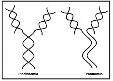
Types of Coiling in Chromonema:
(i) Plectonemic-coiling :- When both the chromonema are inter twined and can not be seperated easily. (in mitotic prophase chromosomes)
(ii) Paranemic coiling :- When both chromonema can be easily seperable. (In meiotic prophase)
Types of Chromosomes on the basis of Number of Centromere
(i) Acentric :- Chromosome without centromere.
(ii) Monocentric :- Chromosome with one centromere.
(iii) Dicentric :- When the number of centromere is two.
(iv) Polycentric chromosome :- When the number of centromere is more than two & diffused in throughout chromosome length.
Some Special Types of Chromosomes:
1. B-Chromosome/Accessory Chromosome/Supernuemerary Chromosome
- Discovered by Wilson in Metapodian Insect. Name supernuemerary chromosome was given by D. Jones -1975.
- These are heterochromatic & small sized chromosome. Thus, no phenotypic effects are known. (Morphological control)
- B-chromosomes also present in plants cells (Maize)
- They are supposed to be involved in ecological adaptation of organisms.
2. Mega Chromosomes
- Found in hybrid species of tobacco.
3. Isochromosomes
- When the both arms of the chromosomes are identical or genetically similar. Then chromosomes called as isochromosomes. If arm of a telocentric chromosome is splitted upto centromere then a metacentric chromosome with two identical arms is formed. such chromosome is called isochromosome.
4. Ring Chromosome
Prokaryotic chromosome are ring chromosome or consists of circular folded DNA without histone.
5. Sex Chromosome
May be XX or XY
6. HACs, MACs, YACs, BACs, etc.
Human Chromosomes
- The normal diploid (2N) chromosome number in human being is 46. It was given by T.H. Tijo and A. Levan in 1956.
- The chromosome complement of a cell depicting the number, size and form of the chromosome as seen in metaphase of mitosis is called karyotype and diagrammatic representation of karyotype is known as Idiogram or Karyogram.
- The chromosomes are morphologically numbered into 7 groups. (size and position of centromere) by conferance in Denver and Colorado.
Group (A) : 1–3 chromosomes of largest size and submetacentric or metacentric centromere.
Group (B) : 4–5 chromosomes with less larger size, submetacentric
Group (C) : 6–12 chromosomes with medium sized and submetacentric centromere.
Group (D) : 13–15 chromosomes, shorter than group ‘C’ with centromere near the end (Acrocentric). They are SAT chromosomes or satellite.
Group (E) : 16–18 chromosomes, short sized, with median (Metacenteric) or submedian centromere (Submetaceteric).
Group (F) : 19–20 chromosomes, short sized with median centromere.
Group (G) : 21–22 chromosomes, smallest in size, acrocentric and are also posess satellites.
- X chromosome is placed in ‘C’ group due to its larger size and submedian centromere.
- Y–chromosome is placed in group ‘G’ due to its short size but satellite absent and is acrocentric type.
|
221 videos|172 docs|126 tests
|
FAQs on Nucleus & Chromosomes - Biology for JAMB
| 1. What is the structure of the nucleus? |  |
| 2. What are the functions of the nucleus? | |
| 3. How are chromosomes related to the nucleus? | |
| 4. What is the significance of the nuclear envelope? | |
| 5. How does the nucleus contribute to cell division? | |


Exam
,Important questions
,mock tests for examination
,Free
,Viva Questions
,video lectures
,practice quizzes
,Semester Notes
,Nucleus & Chromosomes | Biology for JAMB
,Nucleus & Chromosomes | Biology for JAMB
,Sample Paper
,MCQs
,ppt
,shortcuts and tricks
,past year papers
,Previous Year Questions with Solutions
,Nucleus & Chromosomes | Biology for JAMB
,Summary
,Objective type Questions
,Extra Questions
,study material
;


Nucleus & Chromosomes Free PDF Download
Importance of Nucleus & Chromosomes
Nucleus & Chromosomes Notes
Nucleus & Chromosomes JAMB Questions
Study Nucleus & Chromosomes on the App
|
© EduRev
|
Education Revolution
|


|


