Human Circulatory System | Biology Class 11 - NEET PDF Download
| Table of contents |

|
| Human Circulatory System |

|
| What is Cardiac Cycle? |

|
| Steps in Cardiac Cycle |

|
| Electrocardiograph (ECG) |

|
Human Circulatory System
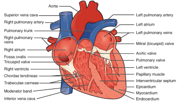
 Section of Human HeartThe human circulatory system, also known as the blood vascular system, is made up of a muscular, chambered heart, a network of closed, branching blood vessels, and blood, which is the fluid being circulated.
- The heart, which comes from the mesoderm during development, is located in the thoracic cavity, between the two lungs and slightly tilted to the left. It is about the size of a clenched fist and is protected by a double-walled membrane called the pericardium, which contains a fluid called pericardial fluid.
- The heart has four chambers:
- Two small upper chambers called atria.
- Two larger lower chambers are called ventricles.
- A thin muscular wall called the interatrial septum separates the right and left atria. A thicker wall called the interventricular septum separates the left and right ventricles.
- The atrium and ventricle on each side are separated by a fibrous tissue known as the atrioventricular septum, which has openings connecting the two chambers.
- The opening between the right atrium and right ventricle is guarded by the tricuspid valve, made of three muscular flaps. The opening between the left atrium and left ventricle is guarded by the bicuspid valve or mitral valve.
- The openings from the right and left ventricles into the pulmonary artery and aorta are equipped with semilunar valves.
- These valves ensure that blood flows in only one direction:
- From the atria to the ventricles
- From the ventricles to the pulmonary artery or aorta
- The heart is made entirely of cardiac muscle, with the walls of the ventricles being thicker than those of the atria.
- Specialized cardiac muscle tissue called nodal tissue isalso found in the heart. This includes:
- The sinoatrial node (SAN) is located in the upper right corner of the right atrium.
- The atrioventricular node (AVN) is situated in the lower left corner of the right atrium near the atrioventricular septum.
- The bundle, which is a bundle of nodal fibres, continues from the AVN, passes through the atrioventricular septum, and divides into right and left branches on top of the interventricular septum. These branches give rise to Purkinje fibres, which spread throughout the ventricular muscle.
- The nodal tissue can generate action potentials on its own without external signals, making it auto-excitable. However, different parts of the nodal system produce different numbers of action potentials per minute. The SAN generates the most, about 70-75 per minute, and is responsible for starting and maintaining the heart's rhythmic contractions, earning it the title of the pacemaker.
- On average, the heart beats 70-75 times per minute.

What is Cardiac Cycle?
The cardiac cycle refers to the sequence of events that occur during one complete heartbeat. It includes both the contraction (systole) and relaxation (diastole) phases of the heart chambers, specifically the atria and ventricles.
 Cardiac Cycle
Steps in Cardiac Cycle
- Initially, all four chambers of the heart are relaxed, known as joint diastole. Blood from the pulmonary veins and vena cava flows into the left and right ventricles through the open tricuspid and bicuspid valves, while the semilunar valves remain closed.
- Then, the sinoatrial node (SAN) generates an electrical signal, prompting both atria to contract simultaneously (atrial systole), increasing blood flow into the ventricles by 30%.
- This signal is relayed to the ventricles, causing them to contract (ventricular systole), while the atria relax (diastole), coordinating with ventricular contraction.
- Ventricular pressure rises, the tricuspid and bicuspid valves close to prevent backflow into the atria. Further pressure opens the semilunar valves, allowing blood to be pumped into the pulmonary artery and aorta.
- After contraction, the ventricles relax (ventricular diastole), and their pressure decreases, causing the semilunar valves to close, preventing backflow. As ventricular pressure continues to drop, the tricuspid and bicuspid valves reopen due to pressure from the blood in the atria, and the cycle restarts.
- This repeating sequence is called the cardiac cycle, comprising systole (contraction) and diastole (relaxation) of both atria and ventricles.
- Typically, the heart beats 72 times per minute, indicating a cardiac cycle duration of 0.8 seconds.
- Each ventricle pumps about 70 mL of blood per beat (stroke volume), and multiplying this by the heart rate gives the cardiac output, averaging 5000 mL or 5 liters per minute in a healthy person.
- The body can adjust stroke volume and heart rate, affecting cardiac output, as seen in athletes with higher cardiac outputs.
- During each cycle, two distinct sounds are produced and can be heard with a stethoscope: the first heart sound (lub) coincides with tricuspid and bicuspid valve closure, while the second sound (dub) accompanies semilunar valve closure. These sounds have diagnostic significance in clinical settings.
Electrocardiograph (ECG)
Electrocardiography (ECG) is a method used to obtain a graphical representation of the heart's electrical activity during a cardiac cycle.
 Electrocardiogram
- During a standard ECG, a patient is connected to an electrocardiograph machine with three electrical leads placed on the wrists and left ankle to monitor heart activity continuously.
- For a detailed evaluation, multiple leads can be attached to the chest region, but we'll focus on the standard ECG here.
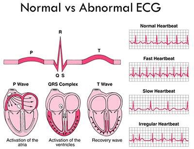
- Peaks in the ECG are labelled from P to T and correspond to specific electrical events in the heart.
- The P-wave indicates the electrical excitation (depolarization) of the atria, leading to their contraction.
- The QRS complex represents the depolarization of the ventricles, initiating their contraction and marking the beginning of systole.
- The T-wave signifies the repolarization of the ventricles, returning them to their normal state, and marks the end of systole.
- Counting the number of QRS complexes in a given time period allows determination of an individual's heart rate.
- ECGs from different individuals typically have similar shapes for a given lead configuration, so any deviation indicates a potential abnormality or disease, making ECGs clinically significant.
|
169 videos|401 docs|136 tests
|
FAQs on Human Circulatory System - Biology Class 11 - NEET
| 1. What are the main structures of the heart and their functions? |  |
| 2. What is the conducting system of the heart and why is it important? |  |
| 3. How does the regulation of the heartbeat occur? |  |
| 4. What is the cardiac cycle and what are its phases? |  |
| 5. What is an electrocardiograph (ECG) and what does it measure? |  |


