The Tongue & Ear | Biology A-Level - A Level PDF Download

EAR
SENSE ORGAN - EAR :
(B) Statoacoustic organ ear : - These are also called phonoreceptors.
All the vertebrates have one pair of ears back to the eyes,
There are two main functions of ears : -
(1) To receive sound waves, hearing
(2) To maintain body balance. Main function of ear is to maintain the balance of body.
Structurally ear may be divided into three parts : -
(a) External ear
(b) Middle ear
(c) Internal ear
(a) External ear : -
It is the outer part of ear. It is well developed in mammals only. External ear may be divided again into 2 parts
(i) ear pinna
(ii) ear canal
(i) Ear pinna : - These may be small or large, fan like structure, important featrue of mammals, but absent in whale, seal, Ornithorhynchus etc. The skin of ear pinna in hairy. These are having yellow elastic cartilage in them. A rabbit can move its pinna accroding to its will, just like dog, cat, cow etc. But a man can not move his pinnae. Muscles of man's ear are vestigeal. Pinna covers some of the ear canal, this part is called choncha.
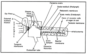
(ii) Ear canal or External auditory meatus : - It is a 24 mm long canal which is expanded from base of pinna to inner side.
Along with mammals, birds and reptiles also have ill or less developed ear canal.
At the end of ear canal a stretched, thin obliquely placed membrane is present, it is called ear drum or tympanic mmebrane.This separates the ear canal to middle ear.
In the wall of external auditory meatus or ear canal there are found modified sweat glands called ceruminous glands. These secrete cerumen or ear wax, which moisten the ear drum and protects it.
Ear drum remians always in stretched position because malleus ear ossicle pulls it towards tympanic cavity by tensor tympani muscle.
Ear drum is a part middle ear.
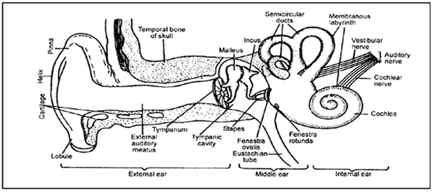
(b) Middle ear : - Middle ear is also called tympanic cavity. It is filled with air. This cavity is covered by a flask like bone called tympanic bulla. This bone is a part of temporal bone of skull.
Middle ear cavity is connected by pharyngeal cavity through a canal. It is called Eustachian duct.
Due to this tube, pressure at both the side of tympanic membrane remains always equal. This duct acts to maintain sound equilibrium. It exples high volume sounds through mouth, to avoid the damage of ear drum.
Tympanic cavity is connected by internal ear cavity by two aperture.
(i)Oval aperture fenestra ovalis (oval window) and
(ii)Spherical aperture fenestra rotundus (round window). A thin and firm membrane covers each aperture.
Three ear ossicle are present arranged in a chain with movable joints connected together in tympanic cavity.
These ear ossicles are : -
(a) Malleus : - It is situated towards outer ear. It is the largest of three and of hammer shaped malleus is formed by the modification of articular bone of jaw. Inner broad part of malleus is connected by incus. Malleus and incus and Joint together by synovial hinge joint.
(b) Incus : - The ossicle is anvil shaped. It is formed by the modification of quadrate bone of jaw. It s outer broad part is connected by malleus and inner thin part is connected by stapes. Incus is joint by stapes by ball and socket joint.
(c) Stapes : - It looks like stirrup of horse. It is formed by the modification of hyomandibular bone of jaw.
It is the smallest bone of body
Stapes is connected to incus at one side and on the other side it is connected to membrane stretched over fenestra ovalis.
[In the tympanic cavity of frog only one ear ossicle is found it is called columella auris. Malleus and incus are absent here.]
All the three ear ossicles are arranged in ear cavity by ligaments. These carry sound wave from ear drum to internal ear through fenestra ovalis.
Internal ear : -
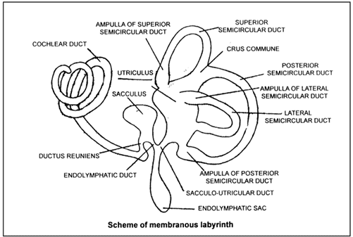
It consist of (1) Bony Labyrinth (2) Membranous Labyrinth.
Internal ear is enclosed in the petrous part temporal bone which form a bony capsule out side the internal ear it is called bony labyrinth. It is the cavity of hearing apparatus.
Internal ear is a complex structure made up of semi transparent membrane. It is called membranous labyrinth.
Bony labyrinth an membranous labyrinth are connected by a cavity called perilymph cavity. Perilymph liquid is filled in it.
Endolymph is filled in membranous labyrinth.
There are two main bag like chambers in membranous labyrinth, utriculus and sacculus.
Both these chambers are connected together by a thin canal called sacculo- utricular duct.
A thin endolymphatic duct opens into sacculo-utricular duct. This endolymphatic duct opens into endolymphatic sac situated at back side of skull on the other side.
Utriculus is comparatively large. Three semicircular canals arise from utriculus at 90° angle to each other and open into utriculus again these are called
(i)Anterior or superior semicircular canal
(ii) Posterior semicircular canal
(iii) External or lateral or horizontal semicircular canal.
Anterior and posterior canals arise in the form of a single canal called ''Crus commune''
The distal end of each semiciruclar canal is some what swollen, called Ampulla.
Sacculus is smaller than utriculus. Its back side is coiled like spring. It is called cochlear canal. it is also known as lagena.
The length of cochlear canal of human, rabbit and whale are , and coils respectively.
Cochlear canal is connected by sacculus by a small duct called ductus reuniens.
All the coils of cochlear canal are connected together by flexible ligaments.
In the centre of coils of cochlea in human, there are present a pillar like structure called modiolus.
(D) Internal structure of inner ear : -
The inner wall of membranous labyrinth is lined by cuboidal epithelium and outer wall is line dby connective tissue richly supplied with blood capillaries.
Membranous labyrinth is empty inside. Its cavity is filled by endolymph which is a milky, mucilagenous fluid.
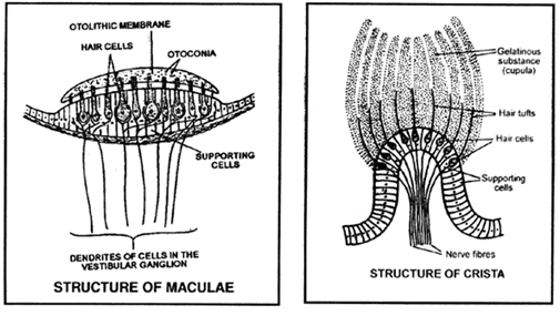
Distal end of each semicircular canal becomes swollen called ampulla. In this ampulla, internal cuboidal epithelium form a ridge like projection called acoustic ridge small immovable microvilli are found at the free edges of sensory cells of acoustic ridge. These microvilli are numerous in number. These are called stereocilia, along with these there are found single movable cilium called kinocillium. Otoconia are absent crista of ampulla. All the microvilli of ridge are bind together like a bag and from cupula.
These sensory cells situated in internal ear are in contact with small nerves. All these thin nerve combine to form vestibular nerve (branch of auditory nerve).
Sensory crista and maculae are related with equilibrium of body
Cristae control and maintain body equilibrium at the time of movement and maculae regulate this at static position.
(E)Internal Structure of Cochlea & Cochlear canal
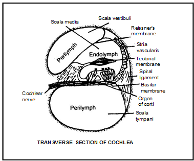
Cochlear duct is connected by bony labyrinth in such a way that it divides the cavity of labyrinth into dorsal and ventral chambers. So in a transverse section of cochlea following three chambers are seen clearly.
(i) Scala vestibuli : - it is situated at dorsal side and is filled with perilymph.
(ii) Scala tympani : - It is situated at the ventral side below the cochlear duct. It is also filled by perilymph.
(iii) Scala media : - It is the triangular cavity of cochlear duct that is situated between scala vestibuli and scala tympani. It is filled with endolymph.
Thin dorsal wall of cochlear duct is called vestibular membrane or Reissner's membrane.
Ventral wall of scala media is thick it is called basilar membrane. Scala vestibuli and scala tympani are connected through a small aperture at the free edge of cochlea. This aperture is called helicotrema.
Scala media is blind (closed) at its both the sides.
(F) Organ of Corti : - A sensory ridge is present at the whole of central line at epithelium lining of basilar membrane of scala media. It is called organ of corti. It has two types of cells (i) Sensory cell (ii) Supporting or suspensory cell and three type of suspensory cell
(i)Cells of Dieter's or basal cells (ii) Pillar cells or rod cells (iii) Hensen's cells or rectangular.
In between the empty spaces of sensory and suspensory cells a lymph like fluid cortilymph is filled.
This space is called tunnel of corti.
Numerous mircrovilli called stereocilia (sensory hair) are present at the free surface of each sensory cell.
At the ventral surface of sensory cells there are present thin fibres of auditory nerve that form cochlear branch.
At the organ of corti a thin jelly like membrane is inclined called tectorial membrane. In this membrane, all the sensory hair's free edges are embeded.
Main credit of hearing goes to ''Organ of corti''.
(G)WORKING OF EAR : -
Ears are stato-acoustic organs of body. Thus these help the body to hear and balancing the body.
(a) Equilibrim: - The first and basic function of ears to maintain balance of body.
This act is done by utriculus, sacculus and three semicircular canals. Equilibrium impulse/sensation is of two types : -
(i) Static balancing : - Its relation is from the point of view of gravity and position of head in static conditions of body and its changes.
The senses of these changes (of head) are produced and carried mainly by utriculus, sacculus and their sensory cristae i.e maculae.
Sensory hair of ridge are sensitized by otoconia or otolith or ear dust. These sensations or impulses are carried to brain by auditory nerve After it messages of appropriate reactions are send through motor fibres to the skeletal muscles of body.
(ii) Dynamic equilibrium : -
It is the action to maintain balance of body during movement.
This act is done by sensory ridges of ampula of semicircular canals.
At the time of movement the endolymph of ampula produces waves in it. Cupula of ampula are effected by these waves and sensory cells cupula are irritated. This sensation or stimulation is carried to brain by auditory nerve and proper messages are send to muscle of legs in reply. Due to this body is balanced at the time of walking.
(b) Hearing : -
This act is done by ''Organ of Corti''.
Sound waves are collected by ear pinnae. These sound waves travel through ear canal and hit the ear drum as a result of it ear drum get vibrated.
These vibrations reach up to stretched membrane of fenestra ovalis through ear ossicles, ear ossicles work as lever.
As a result of this travelling (from ear drum to fenestra ovalis) sound waves become more strong.
When the membrane of fenestra ovalis starts vibrating, perilymph of scala vestibuli also starts vibrating, some vibrations reach up to scala tympani (fenestra rotundus) and its perilymph.
Due to these vibrating waves, Reissner membrane and Basilar membrane of the walls of scala media also start vibrations. These vibrations travel through endolymph reach upto organ of corti. The organ of corti also starts vibrating.
Cochlear nerve carries this impulse to brain through auditory nerve. Appropriate messages are send to receptor organs by brain accordingly.
Vibrations /waves produced by cochlea travel through perilymph, reach up to membrane stretched at fenestra Rotundus and are destroyed.
Some sound waves are also destroyed, when coming from helicotrema.
DIFFERENCES OF RECEPTORS BETWEEN RABBIT AND MAN | ||
RABBIT |
| MAN |
Eye : |
|
|
1. Morocriar vision is present. | 1 | Eianaculsj vision is present |
2. Both eyes aie located or tte dorsal ard lateral side of heed. | 2. | Both eye are located at anterior parr of free |
3. Nictitating nieaihiBiie iipiesait. | 3. | Nictitatina merntrane is vestiaeal which is called as plica-setrilimarij |
4. Eye is red coloried. | 4 | Eye is tlact, hire, trown coloured |
Ear : |
|
|
1. Ear pirra i s fdrral shaped | 1 | Esr pimm is kidney shaped |
2. Ear pirna i s motile | 2. | Ear pinna is nan-motile |
3. No. of coilirs of cochlear is 2 1/2 | 3. | No, of cailinaaf cochlear canal is 2 3/4 |
NOSE
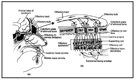
Olfactorecptors :
–Olfactoreceptors are situated in the upper part of nasal chamber in olfactory epithelium.
–This membrane is called as schnederian membrane.
–Olfactoreceptors are relatd with olfactory bulb. It is the extension of limbic system.
–This bulb is situated below the frontal lobe of cerebral hemisphere and above the ethampoid bone of nasalchamber.
–Three types of cells are found in the olfactoreceptors. These are –
(i) Bipolar olfactory nerve cells
(ii) columnar epithelial cells
(iii) Mucous glands
(1)Bipolar olfactory nerve cells : It is special types of nerve cells
– Sensory hair are found at the anterior end of olfactory cells. They contact with external environment in nasal chamber.
–Sensory hairs are related with dendrites of bipolar nerve cells.
–Middle part of olfactory cell is cyton.
–Posterior part of olfactory cell is axon which is nonmyleinated.
(2) Columnar epithelial cells : It is also called as supporting cells. They are present arounds the bipolar olfactory cells.
–They provides support to the olfactory cells.
–Some small conical cells are also found at the basal part of olfactoreceptor and provide base to the olfactoreceptor.
–A layer of connective tissue lies below the olfactoreceptor. It is also called as Lamina propria.
(3) Mucous glands : It is called as Bowman's gland. It is situated in the Lamina propria. It opens at the outer part of olfacto receptor through fine duct. Their secretory mucous substance dissolve the smell particle and carry to the sensory hair of olfactory cells. Unmyleinated axons of all olfacto sensory cells makes the synapse with dendrites of multipolar neurone of olfactory bulb. The number of receptors stimulated indicates the strength of smell.
In addition to smell receptor, a network of nerves is found in the nose, mouth and tongue.
The network formed by trigeminal nerve of V cranial nerve. It is also known as Dentist's nerve, reacts to messages of pain of teeth. It also convey the message of smell to brain. Such as ammonia, vinegar etc.
The trigeminal can protect by warning about harmful chemical in the air. Bowman's glands inside the nose release mucous fluid to get rid of the irritating susbtances.
Loss of the sense of smell is known as anosmia.
TONGUE OR ORGAN OF TASTE
A thick, muscular and movable organ, the tongue is found in the mouth cavity. Tongue bears four types of small papillae which are provided with taste buds. Taste buds are much numerous in the circumvallate and foliate papillae. Taste buds are formed by the transformation of epithelial cells of the tongue. A taste bud possesses two types of cells
1.Supporting cells : These cells are elongated in middle region they do not bear hairs at their free ends.
2.Sensory cells : These cells are alongated, buldge in middle part, they bear sensory hair at their free ends.
Each taste bud is flask or barrel shaped. It's size is 70 m × 50 m it's upper part opens at the epithelial surface of the tongue through a fine pore. These sensory hairs, exposed to outside through the gustatory pore are stimulated by the food substances. The sensory cells are chemoreceptor in nature and taste the food while it is dissolved in saliva. Food substances get mixed with saliva to enter into the pores of taste buds.
In human different regions of the tongue are sensitive to different taste. Anterior and free end of the tongue are sensitive to sweet and salty, lateral sides to sour taste, while the posterior part is particulary sensitive to bitter taste.
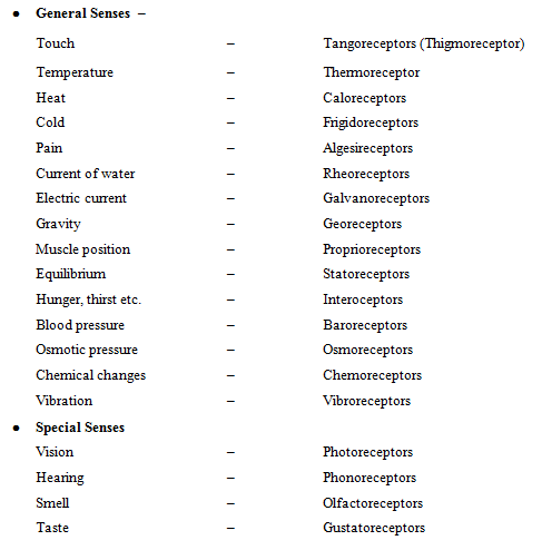
Types of receptors
(1)Receptors of vision, hearing an smell receive stimuli from distance, hence called teleoreceptors.
(2)Tangoreceptors or mechanoreceptors
(i) Merkel's disc (Corpuscles) : Epidermis of non hairy (glabrous) skin, shallow cup shaped disc.
(ii) Meissner's corpulse : Dermis of skin of the finger tip, lips and nipples. Sense of touch and gentle pressure.
(iii) Pacinian corpuscle : Present in subcutaneous tissue of palm, sole of finger etc. stimulated by strong pressure contact.
(iv) Corpuscle of golgi : Sucutaneous tissue of fingers.
(v) Corpuscle of mazzoni : Sub cutaneous tissue of fingers.
(vi) Grandy's corpuscles : Beak of birds
(vii) Herbst corpuscles : Mouth part of birds
(viii) Free never ending : Present of skin, perceive the sensation of touch.
(3)Thermoreceptors
(1) Ampullae of Lorenzini : Scoliodon (Fishes)
(2) Organ of ruffini : Caloreceptor - Heat
(3) End bulb of krause : Frigidoreceptor - cold
(4) Tactile receptors in mammals are maximum on face
(5) Current of water : Rheoreceptors lateral line sense organ in fishes and amphibian of tadpole detect the water current
POINTS TO REMEMBER :
1.Red green colour blindness is hereditory
2.Minimum distance for proper vision of eyes is 25 cm.
3.Anterior - posterior diameter of eyeball is 17.5mm at the time of birth normally and in adults it is 20-21mm.
4.The best colour differentiation is found in primates (Advanced mammals)
5.In the retina of man's eyes there are found 110-1125 lacs rods and 65 lacs cones.
6.Healthy eye of a person can see clearly from 12 inch to 20 feet.
7.Image of object is formed on retina and it is always inverted & real
8.Hyalocytes cells are found in vitreous humor.
9.Cilliary body secretes aqueous humor and vitreous humor.
10.In frog and other amphibians sclerotic layer of eyeball is Cartilaginous
11.The largest eyes are found in deers in vertebrates with respect to body surface area.
12.Owls and cats see only with the help of available light from starts or moon at night
13.The lens of man's eye ball has its diameter of 11 mm.
14.Atropine, Belladona and Cocane medicines are used to dilate the pupil
15.In a newlyborn child, eye balls are very small, i.e. babies ar always very much hypermatropic.
17.Cornea and lens of eye lack blood supply.
18.Eyes are most sensitive to the light having approx 5000 Å wavelength.
19.Internal or inner ear of rabbit is originated by ectoderm of embryo and middle ear (Bony part-mesodermal) and eustachian tube are originated by endoderm layer of embryo.
20.Frog's vision is hypermatropic in water and myopic on land.
21.Light sensitive organ was discovered by Steven.
22.Phaco-emulsification techique in cataract surgery – ''Stichlesss'' technique. Foldable IOL (Intra – ocular Lens) is used..
23.Gland of moll are modified sweat gland.
24.Stye is infection of gland of zeis
25.Hordeolum is inflammation sebaceous gland of eyelid
26.The relationship of receptor to bipolar cells to ganglion cells is 1 : 1 : 1 with in the fovea.
27.From the fovea to the periphery, cones dimimish and rods increase in number.
28.Electronic activity of retina is record sequence of potential change known as elctroretinogram.
29.The horizontal cells which transmit signals horizontally in the outer plexiform layer from the rods and cones to the bipolar cell dendrites.
30.The bipolar cells which transmit signals from the rods, cones and horizontal cells to the inner plexiform layer where they synapse with ganglion cell and amacrine cells.
31.The amacrine cells which transmit signals in two direction directly from bipolar cells to ganglion.
32.Lack of red cones – Protanope
33.Lack of green cones – Deuteranope
34.lack of blue cones – Tritanope
35.Unlike nerve and muscle rods and cones do not show action potential by depolarization but by electronic conduction.
|
279 videos|162 docs|142 tests
|
FAQs on The Tongue & Ear - Biology A-Level - A Level
| 1. What is the NEET exam? |  |
| 2. How can I apply for the NEET exam? | |
| 3. What is the eligibility criteria for the NEET exam? | |
| 4. What is the syllabus for the NEET exam? | |
| 5. How is the NEET exam conducted? | |


Objective type Questions
,mock tests for examination
,Sample Paper
,The Tongue & Ear | Biology A-Level - A Level
,The Tongue & Ear | Biology A-Level - A Level
,Important questions
,MCQs
,past year papers
,Free
,Semester Notes
,Viva Questions
,Extra Questions
,video lectures
,Previous Year Questions with Solutions
,study material
,ppt
,Summary
,Exam
,The Tongue & Ear | Biology A-Level - A Level
,shortcuts and tricks
,practice quizzes
;


The Tongue & Ear Free PDF Download
Importance of The Tongue & Ear
The Tongue & Ear Notes
The Tongue & Ear A Level Questions
Study The Tongue & Ear on the App
|
© EduRev
|
Education Revolution
|


|


