Cardiac Cycle & ECG | Biology A-Level - A Level PDF Download
Introduction of Cardiac Cycle
The cardiac events that occur from the beginning of one heartbeat to the beginning of the next are called cardiac cycle. The action potential travels rapidly through both atria and then through the AV bundle into the wall of ventricles. Because of special arrangement of the conducting system from the atria to the ventricles, there is a delay of more than 1/10th of a second between passage of the cardiac impulse from the atria into the ventricles.
This allows the atria to contract ahead of the ventricles, there by pumping blood into the ventricles before the strong ventricular contraction begins.
Thus the atria are the primer pumps for the ventricles, and ventricles then provide the major source of power for moving blood through the vascular system.
Cardiac-Cycle
The process of heart beat begins from the time of embryonal development. Once the heart beat starts, it continues through out the life (inherent capacity). In resting stage of man in 1 minute the heart beats around 72 times and during this 1 minute, 5 litres of blood is pumped to different parts of the body through heart through left ventricle.The serial wise or sequential changes which take place in the heart are called cardiac-cycle.
The contraction of the auricles is termed as auricular systole or atrial-systole, and their relaxation is called atrial diastole.
Same way the contraction and relaxation of ventricles is termed as ventricular systole and ventricular diastole.
The time of cardiac -cycle is the reverse ratio of heart beat per minute. If heart beat per minute is 72, then the time of cardiac-cycle is 60/72 = 0.8 seconds.

Joint Diastole: 0.8 – 0.4 = 0.4 sec. (Period during which entire heart is in Diastole)
In a single cardiac cycle of man (1) Auricular systole = 0.1 sec (2) Auricular diastole = 0.7 sec (3) Ventricular systole = 0.3 sec (4) Ventricular diastole = 0.5 sec |
Following events are related to the Cardiac-cycle:

(1) Ventricular-Systole
It is an important process because during it the blood is pumped out of the heart into the arteries. It has four main parts:(a) Isometric-Contraction: Walls of the ventricles start contracting, due to which pressure is more in the ventricles. Due to the increase of this pressure the " Cuspid valves " close producing "LUBB' sound.
(b) Period of Ejection: During this cycle when pressure increases in the ventricles, then the semi-lunar valves of the arches open and blood rapidly enters into the arches pushing the valves on one side.
Oxygenated blood from the left-ventricle enters into the carotico-systemic arch or aorta. and deoxygenated blood from the right-ventricle enter into the pulmonary-arch.
(c) Protodiastole: Due to the ejection of blood from the ventricles now the inter-ventricular pressure decreases and the rate of blood ejection from the ventricles also decreases.
(d) Isometric Relaxation: When due to blood-ejection, the pressure inside the ventricles decreases as compared to the pressure inside the arches.The blood stops moving out and the ventricles prepare for relaxation.
During ventricular systole, the auricles receive blood from the veins.
(2) Ventricular Diastole
Ventricles start relaxing now due to which pressure inside them falls further. As a result of this, closure of semi lunar valves occurs due to which 'DUP' sound is heard at the onset of ventricular diastole.Ventricular-diastole has two sub-stages
(a) Rapid in Flow: After the systole in the ventricles the systolic pressure reduces very much. This pressure becomes very less than the atrial-pressure. Moreover due to relaxation in ventricles the pressure inside them falls further. So, now the cuspid -valves open up and blood flows rapidly from the auricles to the ventricles. S3 heart sound is produced.
(b) Diastasis: After rapid in flow, the auricles transfer the blood to the ventricles at the same rate at which they receive blood from the veins. so the inflow of blood reduces considerably. At this moment pressure inside all four chambers is equal and entire heart is in diastole. Also at this moment of this time, the AV valves are open but semilunar valves are closed.
(3) Auricle Systole
Due to contraction in the auricles the remaining blood comes into the ventricles so the atrial pressure now becomes zero. S4 heart sound is produced.(4) Auricle Diastole
Auricle start relaxing now. Due to the presence of almost zero pressure in the auricles, during diastole the auricles start receiving further blood from the veins.

Volumes of blood related with cardiac cycle. During diastole, filling of the ventricles normally increases the volume of each ventricle to about 120 mililitres. This volume is known as end diastolic volume. Then as the ventricles empty during systole, the volume decreases by about 70 mililitres, which is called the stroke volume (i.e. the volume of blood pumped by each ventricle in the aorta in one stroke or beat). The remaining volume in each ventricle is now about 50 mililitres is called end systolic volume. The fraction of the end diastolic volume which is ejected out is called the ejection fraction. (usually around 60% or 7/12). EF = SV/EDV Cardiac output it is the amount of blood pumped by the each ventricle per minute. Its value in a normal adult is about 5 litre/minute. Cardiac output = stroke volume x heart rate.
Stroke Volume = EDV – ESV = 70 ml (approx)
|
Heart-Sound
(1) 1st Sound - This is a contraction sound which denotes the beginning of ventricle-contraction. It arises due to closing of mitral valve and the tricuspid valve. It is weak and appears in the form of " Lubb " (L - U - B - B)
(2) 2nd Sound - This is a diastolic sound which denotes the beginning of ventricular diastole. This arises due to the closing of the semi-lunar valves of the two arches and is heard in the form of" Dup". It is shrill than the 1st sound and takes less time.
These "Lubb" and "Dup" sounds of the heart can be heard with the help of an instrument called " Stethoscope."
The study of heart-sounds by marking them on a Graph is termed as "Phono-Cardiography"
The measurement of the electrical-activity of the cardiac muscles at the time of heart-beat is necessary for the healthy working of the heart. The transmission of impulses in the sarcolemma of cardiac-muscle fibres is in the form of electro-chemical waves.
The graph which is marked by the machine due to the voltage difference is termed as the " E.C.G." or "
Electro Cardio Gram" and this process is termed as " Electro Cardio Graphy"
It was first of all recorded by " Waller"
"Einthoven" is known as the father of Electro Cardio Graphy.
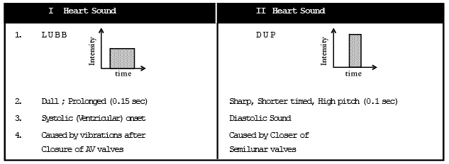
Murmur :- Any abnormal Heart sound other than LUBB or DUP. This may be due to physiological reasons like the increased volume of blood or pathological reason like defects in the valves. Narrowing of valves in called valvular stenosis.
What is an Electrocardiograph?
- An electrocardiograph or ECG is a test used to measure the electrical activity of the heart. The test takes only about a few minutes and is devoid of any pain.
- The electrical activity of the heart causes the heart muscles to contract that results in the pumping of the heart. The ECG is in the form of spikes and dips known as waves. The wave pattern helps in assessing the rate and rhythm of our heartbeat.
- The human heart produces an electrical impulse by itself. As this electrical impulse passes through our heart, it generates an electrical current that spreads over our body and reaches the skin.
- The patient is connected to the Electrocardiograph (ECG) machine with three electrical leads (one each to both wrists and the third to the left ankle of the patient), that is used to monitor the activity of the heart. This is standard ECG testing.
Process
The process of electrocardiograph includes:
- Small sticky electrodes are attached to the arms, chest and legs.
- These electrodes are connected to the ECG machine through wires that help in detecting the electrical impulses occurring at each heartbeat.
- These electrodes usually detect the very minute form of changes in an electrical path on the skin which arises from the heart muscles and the electrophysiologic patterns of the depolarizing during every heartbeat.
Explanation of the Electrocardiograph
- P to T in the graph represents a specific activity of the heart. Let’s break it down.
- The P wave is the electrical excitation of the atria, or depolarization, initiating atrial contraction.
- The QRS complex is the depolarization of ventricles, initiating ventricular contraction. Marking the beginning of the systole.
- T wave means the return of ventricles to the normal state (repolarization). Marking the end of the systole.
- By counting the number of QRS complexes we can evaluate the heartbeat rate of the patient. Any deviations in this shape results in heart diseases or an abnormal heart rhythm which can either be slow, irregular or very fast heartbeats. Hence it is essential equipment in the field of medicine.
Medical Uses of ECG
The main goal of electrocardiography is to obtain information regarding the heart’s electrical impulses. This means it can find evidence of past heart attacks or even any undiagnosed heart disease. The medical uses of such information are very valuable and grant a deeper insight into conditions like :
Seizures
Fainting
Pulmonary embolism
Cardiac dysrhythmias
Myocardial infarction or heart attack
Arrhythmia
Deep vein thrombosis
Ventricular hypertrophy
It also proves itself useful in applications such as:
Biotelemetry of the patient
The testing of Cardiac stress
Diagnosis of structural heart diseases
Monitoring the effects of heart medication
Assessing the severity of the abnormalities in the electrolyte
The monitoring of the form of anaesthesia that is involved
CTA- Computed tomography angiography and also the MRA- Magnetic resonance angiography of the heart
The screening of Hypertrophic cardiomyopathy in adolescents as a part of the sports-related deaths, such as sudden cardiac death.
Why is an ECG done?
An electrocardiograph is done for the following reasons:
1. To check the heart health in case of other diseases such as diabetes, high blood pressure, high cholesterol, etc.
2. To check the thickness of the chambers of the heart wall.
3. To monitor if the medicines are causing any side-effects.
4. To check if the mechanical devices implanted in the heart are working properly or not.
Old NCERT Syllabus
Filling of Heart (Ventricles)
Blood normally flows from the great veins into the atria. About 75% of the blood flows directly through the atria into the ventricles even before the atria contracts. Then atrial contraction usually causes an additional 25% filling of the ventricles.
The period of Atrial Systole – Fills 25% of ventricles.
The period of Atrial Diastole fills 75% of ventricles

Distribution of Blood Flow in various organs at rest in Man
Brain | 700 ml/mt | (14%) approx |
Heart | 300 ml/mt | (7%) |
Muscles | 1200 ml/mt | (20%) |
Skin | 500 ml/mt | (8%) |
Kidney | 1100-1300 ml/mt | (20 - 25%) |
Abdominal organs | 1400 ml/mt | (20-25%) |
Others | 600 ml/mt | (10%) |
Total | 5800 ml/mt |
|
Types of ECG Test
There are three main types of ECG tests:
1. Resting ECG
This type of ECG is used to examine the electrical activity of the heart at rest. While performing this test, the patient is asked to relax and then, their heartbeat is recorded.
2. Exercise ECG
This type of ECG is used to examine the electrical activity of the heart during stress or exercise. In this test, a patient is asked to run or walk on the treadmill or a cycle while the heartbeat is recorded.
3. 24-hour ECG
As the name suggests, this type of ECG is conducted for 24 hours. The heart’s electrical impulses are measured by a device called the Holter Monitor.
|
280 videos|166 docs|147 tests
|
FAQs on Cardiac Cycle & ECG - Biology A-Level - A Level
| 1. What is an electrocardiograph? |  |
| 2. How does an electrocardiograph work? | |
| 3. What is the significance of the cardiac cycle in an electrocardiograph? | |
| 4. Can an electrocardiograph detect all heart conditions? | |
| 5. Is an electrocardiograph safe and non-invasive? | |

|
6.4K Views |

|
4.89/5 Rating |

|
Dec 23, 2024 Last updated |

|
Explore Courses for A Level exam
|
|
Extra Questions
,shortcuts and tricks
,mock tests for examination
,Summary
,practice quizzes
,Free
,Previous Year Questions with Solutions
,Cardiac Cycle & ECG | Biology A-Level - A Level
,past year papers
,Viva Questions
,Cardiac Cycle & ECG | Biology A-Level - A Level
,Cardiac Cycle & ECG | Biology A-Level - A Level
,ppt
,study material
,Objective type Questions
,MCQs
,video lectures
,Exam
,Sample Paper
,Semester Notes
,Important questions
;


Cardiac Cycle & ECG Free PDF Download
Importance of Cardiac Cycle & ECG
Cardiac Cycle & ECG Notes
Cardiac Cycle & ECG A Level Questions
Study Cardiac Cycle & ECG on the App
|
© EduRev
|
Education Revolution
|


|


