Oogenesis & Types of Eggs | Additional Study Material for NEET PDF Download
Oogenesis
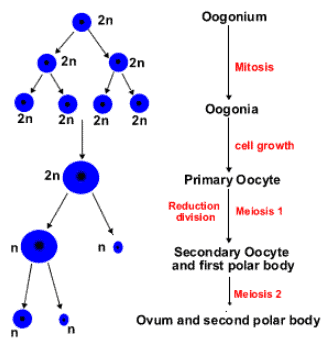
Like spermatogenesis, oogenesis process also can be divided into three stages:
- Multiplication phase
- Growth phase
- Maturation phase

Multiplication phase
- In this stage primordial germ cells or ovum mother cells repeatedly divide by mitosis to form large number of diploid oogonia.
- This process completes in embryo stage of female in most higher animals.
Growth phase
- Like spermatogenesis, in this process oogonia grow in size and form primary oocytes.
- The growth phase is the longest phase in oogenesis (except - in humans maturation is longest phase).
- During growth phase size of egg increases many times.
- During growth phase several changes occur in egg and all these changes are classified in 2 sub-stages:
- Previtellogenesis
- Vitellogenesis
(i) Previtellogenesis: During previtellogenesis, changes occur in nucleus and cytoplasm of egg.
Changes in nucleus
- Amount of nucleoplasm increases in nucleus.
- Number of nucleolus increases in nucleus.
- Formation of lampbrush chromosome starts.
- Activity of DNA increases in nucleus, as a result DNA become highly active and rapidly synthesizes different types of RNA. Increased activity of DNA is called as Gene redundancy / Gene amplification. Due to all these changes, size of nucleus increases and nucleus becomes vesicular. This vasicular nucleus is called germinal vesicle.
Changes in cytoplasm
- In cytoplasm, rate of protein synthesis increases. Cytoplasm rapidly synthesizes different type of protein and enzyme. Due to more availability of protein and enzymes, synthesis of new protoplasm takes place and size of egg increases.
- Number of cell organelles increase in cytoplasm, specially endoplasmic reticulum, golgi-body and mitochondria.
- Mitochondria become very large in number so mitochondrial clouds are found in cytoplasm of egg.

- Later on all these 3 cell organelle (golgi body, endoplasmic reticulum, mitochondria) are arranged in the form of ring around the nucleus, it is called as Balbiani vitelline ring. In this stage golgi body of egg secretes a membrane around the egg which is it called as vitelline membrane. A space appears in between plasma membrane of egg and vitelline membrane called as perivitel line space, Itis filled with a fluid called peri vitelline fluid.
- At the end of previtellogenesis endoplasmic reticulum disappear. Golgi bodies gets converted into corticle granule. Corticle granules are filled with mucopolysacharide. Large number of changes occur in mitochondria also.
(ii) Vitellogenesis: During vitellogenesis egg stores food in the form of yolk. Some part of yolk is synthesised in egg only, but major part of yolk is received from liver. Yolk received from liver is less viscous and is therefore soluble but this type of food cannot be stored for long periods (because it easily gets converted into simple form). So mitochondria of egg with the help of kinase enzyme make the yolk more viscous and insoluble.
2 types of yolk is found:
- Granular yolk occurs in the form of fine granules. eg. Protostomia animals.
- Yolk platelets occur in the form of plate disc like granule. eg. Deuterostomia animals (higher animals)
Chemical composition of yolk
- Most abundant compound in yolk is phospholipid .Most common phospholipid is lecithin.
- Yolk contains different type of protein:
- Simple protein: Albumin, Globin, Globulin
- Phospho-protein: Phosvitene, Ovovitelline
- Lipo-protein: Lipovitelline
- In yolk, least amount of substance found is carbohydrate.
Maturation phase
- Oogenesis takes place in the ovaries. In contrast to males the initial steps in egg production occur prior to birth. By the time the foetus is 25 weeks old, all the oogonia that she will ever produce, are already formed by mitosis. Hundreds of these diploid cells develop into primary oocytes, begin the first steps of the first meiotic division, proceed up to diplotene and then stop any further development. The oocytes grows much larger and completes the meiosis I, forming a large secondary oocyte and a small polar body that receives very little amount of cytoplasm but one full set of chromosomes.
- In humans (and most vertebrates), the first polar body does not undergo meiosis II, whereas the secondary oocyte proceeds as far as the metaphase stage of meiosis II. However, it then stops advancing any further, it awaits the arrival of the spermatozoa for completion of second meiotic division. Entry of the sperm restarts the cell cycle breaking down MPF (M-phase promoting factor and turning on the APC (Anaphase promoting complex).Completion of meiosis II converts the secondary oocyte into a fertilised egg or zygote (and also a second polar body)
- Ova are derived from oogonia present in the cortex of ovary. Some important differences between oogenesis and spermatogenesis are:
- Whereas one primary spermatocyte gives rise to four spermatozoa, one primary oocyte forms only one ovum.
- When the primary spermatocyte divides, its cytoplasm is equally distributed between the two secondary spermatocytes formed. However, when the primary occyte divides, almost all its cytoplasm goes to the daughter cell which forms the secondary oocyte. The other daughter cell (first polar body), receives half the chromosomes of the primary oocyte, but almost no cytoplasm.
- The first polar body is, therefore, formed merely to get rid of unwanted chromosomes.
Types of Eggs
- On the basis of amount of yolk
- Alecithal: In this type of egg, yolk is negligible.
Examples: Human egg. - Microlecithal or Oligolecithal eggs: The amount of yolk is very small in these types of eggs. (oligolecithal , or microlecithal or alecithal).
Examples: Egg of Amphioxus, Eutheria, Metatheria and sea - urchin. - Mesolecithal Eggs: In this type of egg, the amount of yolk is moderate i.e. medium, neither more nor less.
Examples: Eggs of Amphibia , Petromyzon and lung-fishes. - Polylecithal or Macrolecithal or Megalecithal eggs: Eggs are with large amount of yolk.
Examples: Insect's egg, Birds, reptiles and proteotheriam mammals
- Alecithal: In this type of egg, yolk is negligible.
- On the basis of destribution of yolk
- Isolecithal or homolecithal eggs: The yolk is evenly or homogenously distributed in these eggs.
Examples: micro, oligo or alecithal eggs. - Telolecithal eggs: The yolk is concentrated in one part of the egg.
Examples: mesolecithal eggs of amphibia. (Moderately telolecithal) - Discoidal eggs: A type of teloleithal and megalecithal eggs, Where the yolk is in enormous quantity and concentrated in one part of the egg. Thus only a disc of cytoplam called germinal disc remains in the egg which is located at the other pole of egg. (Heavily telolecithal)
Examples: Eggs of reptiles, birds and prototherian mammals. - Centrolecithal eggs: Megalecithal eggs where the enormous amount of yolk is located in the centre and cytoplasm is in the form of superficial layer around the yolk.
Examples: Insects egg.
- Isolecithal or homolecithal eggs: The yolk is evenly or homogenously distributed in these eggs.
- Classification of Eggs on the basis of Shell: On the basis of shell, eggs are of 2 types
- Cleidoic eggs :- eggs surrounded by a hard shell are known as cleidoic eggs. These eggs are found in those animals which have a terrestrial mode of life or which lay eggs on land. These eggs have more amount of yolk. These are adaptations to terrestrial mode of life. Shell prevents the egg from dessication.
Examples: eggs of "Reptiles", "Birds", "Terrestrial Insects " and "Prototherians". Reptilia eggs are called leathery eggs. - Non - Cleidoic eggs :- Eggs which are not surrounded by a hard shell are called non-cleidoic eggs
Examples: all viviparous animals (Mammals) and all oviparous animals which lay eggs in water (Amphibians).
- Cleidoic eggs :- eggs surrounded by a hard shell are known as cleidoic eggs. These eggs are found in those animals which have a terrestrial mode of life or which lay eggs on land. These eggs have more amount of yolk. These are adaptations to terrestrial mode of life. Shell prevents the egg from dessication.
Structure of an Oocyte
- The nucleus of egg is also called germinal vesicle.
- Oocyte is surrounded by membranes termed as the egg-membranes.
- Oocyte / Ovum along with the egg-membrane are termed as the egg.
- Egg = Ovum / Oocyte + Egg membrane.
- Majority eggs are oval but the eggs of insects are long and cylindrical. Smallest eggs are of 50m in polychaeta and the largest eggs are of an Ostrich.
Classification of egg - membranes
On the basis of origin, egg- membranes are of 3 types:
- Primary egg membrane: This membrane is secreted by the oocyte itself. eg. Vitelline membrane, Zona Pellucida (mammals), Zona radiata (Shark and Some amphibians)
- Secondary egg membrane: This is found outside the primary egg membrane and is secreted by the ovary. (eg. Corona radiata, Chorion)
- Tertiary egg membrane: This is present outside the primary egg membrane. It is either secreted by the uterus or the oviduct. (eg. Jelly coat, Shell & Shell membrane)
Functions of Egg-membranes
- To provide protection
- To check polyspermy
- To provide buoyancy to the amphibian eggs
Different types of eggs
1. Insect Egg
- Eggs of insects are megalecithal or polylecithal In them yolk is present in the centre, so the eggs are also centrolecithal.
- Two egg membranes are present here, inner vitelline membrane (primary) and outer chorion (secondary).
- The sperm enters the egg through micropyle because on the head of insect sperm acrosome is absent.
The Cytoplasm here is found in two parts:
- Central cytoplasm :-It is present in a very small amount in the center of the egg. Egg nucleus is located in it.
- Peripheral Cytoplasm :- It is present in a very small amount along the periphery of the egg.

2. Frog's Egg
- Eggs of frog are moderately Telolecithal & Mesolecithal .

Two types of egg membranes are found in frogs egg
- Vitelline membrane : This is primary egg membrane which is secreted by the ovum around itself.
- Jelly coat : This is tertiary egg membrane, secreted by oviduct. Jelly coat has air bubbles trapped in it due to which it floats an water. This group of frogs egg is called spawn, Jelly coat is bitter in taste so enemies do not eat it. Secondary egg membrane is absent in frog's egg.
Internal part of the egg is divided in two parts
- Animal pole
- This part has cytoplasm, egg nucleus in also located in this part. In the cytoplasm melanin granules are found which prevent the egg from harmful radiations. They also help in protection of egg by camouflage.
- A sperm always enters into the ovum at some point in animal hemisphere. This point is normally other than the animal pole itself.
- As the sperm enters into the ovum, taking some pigment granules with it, a grey, crescent shaped region appears in the equatorial zone geometrically opposite to the sperm entrance point. This region is called grey crescent. It is formed due to movement of some pigment granules away from it towards sperm entrance point.
- The area of sperm entrance point marks the anterior side of future embryo. The side diagonally opposite to it in the vegetal hemisphere marks the future posterior side. Thus the sperm entrance establishes the anteroposterior and dorsoventral axis as well as bilateral symmetry of future embryo.
- Vegetal pole
- Yolk is concentrated in this part of egg.
3. Chick Egg
- These eggs are megalecithal or polylecithal and discoidal eggs.
- In these eggs, yolk is present in the centre of the egg in the form of a dense mass. The cytoplasm of the egg is in the form of a disc above the yolk, which is termed as the germinal- disc.
- Yolk is of 2 types i.e. yellow yolk and white yolk.
- Yellow - yolk has more amount of phospholipids. White yolk has less amount of phospholipids. Central white part of yolk is called latebra.
- Both the types of yolk are arranged in alternate and concentric layers.
- Pander was the scientist , who discovered the 3 germinal layers i.e. Ectoderm Mesoderm and Endoderm in chick - egg.

- Around the egg, a porous shell of CaCO3 is present which is secreted by the cells of posterior ligamental part of the oviduct.
- In between the vitelline membrane and the shell membrane albumin is filled which is also called the white of egg. It contains 13 % proteins.
Thick albumin-fibres termed as "Chalaza" are present in the albumin part of egg.
4. Egg of Mammals
- Mammalian eggs have very less amount of yolk, so the eggs are oligolecithal and isolecithal or microlecithal and homolecithal .
The egg has 2 egg-membranes
- Zona pellucida :- This is a transparent membrane like covering and is a primary membrane secreted by the ovum/oocyte itself.
- Corona radiata :- This is a layer of follicular cells" and these cells are attached to the surface of egg through " hyaluronic acid" This is a secondary membrane, which is secreted by the ovary. These eggs don't have tertiary membrane. Mammalian eggs are approx 0.1 mm in size.
|
26 videos|287 docs|64 tests
|
FAQs on Oogenesis & Types of Eggs - Additional Study Material for NEET
| 1. What is oogenesis and how does it differ from spermatogenesis? |  |
| 2. What are the three main stages of oogenesis? |  |
| 3. What are the two types of eggs produced during oogenesis? |  |
| 4. How does oogenesis contribute to genetic diversity? |  |
| 5. What factors can affect oogenesis? |  |

|
Explore Courses for NEET exam
|

|


