MCAT Exam  >  MCAT Tests  >  Test: Vision (Sight) - 1 - MCAT MCQ

Test: Vision (Sight) - 1 - MCAT MCQ


Test Description

10 Questions MCQ Test - Test: Vision (Sight) - 1

Test: Vision (Sight) - 1 for MCAT 2024 is part of MCAT preparation. The Test: Vision (Sight) - 1 questions and answers have been prepared according to the MCAT exam syllabus.The Test: Vision (Sight) - 1 MCQs are made for MCAT 2024 Exam. Find important definitions, questions, notes, meanings, examples, exercises, MCQs and online tests for Test: Vision (Sight) - 1 below.
Solutions of Test: Vision (Sight) - 1 questions in English are available as part of our course for MCAT & Test: Vision (Sight) - 1 solutions in Hindi for MCAT course. Download more important topics, notes, lectures and mock test series for MCAT Exam by signing up for free. Attempt Test: Vision (Sight) - 1 | 10 questions in 10 minutes | Mock test for MCAT preparation | Free important questions MCQ to study for MCAT Exam | Download free PDF with solutions
Test: Vision (Sight) - 1 - Question 1

The tri-chromatic theory of color vision describes the types of cones. Which of these is NOT a type of cone in humans?

Detailed Solution for Test: Vision (Sight) - 1 - Question 1

The trichromatic theory of color vision states that there are three types of cones in humans, each sensitive to different wavelengths of light. These three types are commonly referred to as red, green, and blue cones. They enable us to perceive a wide range of colors by combining the signals from these cones in various proportions. However, yellow cones are not a recognized type of cone in humans. The perception of yellow color arises from a combination of signals from red and green cones in our visual system.

Test: Vision (Sight) - 1 - Question 2

Which of these is the correct order to the steps of the phototransduction cascade in reaction to light?

Detailed Solution for Test: Vision (Sight) - 1 - Question 2

The correct order of steps in the phototransduction cascade in reaction to light is as follows:

  • A rod turns from "on" to "off": When light strikes a rod photoreceptor, it triggers a hyperpolarization response, causing the rod to turn from an "on" state to an "off" state.
  • Turns a bipolar cell on-center: The "off" response of the rod leads to the activation of bipolar cells in the retina. In the on-center configuration, the decrease in neurotransmitter release from the rod stimulates the bipolar cell, resulting in its depolarization.
  • Activates a retinal ganglion cell: The depolarization of the bipolar cell then leads to the activation of a retinal ganglion cell, which sends signals to the brain through the optic nerve, ultimately contributing to visual perception.
1 Crore+ students have signed up on EduRev. Have you? Download the App
Test: Vision (Sight) - 1 - Question 3

The blind spot is the area where the optic nerve connects to the retina. The ‘night blind spot’ occurs under conditions of low light and can extend 5 to 10 degrees from the center of the person’s field of view. What is the cause of the ‘night blind spot’?

Detailed Solution for Test: Vision (Sight) - 1 - Question 3

he 'night blind spot' refers to a reduced ability to see clearly in low-light conditions, particularly in the peripheral vision. This phenomenon occurs because the fovea, which is responsible for high visual acuity and contains a high density of cones, is less active in dim light. Instead, scotopic vision, which primarily relies on the function of rods, is dominant in low-light situations.

Rods are more sensitive to light and better suited for detecting dim light. However, the fovea contains a high concentration of cones, which are responsible for photopic (daytime) vision and color perception but are less sensitive in low-light conditions. Therefore, when the visual system primarily relies on scotopic vision (using rods) in low light, the fovea, with its cone-dominant composition, contributes less to the overall visual perception. This lack of input from the fovea in scotopic vision is the cause of the 'night blind spot' extending 5 to 10 degrees from the center of the person's field of view.

Test: Vision (Sight) - 1 - Question 4

Scientists selectively reared kittens in either an environment with lines of only vertical or only horizontal orientation. After 5 months, the scientists recorded from neurons in the cats’ cortex and found many neurons which responded to vertical lines in vertically reared cats, but no neurons that fired to horizontally oriented lines. The opposite occurred in horizontally reared cats; Horizontally reared cats had many neurons that responded to horizontal lines, but none that responded to vertical lines. What has likely happened to the cats?

Detailed Solution for Test: Vision (Sight) - 1 - Question 4

The observed results suggest that the cats' visual systems underwent changes based on the selective rearing conditions. The neurons in the cats' cortex responded selectively to the orientation of lines that matched the rearing environment. This phenomenon can be attributed to experience-dependent plasticity, which refers to the brain's ability to modify its neural connections and organization in response to environmental experiences.

In this case, the vertically reared cats developed neurons that specifically responded to vertical lines, while the horizontally reared cats developed neurons that specifically responded to horizontal lines. This indicates that the visual system of the cats adapted to process the prevalent orientation of lines in their rearing environment.

The cats' ability to adapt and show specific neural responses to the lines in their environment suggests that their visual systems underwent plastic changes based on their rearing experiences. This phenomenon highlights the brain's remarkable capacity to reorganize and adapt to environmental input, which is a characteristic feature of experience-dependent plasticity.

Test: Vision (Sight) - 1 - Question 5

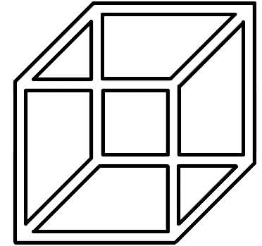
The figure below is called a Necker cube. Opposite sides are parallel, and the cube’s orientation is ambiguous, alternating between the front face being the lower left or the upper right. Which cognitive mechanism creates this illusion?
Detailed Solution for Test: Vision (Sight) - 1 - Question 5

Selective attention is the ability to maintain attention while being presented with masking or interfering stimuli.
Joint attention is the focusing of attention on an object by two separate individuals.
Divided attention occurs when an individual must perform two tasks which require attention, simultaneously.
Directed attention allows attention to be focused sustainably on a single task, in this case a single orientation of the Necker cube.

Test: Vision (Sight) - 1 - Question 6

What is the term for the process of creating electrical energy in response to light?

Detailed Solution for Test: Vision (Sight) - 1 - Question 6

Transduction refers to the process of converting one form of energy into another. In the context of vision, transduction specifically refers to the conversion of light energy into electrical signals by the photoreceptor cells in the retina. When light enters the eye and strikes the photoreceptor cells (rods and cones), it triggers a series of biochemical and electrical changes that ultimately result in the generation of electrical signals, which are then transmitted to the brain for further processing and interpretation. Therefore, the process of creating electrical energy in response to light is called transduction.

Test: Vision (Sight) - 1 - Question 7

Pilots need accurate night vision to see effectively both inside the cockpit and to scan the sky for other aircraft. Pilots are advised to “close one eye when using a light to preserve some degree of night vision”. Why is this?

Detailed Solution for Test: Vision (Sight) - 1 - Question 7

Rhodopsin is a photosensitive pigment found in the rod cells of the retina, which are responsible for vision in low-light conditions. When exposed to light, rhodopsin molecules undergo a process called bleaching, where the pigment molecules change their conformation and become less sensitive to light.

Closing one eye while using a light source helps to preserve some degree of night vision because it allows the closed eye to maintain its dark adaptation. Dark adaptation is the process by which the eyes adjust to low-light conditions, allowing for better sensitivity to faint stimuli. When the eye is exposed to bright light, such as the light from a cockpit instrument or flashlight, it can quickly bleach the rhodopsin in the photoreceptor cells, temporarily reducing the eye's sensitivity to dim light.

By closing one eye while using a light source, pilots can keep one eye adapted to the dark and retain better night vision in that eye. This way, when they need to scan the sky or view the instruments in the cockpit, the eye that has maintained its dark adaptation will have better sensitivity to low light levels, enabling more effective vision in the dark environment.

Test: Vision (Sight) - 1 - Question 8

Many senses can contribute to the sensation of vertigo. Which of these statements describes a sense that is NOT contributing to postural control?

Detailed Solution for Test: Vision (Sight) - 1 - Question 8

Nociceptors are sensory receptors that detect pain or potential tissue damage. While nociceptors play a crucial role in the perception of pain and alerting the body to potential harm, they are not directly involved in postural control or the sensation of vertigo.

The other options describe sensory systems that contribute to postural control:

A. The visual system provides information about the body's position relative to other objects in space, aiding in postural control.

B. The vestibular system, which includes the inner ear structures, provides sensory input that helps individuals maintain balance and spatial orientation.

D. Neurons in skeletal muscle provide proprioceptive feedback, relaying information about the body's position and movements, which is essential for postural control.

Therefore, option C is the statement that describes a sense that does not contribute to postural control.

Test: Vision (Sight) - 1 - Question 9

Which of these is an attribute of the magnocellular pathway?

Detailed Solution for Test: Vision (Sight) - 1 - Question 9

The magnocellular pathway, also known as the M pathway, is one of the two main visual pathways in the visual system, along with the parvocellular pathway (P pathway). The magnocellular pathway is primarily responsible for processing motion and temporal information.

One of the key attributes of the magnocellular pathway is its high temporal resolution. Temporal resolution refers to the ability to perceive and distinguish rapidly changing visual stimuli, such as detecting motion or perceiving dynamic changes in the visual environment. The magnocellular neurons have faster response times compared to the parvocellular neurons, allowing for the detection and analysis of fast-paced visual information.

On the other hand, the parvocellular pathway is associated with attributes such as high spatial resolution and sharp, high feature resolution, allowing for the perception of fine details and color vision. The magnocellular pathway, while important for motion processing, is not specialized for high spatial resolution or detailed feature perception.

Therefore, option A, high temporal resolution, is the attribute that best describes the magnocellular pathway.

Test: Vision (Sight) - 1 - Question 10

Marr’s stages of vision describes how a 2D image on the retina is transformed to a 3D object. Which of these is NOT one of Marr’s 4 stages of vision?

Detailed Solution for Test: Vision (Sight) - 1 - Question 10

Marr's stages of vision is a framework proposed by David Marr to describe the information processing steps involved in transforming a 2D image on the retina into a 3D object representation. Marr's model consists of three main stages:

  1. Primal Sketch: This stage involves extracting low-level image features, such as edges, contours, and basic image properties. The primal sketch provides an initial representation of the image in terms of elementary visual elements.
  2. 2.5D Sketch: In this stage, depth information is recovered from the 2D image by analyzing various cues, such as shading, texture gradients, and motion parallax. The 2.5D sketch represents a partially reconstructed 3D representation of the scene.
  3. 3D Object-centered Description: This stage involves generating a complete and explicit representation of the 3D object. It includes identifying object boundaries, surfaces, and their relationships, enabling a comprehensive understanding of the object's structure.

Therefore, the stage NOT included in Marr's stages of vision is option D, 1D sketch. The model primarily focuses on the progression from primal sketch to 2.5D sketch to a 3D object-centered description, with each stage building upon the previous one to achieve a more detailed and accurate representation of the visual scene.

Information about Test: Vision (Sight) - 1 Page
In this test you can find the Exam questions for Test: Vision (Sight) - 1 solved & explained in the simplest way possible. Besides giving Questions and answers for Test: Vision (Sight) - 1, EduRev gives you an ample number of Online tests for practice

Top Courses for MCAT

Download as PDF

Top Courses for MCAT