


All Exams > NEET > 30-Day Revision Course for NEET > All Questions
All questions of Locomotion and Movement for NEET Exam
Contractile unit of muscle fibres :-- a)H line
- b)Sarcomere
- c)H zone
- d)None
Correct answer is option 'B'. Can you explain this answer?
Contractile unit of muscle fibres :-
a)
H line
b)
Sarcomere
c)
H zone
d)
None
 | Pioneer Academy answered |
A sarcomere is the basic contractile unit of muscle fiber.
Each sarcomere is composed of two main protein filaments, actin and myosin, which are the active structures responsible for muscular contraction.

Cardiac muscles Fibres :-- a)Involuntary
- b)Non-fatigue
- c)Striated like
- d)All
Correct answer is option 'D'. Can you explain this answer?
Cardiac muscles Fibres :-
a)
Involuntary
b)
Non-fatigue
c)
Striated like
d)
All
 | Anand Jain answered |
Cardiac muscle is found only in the walls of the heart. When cardiac muscle contracts, the heart beats and pumps blood. Cardiac muscle contains a great many mitochondria, which produce ATP for energy. This helps the heart resist fatigue. Contractions of cardiac muscle are involuntary, like those of smooth muscle. Cardiac muscle, like skeletal muscle, is arranged in bundles, so it appears striated, or striped.
Hence, the answer is (D)
ATP-ase activity found in :-- a)Myosin filament
- b)Actin filament
- c)Both
- d)None
Correct answer is option 'A'. Can you explain this answer?
ATP-ase activity found in :-
a)
Myosin filament
b)
Actin filament
c)
Both
d)
None
| | Wahid Khan answered |
In all myosins, the head domain is a specialized ATPase that is able to couple the hydrolysis of ATP with motion. A critical feature of the myosin ATPase activity is that it is actin-activated. In the absence of actin, solutions of myosin slowly convertATP into ADP and phosphate.
During contraction of muscles :-- a)Actin Filament slide over actin
- b)Myosin filament slide over actin
- c)Actin filament slide over myosin
- d)none
Correct answer is option 'C'. Can you explain this answer?
During contraction of muscles :-
a)
Actin Filament slide over actin
b)
Myosin filament slide over actin
c)
Actin filament slide over myosin
d)
none
| | Prem Darade answered |
Mechanism of muscle contraction is best explained by the sliding filament theory, which states that contraction of a muscle fibre takes place by the sliding of the thin filaments over the thick filaments. The actin filament slide over myosin filament thus reduces the length of the sarcomere and contracts the muscle fibre.
So, the correct answer is option C.
So, the correct answer is option C.
Smallest bone in human system is- a)stapes
- b)patella
- c)malleus
- d)incus
Correct answer is option 'A'. Can you explain this answer?
Smallest bone in human system is
a)
stapes
b)
patella
c)
malleus
d)
incus
| | Kabir Verma answered |
Stapes, one of the ear ossicles, is the smallest bone in human body.
Which of the following is not a function of vertebral column?- a)Protects spinal cord and supports the head
- b)Serves as the point of attachment for ribs and musculature of the back
- c)Supports tarsals and metacarpals
- d)Both (b) and (c)
Correct answer is option 'C'. Can you explain this answer?
Which of the following is not a function of vertebral column?
a)
Protects spinal cord and supports the head
b)
Serves as the point of attachment for ribs and musculature of the back
c)
Supports tarsals and metacarpals
d)
Both (b) and (c)
| | Sanvi Kapoor answered |
Tarsals and metacarpals are the bones of the limb, therefore, they are the part of appendicular skeleton and not the axial skeleton which consists of vertebral column.
Which of the following bones form a link between axial and appendicular skeleton?- a)First rib
- b)Clavicle
- c)Scapula
- d)Both (a) and (b)
Correct answer is option 'B'. Can you explain this answer?
Which of the following bones form a link between axial and appendicular skeleton?
a)
First rib
b)
Clavicle
c)
Scapula
d)
Both (a) and (b)
| | Vedika Singh answered |
Clavicle is a bone that forms part of the pectoral girdle (part of appendicular skeleton) linking the scapula to the sternum (part of axial skeleton).
A cricket player is fast chasing a ball in the field. Which one of the following groups of bones are directly contributing in this movement?- a)Femur, malleus, tibia, metatarsals
- b)Pelvis, ulna, patella, tarsals
- c)Sternum, femur, tibia, fibula
- d)Tarsals, femur, metatarsals, tibia
Correct answer is option 'D'. Can you explain this answer?
A cricket player is fast chasing a ball in the field. Which one of the following groups of bones are directly contributing in this movement?
a)
Femur, malleus, tibia, metatarsals
b)
Pelvis, ulna, patella, tarsals
c)
Sternum, femur, tibia, fibula
d)
Tarsals, femur, metatarsals, tibia
| | Kabir Verma answered |
Trasals, femur, metatarsals and tibia are bones of the legs which are involved in running during chasing the ball by a cricket player.
Appendicular skeleton includes- a)girdles and their limbs
- b)vertebrae
- c)skull and vertebral column
- d)ribs and sternum
Correct answer is option 'A'. Can you explain this answer?
Appendicular skeleton includes
a)
girdles and their limbs
b)
vertebrae
c)
skull and vertebral column
d)
ribs and sternum
| | Gitanjali Dasgupta answered |
Appendicular skeleton includes
The appendicular skeleton includes the bones of the limbs and their associated girdles. It is one of the two main divisions of the human skeleton, with the other being the axial skeleton. The appendicular skeleton provides support and enables movement of the limbs.
1. Girdles
The appendicular skeleton includes two girdles: the pectoral girdle and the pelvic girdle.
- Pectoral Girdle: The pectoral girdle, also known as the shoulder girdle, consists of the clavicle (collarbone) and the scapula (shoulder blade). It connects the upper limbs to the axial skeleton, allowing for the movement of the arms and shoulders.
- Pelvic Girdle: The pelvic girdle, also known as the hip girdle, consists of two hip bones, also called coxal bones or innominate bones. The pelvic girdle connects the lower limbs to the axial skeleton and supports the weight of the body.
2. Limbs
The appendicular skeleton also includes the bones of the limbs, including the upper limbs (arms) and the lower limbs (legs).
- Upper Limbs: The upper limbs consist of the humerus (upper arm bone), radius and ulna (forearm bones), carpals (wrist bones), metacarpals (hand bones), and phalanges (finger bones). These bones provide support and allow for various movements of the arms, hands, and fingers.
- Lower Limbs: The lower limbs consist of the femur (thigh bone), tibia and fibula (leg bones), tarsals (ankle bones), metatarsals (foot bones), and phalanges (toe bones). These bones provide support and enable movements such as walking, running, and jumping.
3. Function
The appendicular skeleton plays a crucial role in maintaining posture, supporting the body's weight, and facilitating movement. The girdles connect the limbs to the axial skeleton and provide a stable base for the movement of the arms and legs. The bones of the limbs allow for various movements and actions, such as reaching, grasping, walking, running, and performing fine motor skills.
4. Conclusion
In conclusion, the appendicular skeleton includes the girdles and the bones of the limbs. It provides support, stability, and enables movement of the limbs, allowing for a wide range of activities and functions.
Which of the statements given above is/are correct regarding the structure and function of actin filaments?i. Each actin filament is composed of two helically wound 'F' (filamentous) actins.ii. 'G' (Globular) actins are monomers that polymerize to form 'F' actins.iii. Tropomyosin runs parallel to the 'F' actins and does not interact with them.iv. Troponin is attached to tropomyosin at regular intervals and regulates the binding sites for myosin on the actin filaments.- a)i and ii
- b)ii and iv
- c)i, ii and iv
- d)i, ii and iii
Correct answer is option 'C'. Can you explain this answer?
Which of the statements given above is/are correct regarding the structure and function of actin filaments?
i. Each actin filament is composed of two helically wound 'F' (filamentous) actins.
ii. 'G' (Globular) actins are monomers that polymerize to form 'F' actins.
iii. Tropomyosin runs parallel to the 'F' actins and does not interact with them.
iv. Troponin is attached to tropomyosin at regular intervals and regulates the binding sites for myosin on the actin filaments.
a)
i and ii
b)
ii and iv
c)
i, ii and iv
d)
i, ii and iii
| | Harsh Chauhan answered |
Understanding Actin Filaments
Actin filaments are crucial components of the cytoskeleton in eukaryotic cells, playing vital roles in muscle contraction, cell shape, and motility. Let's analyze the statements regarding their structure and function.
Statement Analysis
- i. Each actin filament is composed of two helically wound F (filamentous) actins.
This statement is correct. Actin filaments, or F-actin, are formed by the polymerization of G-actin monomers into long, helical structures.
- ii. G (Globular) actins are monomers that polymerize to form F actins.
This statement is also correct. G-actin monomers assemble to create F-actin, highlighting the dynamic nature of actin filaments.
- iii. Tropomyosin runs parallel to the F actins and does not interact with them.
This statement is incorrect. Tropomyosin binds to F-actin and stabilizes it, playing a significant role in muscle contraction and regulation of myosin binding sites.
- iv. Troponin is attached to tropomyosin at regular intervals and regulates the binding sites for myosin on the actin filaments.
This statement is correct. Troponin interacts with tropomyosin and, in the presence of calcium ions, regulates the exposure of binding sites for myosin, which is essential for muscle contraction.
Conclusion
Given the analysis, the correct statements are ii and iv. Therefore, the correct answer is option 'C' (i, ii, and iv). Understanding these components is crucial for comprehending the mechanisms of muscle contraction and cellular movement.
Out of the following pairs of the human skeletal parts, identify the wrongly matched pair- a)Sternum and ribs - Axial skeleton
- b)Clavicle and glenoid cavity - Pelvic girdle
- c)Humerus and ulna - Appendicular skeleton
- d)Malleus and stapes - Ear ossicles
Correct answer is option 'B'. Can you explain this answer?
Out of the following pairs of the human skeletal parts, identify the wrongly matched pair
a)
Sternum and ribs - Axial skeleton
b)
Clavicle and glenoid cavity - Pelvic girdle
c)
Humerus and ulna - Appendicular skeleton
d)
Malleus and stapes - Ear ossicles
| | Kabir Verma answered |
Clavicle and glenoid cavity are skeletal parts pelvic girdle.
Assertion (A): Myasthenia gravis is primarily characterized by the rapid degeneration of skeletal muscle fibers.
Reason (R): Myasthenia gravis affects the neuromuscular junction, leading to fatigue and weakness in skeletal muscles.- a) If both Assertion and Reason are true and Reason is the correct explanation of Assertion
- b)If both Assertion and Reason are true but Reason is not the correct explanation of Assertion
- c) If Assertion is true but Reason is false
- d) If both Assertion and Reason are false
Correct answer is option 'D'. Can you explain this answer?
Assertion (A): Myasthenia gravis is primarily characterized by the rapid degeneration of skeletal muscle fibers.
Reason (R): Myasthenia gravis affects the neuromuscular junction, leading to fatigue and weakness in skeletal muscles.
Reason (R): Myasthenia gravis affects the neuromuscular junction, leading to fatigue and weakness in skeletal muscles.
a)
If both Assertion and Reason are true and Reason is the correct explanation of Assertion
b)
If both Assertion and Reason are true but Reason is not the correct explanation of Assertion
c)
If Assertion is true but Reason is false
d)
If both Assertion and Reason are false
 | Nipuns Institute answered |
- The Assertion (A) is false because myasthenia gravis does not primarily involve the degeneration of muscle fibers; rather, it is an autoimmune disorder affecting the communication at the neuromuscular junction.
- The Reason (R) is true as it correctly describes the mechanism of myasthenia gravis, which indeed leads to fatigue and weakness in skeletal muscles due to the disruption at the neuromuscular junction.
- Therefore, since the Assertion is false and the Reason is true, the correct answer is Option 4: If both Assertion and Reason are false.
Which of the statements given above is/are correct?i. Muscle contraction begins with a signal from the central nervous system transmitted through a motor neuron.ii. The neuromuscular junction is the site where the motor neuron and muscle fibre communicate.iii. Calcium ions released into the sarcoplasm directly bind to myosin to initiate contraction.iv. Troponin plays a critical role in exposing active sites on actin filaments for myosin binding.- a)i and ii
- b)ii and iv
- c)i, ii, and iv
- d)iii and iv
Correct answer is option 'C'. Can you explain this answer?
Which of the statements given above is/are correct?
i. Muscle contraction begins with a signal from the central nervous system transmitted through a motor neuron.
ii. The neuromuscular junction is the site where the motor neuron and muscle fibre communicate.
iii. Calcium ions released into the sarcoplasm directly bind to myosin to initiate contraction.
iv. Troponin plays a critical role in exposing active sites on actin filaments for myosin binding.
a)
i and ii
b)
ii and iv
c)
i, ii, and iv
d)
iii and iv
 | Lead Academy answered |
- Statement i is correct because muscle contraction is initiated by a neural signal from the CNS through a motor neuron.
- Statement ii is correct as the neuromuscular junction is indeed where the motor neuron connects to the muscle fibre.
- Statement iii is incorrect because calcium ions do not bind to myosin; they bind to troponin, which then allows myosin to attach to actin.
- Statement iv is correct as troponin binds calcium, causing a conformational change that exposes the active sites on actin.
Thus, the correct statements are i, ii, and iv.
Line in NCERT: "Muscle contraction is initiated by a signal sent by the central nervous system (CNS) via a motor neuron. The junction between a motor neuron and the sarcolemma of the muscle fibre is called the neuromuscular junction or motor-end plate. This causes the release of Ca** from sarcoplasmic reticulum. Ca** activates actin which binds to the myosin head to form a cross bridge."
Assertion (A): Muscle fibers are classified as red and white fibers based on the amount of myoglobin present in them.Reason (R): Red fibers contain more myoglobin, which enhances their ability to store oxygen and sustain aerobic metabolism.- a)If both Assertion and Reason are true and Reason is the correct explanation of Assertion
- b)If both Assertion and Reason are true but Reason is not the correct explanation of Assertion
- c)If Assertion is true but Reason is false
- d)If both Assertion and Reason are false
Correct answer is option 'B'. Can you explain this answer?
Assertion (A): Muscle fibers are classified as red and white fibers based on the amount of myoglobin present in them.
Reason (R): Red fibers contain more myoglobin, which enhances their ability to store oxygen and sustain aerobic metabolism.
a)
If both Assertion and Reason are true and Reason is the correct explanation of Assertion
b)
If both Assertion and Reason are true but Reason is not the correct explanation of Assertion
c)
If Assertion is true but Reason is false
d)
If both Assertion and Reason are false
| | Debolina Desai answered |
Understanding Muscle Fiber Classification
Muscle fibers are primarily classified based on their biochemical properties, particularly the presence of myoglobin. This classification is crucial in understanding muscle function and performance.
Assertion (A) Explained
- Muscle fibers are indeed classified as red and white fibers.
- The classification is based on the amount of myoglobin present in them.
Reason (R) Explained
- Red fibers, also known as slow-twitch fibers, contain a higher amount of myoglobin.
- This increased myoglobin allows red fibers to store more oxygen, which is essential for aerobic metabolism.
- Aerobic metabolism is important for endurance activities, as it provides energy over extended durations.
Correctness of Assertion and Reason
- Both Assertion (A) and Reason (R) are true.
- However, while Reason (R) provides a valid explanation for the classification, it does not encompass all aspects of muscle fiber classification. Other factors like fiber type, contraction speed, and fatigue resistance also play roles in differentiating red and white fibers.
Conclusion
- Hence, while both statements are true, the reason does not fully explain the assertion since the classification of muscle fibers is based on multiple criteria.
- Therefore, the correct answer is option 'B': both Assertion and Reason are true, but Reason is not the correct explanation of Assertion.
Which of these is not a characteristic of cardiac muscles?- a)They work continuously
- b) They are not striated
- c)They are branched
- d)They are involuntary
Correct answer is option 'B'. Can you explain this answer?
Which of these is not a characteristic of cardiac muscles?
a)
They work continuously
b)
They are not striated
c)
They are branched
d)
They are involuntary
| | Lead Academy answered |
- Cardiac muscles are the muscles of the heart.
- Cardiac muscles are involuntary muscles that work continuously to pump blood throughout the body.
- They are branched and are striated in appearance.
Line in NCERT: "Based on appearance, cardiac muscles are striated."
Which of these structures has alternate dark and light bands on it?- a)Fascicles
- b)Sarcolemma
- c) Fascia
- d)Myofibrils
Correct answer is option 'D'. Can you explain this answer?
Which of these structures has alternate dark and light bands on it?
a)
Fascicles
b)
Sarcolemma
c)
Fascia
d)
Myofibrils
| | Nipuns Institute answered |
Each muscle cell or muscle fibre contains filaments in its sarcoplasm which are arranged in a parallel manner. These filaments are known as myofibrils and they have alternate dark and light bands.
The shoulder blade is made of- a)clavicle
- b)humerus
- c)ilium
- d)scapula
Correct answer is option 'D'. Can you explain this answer?
The shoulder blade is made of
a)
clavicle
b)
humerus
c)
ilium
d)
scapula
| | Kabir Verma answered |
Scapula or shoulder blade is a bone of the pectoral girdle. It is a flat triangular bone, providing anchoroge for the muscle of the forelimb and an articulation for the humerus at the glenoid cavity.
The coxal of the pelvic girdle is formed by the fusion of- a)ilium, ischium and pubis
- b)scapula and clavicle
- c)ilium and scapula
- d)ilium, scapula and ischium
Correct answer is option 'A'. Can you explain this answer?
The coxal of the pelvic girdle is formed by the fusion of
a)
ilium, ischium and pubis
b)
scapula and clavicle
c)
ilium and scapula
d)
ilium, scapula and ischium
| | Sanvi Kapoor answered |
The pelvic girdle is composed of two coxal (hip) bones. Each coxal bone consists of three separate parts: the ilium (short and straight bone), the ischium (lower elongated bone running parallel to vertebral column) and the pubis (inner smaller bone).
Acromion process is characteristically found in the __________ of mammals.- a)pectoral girdle
- b)sperm
- c)pelvic girdle
- d)skull
Correct answer is option 'A'. Can you explain this answer?
Acromion process is characteristically found in the __________ of mammals.
a)
pectoral girdle
b)
sperm
c)
pelvic girdle
d)
skull
| | Kabir Verma answered |
Each pectoral girdle consists of two bones -1 clavicle and 1 scapula. The scapula consists of a sharp ridge, the spine and a triangular body. The end of the spine projects as a flattened and expanded process called acromion. This process articulates with the clavicle
Humerus with Its rounded upper end (head) articulates into:- a)acromion process
- b)deltoid cavity
- c)glenoid cavity
- d)acetabulum
Correct answer is option 'C'. Can you explain this answer?
Humerus with Its rounded upper end (head) articulates into:
a)
acromion process
b)
deltoid cavity
c)
glenoid cavity
d)
acetabulum
| | Sharmila Khanna answered |
The correct answer for this question is option 'C' - glenoid cavity. Let's understand why the humerus articulates with the glenoid cavity.
Head of the Humerus:
The humerus is the long bone that forms the upper arm. It consists of a rounded upper end called the head, which is located proximally.
Articulation with the Glenoid Cavity:
The glenoid cavity is a shallow, concave socket located on the lateral side of the scapula. It forms the glenohumeral joint, commonly known as the shoulder joint. The head of the humerus articulates with the glenoid cavity to form this joint.
Explanation:
When the rounded head of the humerus articulates with the glenoid cavity, it allows for a wide range of movement in the shoulder joint. The shallow nature of the glenoid cavity provides stability to the joint while allowing for flexibility and mobility.
The articulation between the humerus and the glenoid cavity is a synovial joint, which means it is surrounded by a joint capsule filled with synovial fluid. This fluid lubricates the joint, reducing friction and allowing for smooth movement.
Importance of the Glenohumeral Joint:
The glenohumeral joint is one of the most mobile joints in the human body. It allows for movements such as flexion, extension, abduction, adduction, medial and lateral rotation, and circumduction of the arm. This joint plays a crucial role in activities that involve reaching, lifting, throwing, and other arm movements.
Conclusion:
In summary, the rounded upper end (head) of the humerus articulates with the glenoid cavity of the scapula to form the glenohumeral joint. This joint provides stability and allows for a wide range of movements in the shoulder.
Consider the following four statements (i) - (iv) and select the correct option.
(i) Actin is present in thin filament.
(ii) H-zone of striated muscle fibre represents both thick and thin filaments.
(iii) There are 11 pairs of ribs in man.
(iv) Sternum Is present on ventral side of the body.- a)(i) F, (ii) F, (iii) T, (iv) F
- b)(i) F, (ii) F, (iii) F, (iv) T
- c)(i) T,, (ii) F (iii) F, (iv) T
- d)(i) T, (ii) F, (iii) T, (iv) F
Correct answer is option 'C'. Can you explain this answer?
Consider the following four statements (i) - (iv) and select the correct option.
(i) Actin is present in thin filament.
(ii) H-zone of striated muscle fibre represents both thick and thin filaments.
(iii) There are 11 pairs of ribs in man.
(iv) Sternum Is present on ventral side of the body.
(i) Actin is present in thin filament.
(ii) H-zone of striated muscle fibre represents both thick and thin filaments.
(iii) There are 11 pairs of ribs in man.
(iv) Sternum Is present on ventral side of the body.
a)
(i) F, (ii) F, (iii) T, (iv) F
b)
(i) F, (ii) F, (iii) F, (iv) T
c)
(i) T,, (ii) F (iii) F, (iv) T
d)
(i) T, (ii) F, (iii) T, (iv) F
| | Yash Patel answered |
Actin is a globular protein present in thin filaments. There are 12 pairs of ribs in man. Sternun is a flat bone present on the ventral side of the body in the middle of the front of the chest.
What is fascia made of?- a)Collagen
- b)Keratin
- c)Microtubules
- d)Muscle fibresView Answer
Correct answer is option 'A'. Can you explain this answer?
What is fascia made of?
a)
Collagen
b)
Keratin
c)
Microtubules
d)
Muscle fibresView Answer
| | Nipuns Institute answered |
Fascia is a layer of connective tissue that surrounds the fascicles or muscle bundles in a muscle. It is made out of collagen. Each muscle bundle contains a number of muscle fibres or muscle cells.
What sequence of events correctly describes the role of calcium ions in muscle contraction?- a)Calcium ions bind directly to myosin, initiating muscle contraction without the formation of cross bridges.
- b)Calcium ions are released from the sarcoplasmic reticulum, activating myosin to form cross bridges with actin.
- c)Upon an action potential in the muscle fiber, calcium ions are released from the sarcoplasmic reticulum, activating actin to bind with myosin and form cross bridges, resulting in muscle contraction.
- d)Calcium ions activate the motor neuron to generate an action potential that leads to muscle contraction.
Correct answer is option 'E'. Can you explain this answer?
a)
Calcium ions bind directly to myosin, initiating muscle contraction without the formation of cross bridges.
b)
Calcium ions are released from the sarcoplasmic reticulum, activating myosin to form cross bridges with actin.
c)
Upon an action potential in the muscle fiber, calcium ions are released from the sarcoplasmic reticulum, activating actin to bind with myosin and form cross bridges, resulting in muscle contraction.
d)
Calcium ions activate the motor neuron to generate an action potential that leads to muscle contraction.
 | Stepway Academy answered |
The sequence begins when a motor neuron's signal generates an action potential in the muscle fiber. This event triggers the release of calcium ions from the sarcoplasmic reticulum. These calcium ions then activate actin, allowing it to bind to myosin heads, forming cross bridges. The formation of these cross bridges enables the actin filaments to slide over the myosin filaments, causing the muscle to contract. Once contraction is completed, calcium ions are reabsorbed into the sarcoplasmic reticulum, actin becomes inactivated, cross bridges are broken, and the muscle relaxes.
Which of the statements given above is/are correct?i. Each myosin filament is composed of many monomeric proteins known as Meromyosins.ii. The heavy meromyosin (HMM) consists of a tail and a globular head.iii. The cross arm of the myosin filament projects outwards at a regular distance and angle.iv. The globular head of myosin serves as an inactive enzyme with no binding sites for ATP.- a)i and iii
- b)ii and iv
- c)i and ii
- d)iii and iv
Correct answer is option 'A'. Can you explain this answer?
Which of the statements given above is/are correct?
i. Each myosin filament is composed of many monomeric proteins known as Meromyosins.
ii. The heavy meromyosin (HMM) consists of a tail and a globular head.
iii. The cross arm of the myosin filament projects outwards at a regular distance and angle.
iv. The globular head of myosin serves as an inactive enzyme with no binding sites for ATP.
a)
i and iii
b)
ii and iv
c)
i and ii
d)
iii and iv
 | Infinity Academy answered |
- Statement i is correct. Myosin filaments are indeed composed of multiple monomeric proteins called Meromyosins.
- Statement ii is incorrect. The heavy meromyosin (HMM) refers specifically to the globular head and short arm, while the light meromyosin (LMM) refers to the tail.
- Statement iii is correct. The cross arm of the myosin filament does project outwards at a regular distance and angle.
- Statement iv is incorrect. The globular head of myosin is an active ATPase enzyme with binding sites for ATP and active sites for actin.
Thus, the correct statements are i and iii, making the correct answer Option A.
Line in NCERT: "Each myosin (thick) filament is also a polymerised protein. Many monomeric proteins called Meromyosins constitute one thick filament. Each meromyosin has two important parts, a globular head with a short arm and a tail, the former being called the heavy meromyosin (HMM) and the latter, the light meromyosin (LMM). The HMM component, i.e.; the head and short arm projects outwards at regular distance and angle from each other from the surface of a polymerised myosin filament and is known as cross arm."
Which of the following statements regarding muscle structure and contraction are correct?i. The light bands in muscle fibers, known as I-bands, contain actin filaments.ii. Cardiac muscles are voluntary muscles controlled directly by the nervous system.iii. Each functional unit of contraction in a muscle fiber is called a sarcomere, defined by Z-lines.iv. Myosin filaments are thinner than actin filaments and are referred to as thick filaments.- a)i and iii
- b)ii and iv
- c)i and iv
- d)i and iii and iv
Correct answer is option 'A'. Can you explain this answer?
Which of the following statements regarding muscle structure and contraction are correct?
i. The light bands in muscle fibers, known as I-bands, contain actin filaments.
ii. Cardiac muscles are voluntary muscles controlled directly by the nervous system.
iii. Each functional unit of contraction in a muscle fiber is called a sarcomere, defined by Z-lines.
iv. Myosin filaments are thinner than actin filaments and are referred to as thick filaments.
a)
i and iii
b)
ii and iv
c)
i and iv
d)
i and iii and iv
 | Ciel Knowledge answered |
- Statement i is correct because the I-band is indeed composed of actin filaments, which are thinner than myosin filaments.
- Statement ii is incorrect; cardiac muscles are involuntary and are not directly controlled by the nervous system.
- Statement iii is correct as the sarcomere is defined as the segment between two Z-lines and is the functional unit of contraction.
- Statement iv is incorrect; myosin filaments are actually thicker than actin filaments.
Thus, the correct statements are i and iii, making Option A the correct answer.
Line in NCERT: "The light bands contain actin and is called I-band or Isotropic band, whereas the dark band called 'A' or Anisotropic band contains myosin." "Each functional unit of contraction and is called a sarcomere (Figure 17.2)." "Actin filaments are thinner as compared to the myosin filaments, hence are commonly called thin and thick filaments respectively."
Cranium of human contains- a)8 bones
- b)12 bones
- c)14 bones
- d)20 bones
Correct answer is option 'A'. Can you explain this answer?
Cranium of human contains
a)
8 bones
b)
12 bones
c)
14 bones
d)
20 bones
| | Sanvi Kapoor answered |
Cranium consists of 8 bones-1 frontal, 2 parietal, 2 temporal, 1 occipital, 1 sphenoid and 1 ethmoid.
Which of the following statements are incorrect regarding a normal human?
(i) The skull is dicondylic.
(ii) Metacarpals are five in numbers.
(iii) Patella is a cup-shaped bone covering the knee dorsally.
(iv) Scapula is a large triangular flat bale, situated on the ventral side of the thorax.
(v) The pelvic girdle has two coxal bones.- a)(i) and (v)
- b)(i) and (ii)
- c)(ii) and (v)
- d)(iii) and (iv)
Correct answer is option 'D'. Can you explain this answer?
Which of the following statements are incorrect regarding a normal human?
(i) The skull is dicondylic.
(ii) Metacarpals are five in numbers.
(iii) Patella is a cup-shaped bone covering the knee dorsally.
(iv) Scapula is a large triangular flat bale, situated on the ventral side of the thorax.
(v) The pelvic girdle has two coxal bones.
(i) The skull is dicondylic.
(ii) Metacarpals are five in numbers.
(iii) Patella is a cup-shaped bone covering the knee dorsally.
(iv) Scapula is a large triangular flat bale, situated on the ventral side of the thorax.
(v) The pelvic girdle has two coxal bones.
a)
(i) and (v)
b)
(i) and (ii)
c)
(ii) and (v)
d)
(iii) and (iv)
| | Kabir Verma answered |
Patella is a cup-shaped bone covering and protecting the anterior surface of the knee joint. Scapula is a large triangular flat bone, situated on the dorsal side of the thorax.
Total number of bones in a hindlimb of a man is- a)24
- b)30
- c)14
- d)21
Correct answer is option 'B'. Can you explain this answer?
Total number of bones in a hindlimb of a man is
a)
24
b)
30
c)
14
d)
21
| | Sanvi Kapoor answered |
Each hind limb consists of 30 bones 1 femur, 1 patella, 1 tibia, 1 fibula, 7 tarsals, 5 metatarsals and 14 phalanges.
In an adult human, how many bones are present as ear ossicles?- a)4
- b)6
- c)3
- d)None of these
Correct answer is option 'B'. Can you explain this answer?
In an adult human, how many bones are present as ear ossicles?
a)
4
b)
6
c)
3
d)
None of these
| | Yash Patel answered |
Six ear ossicles are present, three in each ear. They are malleus, incus and stapes.
Lumbar vertebra are found in- a)neck region
- b)abdominal region
- c)hip region
- d)thorax
Correct answer is option 'B'. Can you explain this answer?
Lumbar vertebra are found in
a)
neck region
b)
abdominal region
c)
hip region
d)
thorax
| | Arka Patel answered |
Lumbar vertebrae are found in the abdominal region.
Explanation:
The lumbar vertebrae are a set of five vertebrae located in the lower back region of the spinal column. They are the largest and strongest vertebrae in the spinal column and are responsible for supporting the weight of the upper body. The lumbar vertebrae are numbered L1 to L5, with L1 being the topmost lumbar vertebra and L5 being the bottommost.
Here is a detailed explanation of why the correct answer is option 'B' - abdominal region:
1. Structure of the lumbar vertebrae:
- The lumbar vertebrae are larger and thicker compared to the other vertebrae in the spinal column.
- They have a robust body and a wide, thick vertebral arch that protects the spinal cord.
- The spinous processes of the lumbar vertebrae are relatively short and project posteriorly.
- The transverse processes are strong and project laterally.
2. Location of the lumbar vertebrae:
- The lumbar vertebrae are situated between the thoracic vertebrae (in the thoracic region) and the sacral vertebrae (in the sacral region).
- They are located in the lower back region, just above the pelvis.
- The lumbar vertebrae are positioned below the thoracic vertebrae, which are in the thorax region.
- This makes option 'D' - thorax, incorrect.
3. Function of the lumbar vertebrae:
- The lumbar vertebrae provide support to the upper body and help maintain an upright posture.
- They play a crucial role in weight-bearing and are subjected to significant forces during activities such as lifting, bending, and twisting.
- The lumbar vertebrae also contribute to the flexibility and mobility of the lower back.
Based on the above points, it is clear that lumbar vertebrae are found in the abdominal region (option 'B'). They are located below the thoracic vertebrae and above the sacral vertebrae, playing a vital role in supporting the upper body and providing flexibility to the lower back.
Match column I with column II and select the correct option from the codes given below.
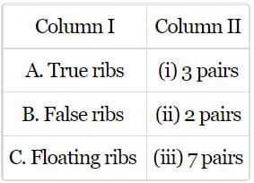
- a)A-(i), B-(ii), C-(iii)
- b)A-(iii), B-(i), C-(ii)
- c)A-(iii), B-(ii), C-(i)
- d)A-(ii), B-(i), C-(iii)
Correct answer is option 'B'. Can you explain this answer?
Match column I with column II and select the correct option from the codes given below.
a)
A-(i), B-(ii), C-(iii)
b)
A-(iii), B-(i), C-(ii)
c)
A-(iii), B-(ii), C-(i)
d)
A-(ii), B-(i), C-(iii)
| | Yash Patel answered |
The first 7 pairs of ribs are called true ribs as their anterior ends are attached directly to the sternum. The 8th, 9th and 10th pairs are called false ribs as they are attached indirectly to the sternum. The 11th and 12th pairs are called floating ribs as their anterior ends are not attached to sternum.
Fill in the blanks to complete the sequence of vertebral counts starting from the skull:
Cervical (), Thoracic (), Lumbar (), Sacral ( ), Coccygeal ( ).- a)7, 12, 5, 1, 1
- b)7, 10, 5, 2, 1
- c)5, 12, 5, 1, 1
- d)7, 12, 4, 1, 2
Correct answer is option 'A'. Can you explain this answer?
Cervical (), Thoracic (), Lumbar (), Sacral ( ), Coccygeal ( ).
a)
7, 12, 5, 1, 1
b)
7, 10, 5, 2, 1
c)
5, 12, 5, 1, 1
d)
7, 12, 4, 1, 2
 | Top Rankers answered |
The human vertebral column is divided into five regions with the following typical vertebrae counts:
Cervical: 7 vertebrae
Thoracic: 12 vertebrae
Lumbar: 5 vertebrae
Sacral: 1 segment formed by the fusion of 5 sacral vertebrae
Coccygeal: 1 segment formed by the fusion of 4 coccygeal vertebrae
Thus the correct sequence is 7, 12, 5, 1 (fused), 1 (fused), matching Option A.
Cervical: 7 vertebrae
Thoracic: 12 vertebrae
Lumbar: 5 vertebrae
Sacral: 1 segment formed by the fusion of 5 sacral vertebrae
Coccygeal: 1 segment formed by the fusion of 4 coccygeal vertebrae
Thus the correct sequence is 7, 12, 5, 1 (fused), 1 (fused), matching Option A.
11th and 12th pair of ribs which are imperfectly formed and do not reach the sternum are called- a)pseudo ribs
- b)false ribs
- c)floating ribs
- d)visceral ribs
Correct answer is option 'C'. Can you explain this answer?
11th and 12th pair of ribs which are imperfectly formed and do not reach the sternum are called
a)
pseudo ribs
b)
false ribs
c)
floating ribs
d)
visceral ribs
| | Kabir Verma answered |
The last two pairs of ribs (11th and 12th) are called floating ribs as their anterior ends are not attached to either the sternum or the cartilage of another rib. The floating ribs provide protection to kidneys.
Choose the letter from the figure that most appropriately corresponds to the structure: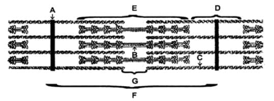 I.A-band II. I-band III. Sarcomere IV. H-zoneV.Myosin VI.Actin, Troponin, Tropomyosin VII. Z –line
- a)I - E, II - D, III - F, IV - G, V - B, VI- C, VII - A
- b)I - E, II - D, III - C, IV - G, V - B, VI - A, VII - F
- c) I - E, II - D, III - F, IV - G, V - C, VI - A, VII - B
- d)I - E, II - D, III - F, IV - A, V - B, VI - C, VII - G
Correct answer is option 'A'. Can you explain this answer?
Choose the letter from the figure that most appropriately corresponds to the structure:
I.A-band II. I-band III. Sarcomere IV. H-zone
V.Myosin VI.Actin, Troponin, Tropomyosin VII. Z –line
a)
I - E, II - D, III - F, IV - G, V - B, VI- C, VII - A
b)
I - E, II - D, III - C, IV - G, V - B, VI - A, VII - F
c)
I - E, II - D, III - F, IV - G, V - C, VI - A, VII - B
d)
I - E, II - D, III - F, IV - A, V - B, VI - C, VII - G
 | Ambition Institute answered |
Sarcomere Components
- A-Band (I): Represents the length of the myosin filaments, including areas where actin and myosin overlap, and appears as the darker region.
- I-Band (II): Contains only actin filaments and is the lighter area that appears to shorten during muscle contraction.
- Sarcomere (III): The entire unit from one Z-line to the next.
- H-Zone (IV): The lighter region in the middle of the A-band where only myosin filaments are present when the muscle is relaxed.
- Myosin (V): The thick filament.
- Actin, Troponin, Tropomyosin (VI): Components of the thin filaments.
- Z-line (VII): The dark line to which actin filaments are attached, marking the boundary of each sarcomere.
Label Correspondence
- A: Appears to be the Z-line, marking the boundary of the sarcomere.
- B: Thick filaments (myosin) as seen in the center part of the sarcomere.
- C: Contains elements of both myosin and actin filaments, typical of the A-band.
- D: Shows the I-band, where only actin filaments are present.
- E: Another part of the A-band with overlapping actin and myosin filaments.
- F: The entire unit from one Z-line to the next Z-line, representing the sarcomere.
- G: The lighter middle region within the A-band where only myosin filaments are visible, representing the H-zone.
The figure given here is of rib cage. Identify the parts labelled as A, B and C and select the correct option.
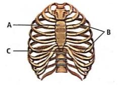
- a)A - Coccyx, B - Ribs, C - Vertebral column
- b)A - Sternum, B - Ribs, C - Vertebral column
- c)A - Scapula, B - Ribs, C - Vertebral column
- d)A - Tarsal, B - Ribs, C - Vertebral column
Correct answer is option 'B'. Can you explain this answer?
The figure given here is of rib cage. Identify the parts labelled as A, B and C and select the correct option.
a)
A - Coccyx, B - Ribs, C - Vertebral column
b)
A - Sternum, B - Ribs, C - Vertebral column
c)
A - Scapula, B - Ribs, C - Vertebral column
d)
A - Tarsal, B - Ribs, C - Vertebral column
| | Kabir Verma answered |
A - Sternum
B - Ribs
C - Vertebral column
B - Ribs
C - Vertebral column
Chapter doubts & questions for Locomotion and Movement - 30-Day Revision Course for NEET 2026 is part of NEET exam preparation. The chapters have been prepared according to the NEET exam syllabus. The Chapter doubts & questions, notes, tests & MCQs are made for NEET 2026 Exam. Find important definitions, questions, notes, meanings, examples, exercises, MCQs and online tests here.
Chapter doubts & questions of Locomotion and Movement - 30-Day Revision Course for NEET in English & Hindi are available as part of NEET exam. Download more important topics, notes, lectures and mock test series for NEET Exam by signing up for free.
30-Day Revision Course for NEET85 videos|301 docs|171 tests |