NEET PG Exam  >  NEET PG Tests  >  Test: Abdomen - NEET PG MCQ

Test: Abdomen - NEET PG MCQ


Test Description

25 Questions MCQ Test - Test: Abdomen

Test: Abdomen for NEET PG 2025 is part of NEET PG preparation. The Test: Abdomen questions and answers have been prepared according to the NEET PG exam syllabus.The Test: Abdomen MCQs are made for NEET PG 2025 Exam. Find important definitions, questions, notes, meanings, examples, exercises, MCQs and online tests for Test: Abdomen below.
Solutions of Test: Abdomen questions in English are available as part of our course for NEET PG & Test: Abdomen solutions in Hindi for NEET PG course. Download more important topics, notes, lectures and mock test series for NEET PG Exam by signing up for free. Attempt Test: Abdomen | 25 questions in 25 minutes | Mock test for NEET PG preparation | Free important questions MCQ to study for NEET PG Exam | Download free PDF with solutions
Test: Abdomen - Question 1

Transpyloric plane separates: 

Detailed Solution for Test: Abdomen - Question 1

Transpyloric plane separates hypochondrium from lumbar region and epigastrium from umbilical region.

Test: Abdomen - Question 2

Which of the following structure is NOT present in transpyloric plane?

Detailed Solution for Test: Abdomen - Question 2
  • Transpyloric plane is present at the lower border of L1 vertebra and superior (not inferior) mesenteric artery lies here.
  • The pylorus of stomach may be found in the transpyloric plane but is not a constant feature.
  • Hila of each kidney is present at this plane.
  • Fundus of gallbladder can be touched at this plane (Murphy’s sign).
Test: Abdomen - Question 3

Which of the following is the correct matching, regarding the attachments of external oblique muscle? 

Detailed Solution for Test: Abdomen - Question 3
  • External oblique muscle takes origin from the external surfaces of lower 8 (5-12th) ribs.
  • It inserts into the linea alba (marker ‘I’), pubic tubercle, and outer lip of the anterior two-thirds of the iliac crest (marker ‘III’).
  • At linea semilunaris (marker ‘II’) the muscular fibers of muscle become aponeurotic.
  • The free lower border of the aponeurosis is rolled inward on itself to form the inguinal ligament (marker ‘IV’).
Test: Abdomen - Question 4

Function of external oblique muscle: 

Detailed Solution for Test: Abdomen - Question 4
  • External oblique contributes to the maintenance of abdominal tone, increasing intra-abdominal pressure (as in active expiration), and lateral flexion of the trunk against resistance. Bilateral contraction flexes the trunk forward.
  • Contraction of external oblique muscle approximates the two crura (medial and lateral) of superficial inguinal ring like a slit valve to maintain the integrity of inguinal canal.
Test: Abdomen - Question 5

Rectus abdominis is inserted into:

Detailed Solution for Test: Abdomen - Question 5

Rectus abdominis takes origin from the crest of pubis and Inserts into costal cartilages of ribs 5-7 and Xiphisternum.
Note: Rectus abdominis muscle is NOT attached to the median raphe- linea alba.

Test: Abdomen - Question 6

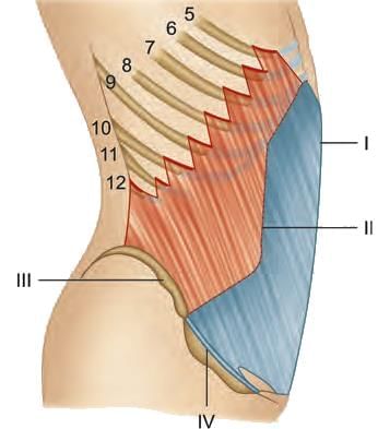
Which of the following marker is conjoint tendon in the following diagram of transversus abdominis? 

Detailed Solution for Test: Abdomen - Question 6
  • Conjoint tendon (marker D) is formed from the lower part of the common aponeurosis of the internal abdominal oblique and the transverse abdominal as it inserts into the crest of the pubis and pectineal line.
  • Key- A: Thoracolumbar fascia; B: Line of demarcation between muscular and aponeurotic parts of muscle; C: Origin from lateral 1/3 of inguinal ligament; D: Conjoint tendon.
Test: Abdomen - Question 7

Flexors of lumbar spine are all EXCEPT: 

Detailed Solution for Test: Abdomen - Question 7
  • Forward flexors: Rectus abdominis, external abdominal oblique, internal abdominal obliquus, transversus abdominis and iliacus-psoas.
  • Lateral flexors: Ipsilateral contraction of the oblique and transversus abdominal muscles and quadratus lumborum.
Test: Abdomen - Question 8

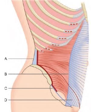
Posterior wall of rectus sheath below the level of anterior superior iliac spine is formed by:

Detailed Solution for Test: Abdomen - Question 8

Rectus abdominis lies on the transversalis fascia below the arcuate line.

Test: Abdomen - Question 9

Rectus sheath contains all of the following EXCEPT:

Detailed Solution for Test: Abdomen - Question 9

Rectus sheath contains the muscles (rectus abdominis and pyramidalis); vessels (superior epigastric and inferior epigastric artery and veins) and terminal parts of lower six thoracic nerves.

Test: Abdomen - Question 10

TRUE statement about lower 1/4 anterior abdominal wall: 

Detailed Solution for Test: Abdomen - Question 10
  • Linea alba is wider and more obvious above the umbilicus and is almost linear and less visible below this level.
  • External oblique muscle is aponeurotic in the lower part.
  • Tendinous intersections divide rectus abdominis muscle, but this is not a feature in the lower part.
  • Posterior layer of rectus sheath is deficient in the lower part.
Test: Abdomen - Question 11

Lymphatic drainage of umbilicus is towards: 

Detailed Solution for Test: Abdomen - Question 11
  • Umbilicus drains into both the directions, supra-umbilically into axillary lymph nodes and infra-umbilically into inguinal lymph nodes.
  • The deep lymph vessels follow the arteries and drain into the internal thoracic, external iliac, posterior mediastinal, and para-aortic (lumbar) nodes.
Test: Abdomen - Question 12

Corona mortis is: 

Detailed Solution for Test: Abdomen - Question 12
  • A collateral circulation between the external and internal iliac system is known as corona mortis.
  • It is a common variant vascular anastomosis between the external iliac artery or deep inferior epigastric artery with the obturator artery.
Test: Abdomen - Question 13

In rectus sheath which branch of aorta make anastomosis with superior epigastric artery: 

Detailed Solution for Test: Abdomen - Question 13

In rectus sheath superior epigastric artery (branch of internal thoracic artery) anastomoses with inferior epigastric artery (branch of external iliac artery).

Test: Abdomen - Question 14

Which nerve supplies celiac plexus? 

Detailed Solution for Test: Abdomen - Question 14
  • Celiac plexus receives preganglionic sympathetic fibers contributed by the greater and lesser splanchnic nerves.
  • Parasympathetic fibers are contributed by vagus nerve. Postganglionic fibers accompany the respective blood vessels to the target organs.
Test: Abdomen - Question 15

RUE about the autonomic nervous system is:

Detailed Solution for Test: Abdomen - Question 15
  • Superior hypogastric plexus is chiefly sympathetic but some parasympathetic fibers from pelvic splanchnic nerves ascend from the inferior hypogastric plexus via the right and left hypogastric nerves to reach the superior hypogastric plexus.
  • The sympathetic outflow is thoracolumbar ( T1-L2) and doesn’t involve cranial nerves.
  • Parasympathetic flow is craniosacral and involves cranial nerves (3,7,9,10) and sacral nerves (S2,3,4).
Test: Abdomen - Question 16

All are true about Celiac trunk EXCEPT: 

Detailed Solution for Test: Abdomen - Question 16
  • Celiac trunk lies at the upper aspect of pancreas (not lower)
  • Celiac trunk is a ventral branch of abdominal aorta, is surrounded by the nerve plexus called celiac plexus.
  • It gives three branches, including left gastric artery.
Test: Abdomen - Question 17

Which of the following ar teries is a direct branch of the gastroduodenal artery? 

Detailed Solution for Test: Abdomen - Question 17
  • Right gastroepiploic (gastro-omental artery) artery is one of the two terminal branches of the gastroduodenal artery.
  • It runs from right to left along the greater curvature of the stomach, between the layers of the greater omentum, anastomosing with the left gastroepiploic artery, a branch of the splenic artery.
Test: Abdomen - Question 18

Which of the following is TRUE about splenic artery?

Detailed Solution for Test: Abdomen - Question 18

Splenic artery is the largest branch of Celiac trunk, runs posterior to stomach (in stomach bed) and send short gastric arteries in gastrosplenic ligament to reach the fundus of stomach.

Test: Abdomen - Question 19

All are branches of splenic artery EXCEPT: 

Detailed Solution for Test: Abdomen - Question 19
  • Right gastroepiploic artery is a branch of gastroduodenal artery. Splenic artery gives the left gastroepiploic artery.
  • Short gastric arteries are the branches of splenic artery, which travel in the gastrosplenic ligament and supply the fundus of stomach.
  • Splenic artery runs posterior to the superior border of pancreas and supplies the organ by giving pancreatic branches) like arteria pancreatica magna.
  • It reaches the hilum of the kidney and gives the hilar branches, which enter the spleen as end arteries.
Test: Abdomen - Question 20

All of the following statements about the splenic artery are true EXCEPT that it: 

Detailed Solution for Test: Abdomen - Question 20
  • Splenic artery branches do not anastomose within the substance of spleen (end arteries).
  • Splenic artery is the largest branch of celiac trunk, with a tortuous course to allow for movement of spleen.
  • It supplies body and tail of pancreas via pancreatic branches; greater curvature of stomach by giving short gastric and left gastroepiploic branches.
Test: Abdomen - Question 21

Most important arterial supply to stomach is:

Detailed Solution for Test: Abdomen - Question 21

The consistently largest artery to the stomach is left gastric artery.

*Multiple options can be correct
Test: Abdomen - Question 22

Arterial supply of stomach: 

Detailed Solution for Test: Abdomen - Question 22

Inferior phrenic artery supplies diaphragm. Superior mesenteric artery supplies mid-gut derivatives.

*Multiple options can be correct
Test: Abdomen - Question 23

TRUE about inferior vena cava:

Detailed Solution for Test: Abdomen - Question 23

Inferior vena cava passes through the central tendon of diaphragm at D8 (not D10) vertebra level.

Test: Abdomen - Question 24

Tributaries of left renal vein are all EXCEPT: 

Detailed Solution for Test: Abdomen - Question 24
  • Left inferior phrenic (diaphragmatic) vein, left adrenal vein and left gonadal vein are tributaries of left renal vein (but not the left lumbar vein).
  • On the right side the corresponding veins drain directly into IVC (and not into the right renal vein).
Test: Abdomen - Question 25

Vein that doesn’t cross the midline is: 

Detailed Solution for Test: Abdomen - Question 25
  • Left gonadal vein drains into left renal vein and remains on the left side of midline (doesn’t cross it).
  • Veins crossing the midline to join the drainage structures lying on right side of midline are:
    • Left brachiocephalic vein crosses the midline to join right counterpart and form SVC.
    • Hemiazygous vein crosses the midline at T8 vertebra level to drain into azygous vein.
    • Left renal vein crosses the midline to enter the IVC.
  • Note: Left suprarenal, and left inferior phrenic vein enter the left renal vein (do not cross the midline).
Information about Test: Abdomen Page
In this test you can find the Exam questions for Test: Abdomen solved & explained in the simplest way possible. Besides giving Questions and answers for Test: Abdomen, EduRev gives you an ample number of Online tests for practice
Download as PDF