NEET PG Exam  >  NEET PG Tests  >  Test: Anatomy - 1 - NEET PG MCQ

Test: Anatomy - 1 - NEET PG MCQ


Test Description

25 Questions MCQ Test - Test: Anatomy - 1

Test: Anatomy - 1 for NEET PG 2025 is part of NEET PG preparation. The Test: Anatomy - 1 questions and answers have been prepared according to the NEET PG exam syllabus.The Test: Anatomy - 1 MCQs are made for NEET PG 2025 Exam. Find important definitions, questions, notes, meanings, examples, exercises, MCQs and online tests for Test: Anatomy - 1 below.
Solutions of Test: Anatomy - 1 questions in English are available as part of our course for NEET PG & Test: Anatomy - 1 solutions in Hindi for NEET PG course. Download more important topics, notes, lectures and mock test series for NEET PG Exam by signing up for free. Attempt Test: Anatomy - 1 | 25 questions in 25 minutes | Mock test for NEET PG preparation | Free important questions MCQ to study for NEET PG Exam | Download free PDF with solutions
Test: Anatomy - 1 - Question 1

Meiosis occurs in: (NEET-PG 2020)

Detailed Solution for Test: Anatomy - 1 - Question 1

Meiosis is a type of reduction division that takes place in the gonads (ovaries and testes).

  • In females, oogenesis starts before birth (in utero).
  • Spermatogenesis in males commences after puberty.
  • Until that point, primordial germ cells in the pre-pubertal testes remain inactive.

Note: Meiosis is a form of reduction cell division that halves the chromosome count. Cells in the adrenal gland and hypothalamus undergo mitosis, which is an equational division process.

Test: Anatomy - 1 - Question 2

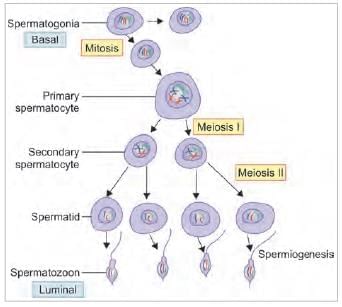
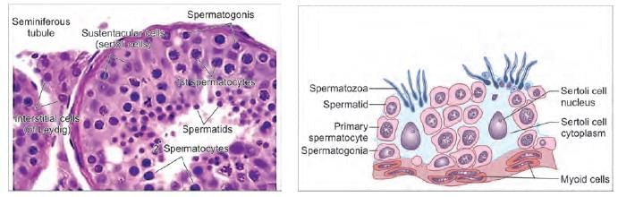
Arrange the steps of spermatogenesis in a sequence: (AIIMS May 2019)
1. Spermatid
2. Spermatocyte
3. Spermatogonium
4. Spermatozoa

Detailed Solution for Test: Anatomy - 1 - Question 2

Spermatogonium → Spermatocyte → Spermatid → Spermatozoa. Spermatogonium develops into a primary spermatocyte, which then undergoes meiosis I to create a secondary spermatocyte. This secondary spermatocyte proceeds through meiosis II, resulting in a spermatid that eventually matures into spermatozoa.

Test: Anatomy - 1 - Question 3

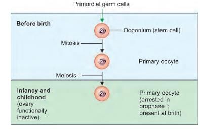
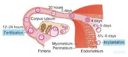
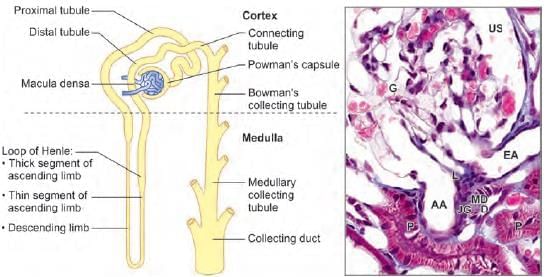
Meiosis Fertilization occurs in which part of the fallopian tube: (NEET-PG 2020)

Detailed Solution for Test: Anatomy - 1 - Question 3

Fertilisation of the secondary oocyte by sperm takes place in the ampullary section of the fallopian tube. The secondary oocyte is expelled from the ovary during ovulation (Day 14 of the menstrual cycle). Fertilisation happens in the ampulla of the fallopian tube within a day (24 hours) following ovulation.

  • Note: The day of ovulation coincides with the day of fertilisation.

An advanced morula (with more than 16 cells) enters the uterine tube on Day 4 after ovulation/fertilisation.

Implantation occurs in the endometrium of the uterus between Days 7 and 9. This process spans a week, starting on Day 5 and concluding on Day 12.

Test: Anatomy - 1 - Question 4

Double bleb sign on USG is suggestive of: (INI-CET Nov 2021)

Detailed Solution for Test: Anatomy - 1 - Question 4

The double bleb sign is an ultrasonographic characteristic that shows a gestational sac containing both a yolk sac and an amniotic sac, creating the appearance of two small bubbles. The embryonic disc is situated between these two bubbles. This sign is significant for identifying an intrauterine pregnancy, helping to differentiate it from a pseudo-gestational sac or a decidual cast cyst.

Heterotopic pregnancy is defined as having multiple gestations, with one occurring within the uterine cavity and another located outside the uterus, typically in the fallopian tube, and less frequently in the cervix or ovary.

*Multiple options can be correct
Test: Anatomy - 1 - Question 5

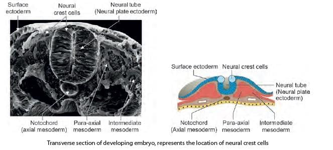
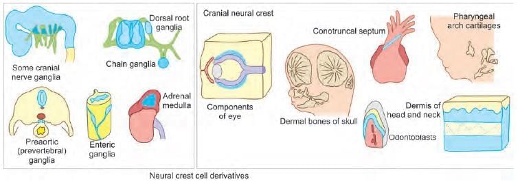
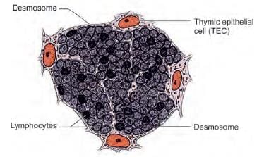
Which of the following are derived from the star marked region? (INI-CET Nov 2022)

Detailed Solution for Test: Anatomy - 1 - Question 5

The star-marked area illustrated in this transverse section of the developing embryo indicates the position of neural crest cells, which are situated dorsal and lateral to the neural tube. These cells originate from epiblast cells (considered the fourth germ layer), possess migratory characteristics, and play a role in forming various body components, including neurons in the peripheral nervous system. It is noteworthy that most ganglia develop from neural crest cells, such as:

  • Parasympathetic/sympathetic ganglia
  • Dorsal root ganglia
  • Enteric ganglia

Additionally, the nephrogenic cord, which contributes to the genitourinary system, is derived from intermediate mesoderm rather than neural crest cells. Other derivatives of neural crest cells include:

  • Odontoblasts (which form the dentine of teeth)
  • Cono-truncal septum (which separates the truncus arteriosus into the aorta and pulmonary trunk)
  • Melanoblasts (cells that produce melanin)

Neural crest cells provide secondary mesenchyme and contribute to the connective tissue in the head and neck region, particularly on the anterolateral aspect. They also form various mesodermal components such as:

  • Parts of the eyeball
  • Facial skeleton bones
  • Pharyngeal arch bones (including the malleus, incus, stapes, and hyoid bone)
  • Dermis of the skin in the head and neck region

DiGeorge syndrome occurs due to the failure of neural crest cells to migrate properly in the head and neck region and proximal vessels of the heart. This results in:

  • Pharyngeal arch anomalies (micrognathia)
  • Pouch anomalies (such as an absent thymus)
  • Cardiovascular defects like cono-truncal septum anomalies (including persistent truncus arteriosus)

Hirschsprung disease is caused by the failure of neural crest cells to migrate into the distal intestine, leading to congenital aganglionic megacolon and subsequent failure to pass meconium.


Test: Anatomy - 1 - Question 6

Transitional epithelium lines all, except: (AIIMS June 2020)

Detailed Solution for Test: Anatomy - 1 - Question 6

Membranous urethra is covered by stratified columnar epithelium. Transitional epithelium starts at the end of the collecting ducts and extends to:

  • minor calyx
  • major calyx
  • renal pelvis
  • ureter
  • urinary bladder
  • proximal prostatic urethra

Male Urethra (Epithelium)
Linin : Siteg

  • Proximal: Transitional (Urothelium)
  • Major lining: Pseudo-stratified or stratified columnar epithelium
  • Terminal: Stratified squamous (non-keratinized) epithelium
Test: Anatomy - 1 - Question 7

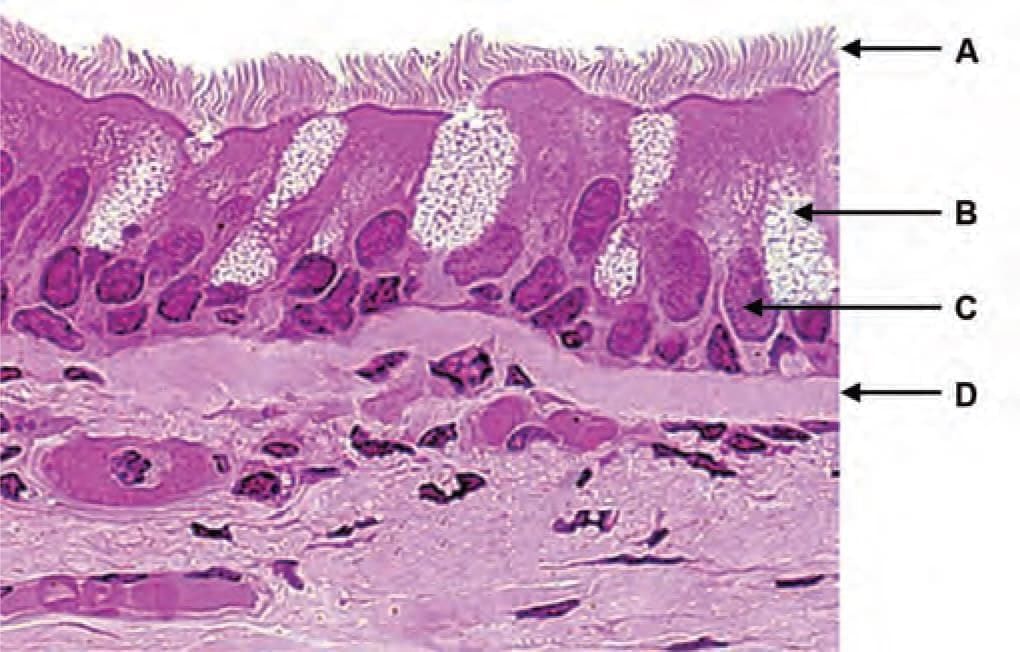
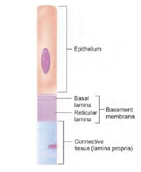
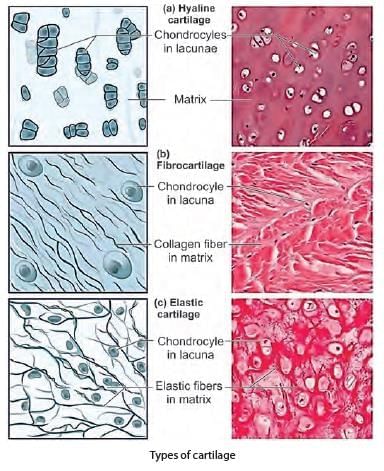
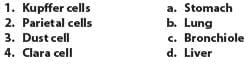
Which of the following pair is WRONGLY matched for the following microscopic picture? (INI-CET Nov 2021)

Detailed Solution for Test: Anatomy - 1 - Question 7

This slide displays respiratory epithelium (pseudo-stratified ciliated columnar epithelium). ‘Marker D’ indicates the basement membrane (not the lamina propria).

  • The lamina propria is the sub-epithelial connective tissue, typically comprising ‘loose areolar connective tissue’, which is abundant in cells.
  • In light microscopy, the term basement membrane actually refers to the combined basal lamina and the underlying reticular lamina.
  • The basal lamina originates from epithelial cells and is composed of collagen fibres.
  • The reticular lamina is produced by connective tissue cells and mainly consists of reticular fibres.


Test: Anatomy - 1 - Question 8
Arrow marked surface projections are ABSENT in: (INI-CET Nov 2020)
Detailed Solution for Test: Anatomy - 1 - Question 8
The structure indicated by the arrow is cilia, which are absent in the vas deferens. Instead, the vas deferens contains stereo-cilia.
  • Identification point: Cilia are slender, microscopic, hair-like organelles that protrude from the apical surfaces of cells.
  • Cilia typically appear in large quantities on a cell’s apical surface and beat in synchronised waves to facilitate the movement of fluids and structures along the luminal surface.
  • For instance, in the respiratory epithelium (bronchus), they assist in the muco-ciliary clearance by sweeping mucus and foreign particles.
  • In the oviduct (fallopian tubes), cilia transport the fertilised oocyte towards the uterus.
  • In the ependymal lining of the ventricles of the CNS, they aid in the movement of cerebrospinal fluid (CSF).
Kartagener syndrome is a condition characterised by immotile cilia syndrome, leading to chronic sinusitis, bronchiectasis, and reduced fertility. A deficiency of functional cilia in the fallopian tubes can result in ectopic pregnancy. If the cilia are unable to transport the fertilised ovum to the uterus, it may implant in the fallopian tubes, resulting in a tubal pregnancy, which is the most prevalent type of ectopic pregnancy. Stereocilia are found in the inner ear (hair cells) and in male reproductive structures such as the vas deferens and epididymis. They are non-motile and serve to increase the surface area for absorption.
Test: Anatomy - 1 - Question 9

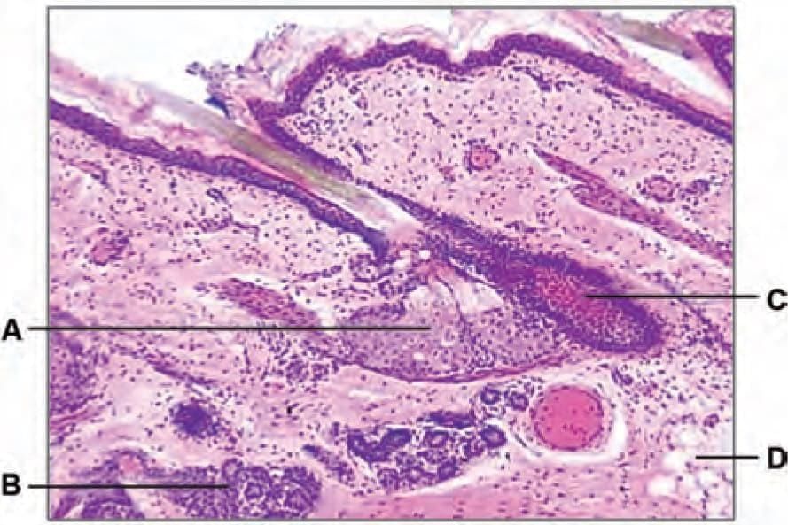
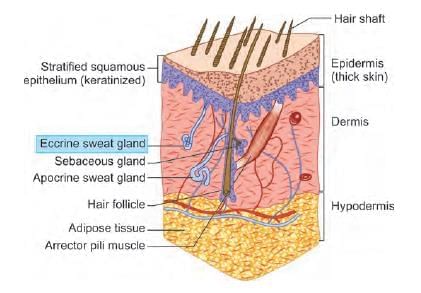
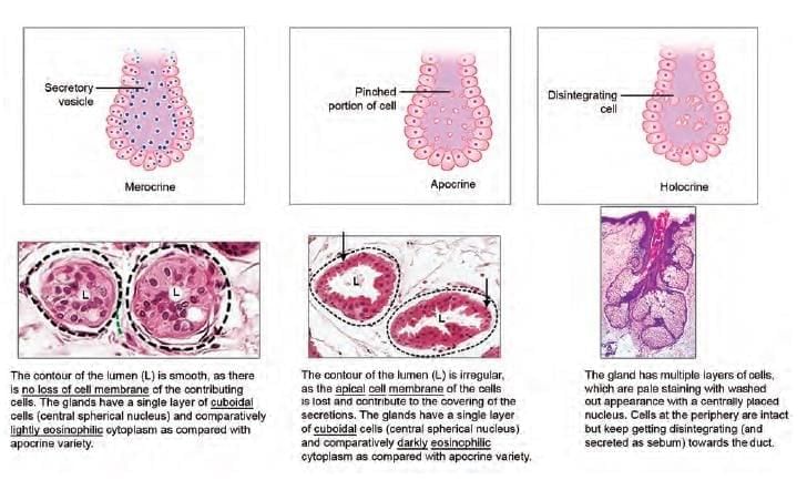
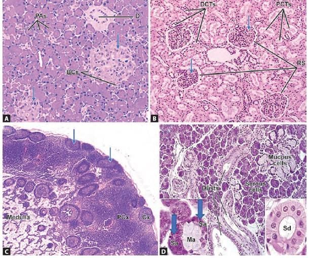
Which of the following marked structures represent merocrine gland? (INI-CET May 2022)

Detailed Solution for Test: Anatomy - 1 - Question 9

This figure presents a histological section of skin. The sweat gland (marker B) is classified as a merocrine gland. It can be identified on a histology slide as several sections of the gland and ducts, located within the dermis. Note that the skin in this slide is covered by stratified squamous epithelium (keratinised) and contains hair follicles, a few glands, and adipose tissue.

  • A: Sebaceous gland (holocrine)
  • B: Sweat gland (Merocrine/Eccrine)
  • C: Hair follicle
  • D: Adipose tissue

According to the method of secretion, there are three categories of glands:

  • Merocrine/Eccrine: Secretions are released through exocytosis from secretory cells into a duct with an epithelial lining, reaching the lumen or body surface. The gland discharges its product without losing any part of itself. For instance, most sweat glands are of the merocrine type.
  • Apocrine: The cell's secretions involve a portion of the plasma membrane, forming membrane-bound vesicles within the lumen. The apical segment of the secretory cell pinches off and enters the lumen, resulting in the loss of some cytoplasm during secretion. A few sweat glands, such as ceruminous glands and mammary glands, are modified sweat glands of the apocrine type.
  • Holocrine: The secretory product is expelled through the complete destruction of the cell. In this case, as lipid droplets build up, the cytoplasm becomes congested and disrupted, leading to necrosis. Ultimately, the total breakdown of the cell results in the release of sebum along with cellular debris into the duct near the hair follicle.


Test: Anatomy - 1 - Question 10

Reticular fibres are ABSENT in:

Detailed Solution for Test: Anatomy - 1 - Question 10

The thymus gland, although classified as a lymphoid organ, lacks reticular fibres as a supportive network. Instead, it features a cytoreticulum that creates the structural framework.

  • The thymic epithelial reticular cells (TEC) throughout the thymus cortex function as APCs (Antigen Presenting Cells).
  • These cells extend long processes that are interconnected by desmosomes, forming the cytoreticulum that supports lymphoblasts.

In this environment, the lymphoblasts are supported by a cytoreticulum made up of APC cellular processes rather than the typical network of simple reticulin fibres. This arrangement facilitates the regulated specificity of lymphocyte binding through the changing antigens presented on MHC proteins.

Reticular tissue comprises delicate networks of type III collagen and is particularly prevalent in specific lymphoid organs, where the fibres create attachment sites for lymphocytes and other immune cells.

Red bone marrow contains a stroma of reticular connective tissue, which includes hemopoietic cords or islands of cells, as well as sinusoidal capillaries.

  • The stroma is a meshwork of specialised fibroblastic cells known as stromal cells.
  • It also consists of a delicate web of reticular fibres that support the hemopoietic cells and macrophages.

Both the spleen and lymph nodes exhibit the distinctive reticular fibre framework characteristic of lymphoid organs.

Test: Anatomy - 1 - Question 11

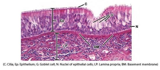
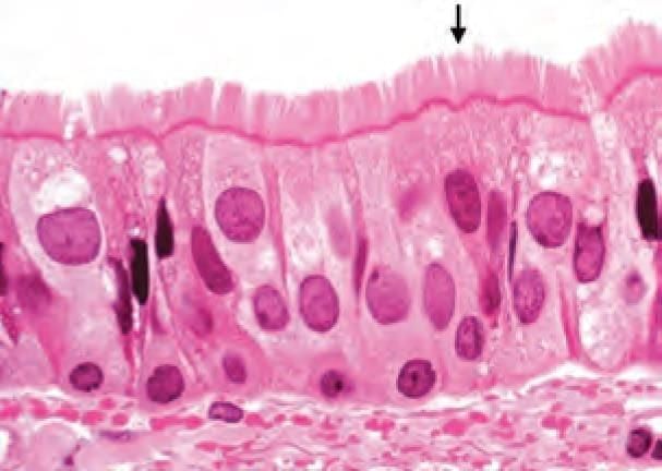
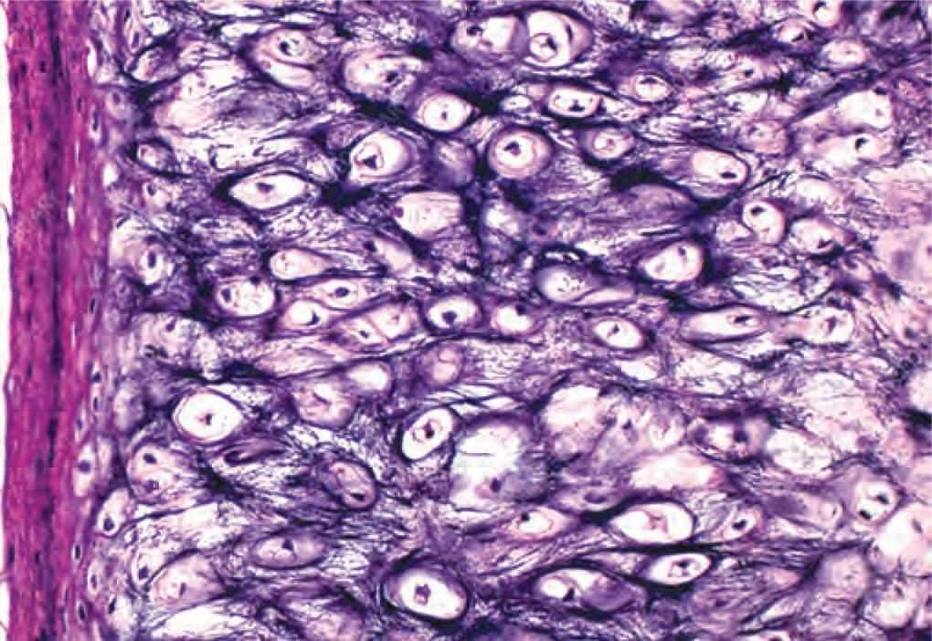
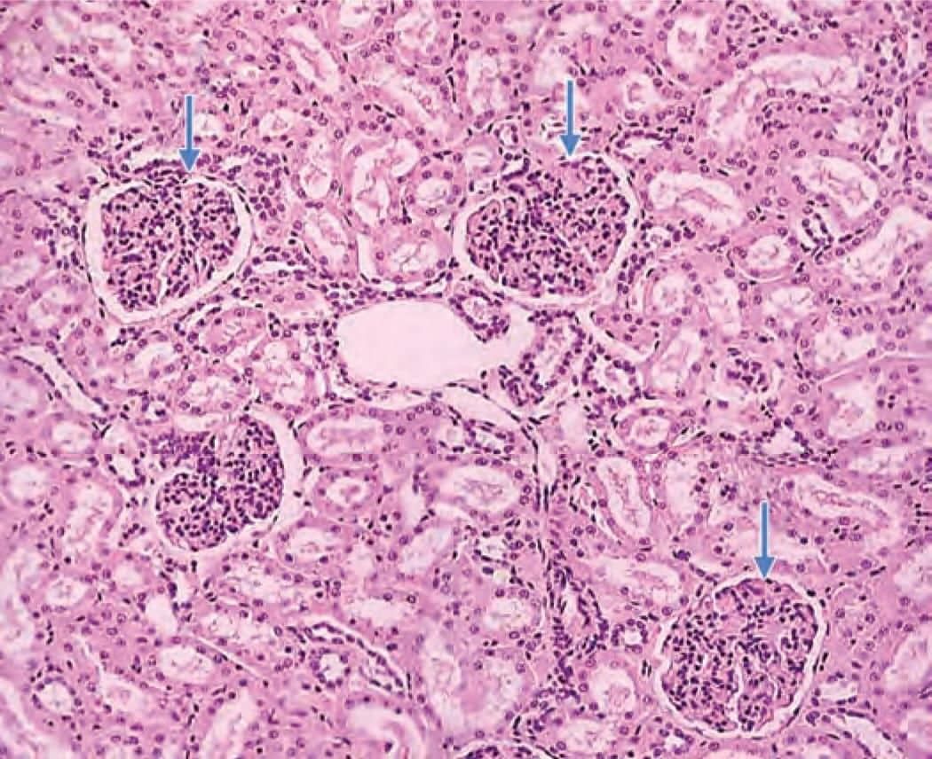
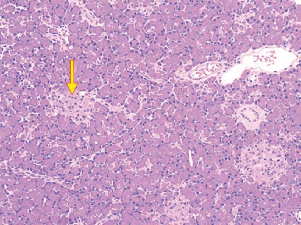
Identify the structure in the given microscopic picture: (NEET-PG 2022)

Detailed Solution for Test: Anatomy - 1 - Question 11

This microscopic slide of fibrocartilage is characterised by an abundance of chondrocytes, which are organised in a linear arrangement, interspersed with numerous bundles of collagen fibres, predominantly type I, alternating with rows of chondrocytes. The perichondrium is not present.

Elastic cartilage is recognised by the presence of chondrocytes mixed with a significant amount of elastic fibres and the existence of perichondrium on both sides.

Non-articular hyaline cartilage features chondrocytes arranged in nests or groups, embedded in a hyalos (glass-like) matrix. In this matrix, collagen fibres, mainly type II, are not visible to the naked eye because their refractive index is similar to that of the matrix, creating an optical illusion. The perichondrium is present on either side.

Articular cartilage is primarily composed of hyaline cartilage, which covers the articular surfaces of synovial joints, ensuring a smooth surface for movement within these joints. The perichondrium is absent in this type. It exhibits zonation:

  • Superficial tangential zone − Small, elongated cells aligned parallel to the surface.
  • Transitional intermediate zone − Larger, rounded chondrocytes; collagen fibrils (T-II) arranged in an oblique orientation.
  • Deep radial zone − Vertical fibres arranged parallel; cells are also oriented vertically.
  • Tidemark − The deepest zone consisting of calcified cartilage, which increases in thickness with age.

Test: Anatomy - 1 - Question 12
Identify the type of cartilage: (NEET-PG 2020)
Detailed Solution for Test: Anatomy - 1 - Question 12

The image displays a histological sample of elastic cartilage, characterised by the presence of chondrocytes, which are intermingled with distinct elastic fibres. The perichondrium, containing fibrous and cellular layers, is clearly visible on the left side of the image.

*Multiple options can be correct
Test: Anatomy - 1 - Question 13

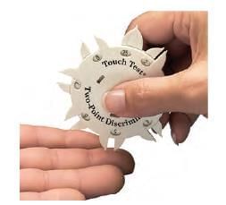
Receptor responsible for tactile discrimination is: (INI-CET July 2021)

Detailed Solution for Test: Anatomy - 1 - Question 13

Meissner corpuscles are highly responsive to touch and act as rapidly adapting mechanoreceptors. They allow a person to differentiate between two pointed objects when placed closely together on the skin (two-point tactile discrimination – 2PD).

  • Both Meissner’s corpuscles and Merkel’s discs possess small receptive fields, assisting in discerning the spatial arrangement of stimuli.
  • This discriminative ability is particularly well developed in the fingertips.

The subcutaneous layers contain Pacinian corpuscles and Ruffini endings, which have larger receptive fields, making them less effective for discrimination. Tactile discrimination (fine touch), assessed by 2PD, is mediated by Meissner’s corpuscles and transmitted via the Dorsal Column-Medial Lemniscal system. In contrast, Merkel Tactile Discs detect crude touch, evaluated by lightly touching the skin with a cotton wisp, and are conveyed through the anterior Spinothalamic tract. Two-point discrimination (2PD) serves as an indicator of the innervation density of the body surface, such as the fingertips. There are two types of discrimination:

  • Static 2PD: Involves slowly adapting receptors, like the Merkel Tactile disc. Range: Points 2-6 mm apart on the fingertip.
  • Moving 2PD: Involves rapidly adapting receptors, such as Meissner’s corpuscles. More sensitive and responds quicker. Range: Points 2-4 mm apart on the fingertip.

Applied Anatomy: In conditions like scleroderma, there is a loss of two-point discrimination.

Test: Anatomy - 1 - Question 14

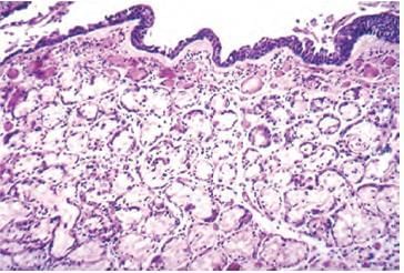
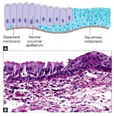
A 45-year-old chronic smoker presented with the complaints of cough. A biopsy was taken after the preliminary examination. What is cellular change visible on the biopsy? (NEET-PG 2020p)

Detailed Solution for Test: Anatomy - 1 - Question 14

The provided diagram illustrates a histopathological specimen depicting the lumen of the bronchus featuring significant thickening of the mucous gland layer (approximately double the normal size) along with squamous metaplasia of the lung epithelium.

  • Squamous metaplasia and dysplasia of the bronchial mucosa are evident.
  • This condition is commonly observed in habitual smokers, which is a risk factor for lung carcinoma.
Test: Anatomy - 1 - Question 15

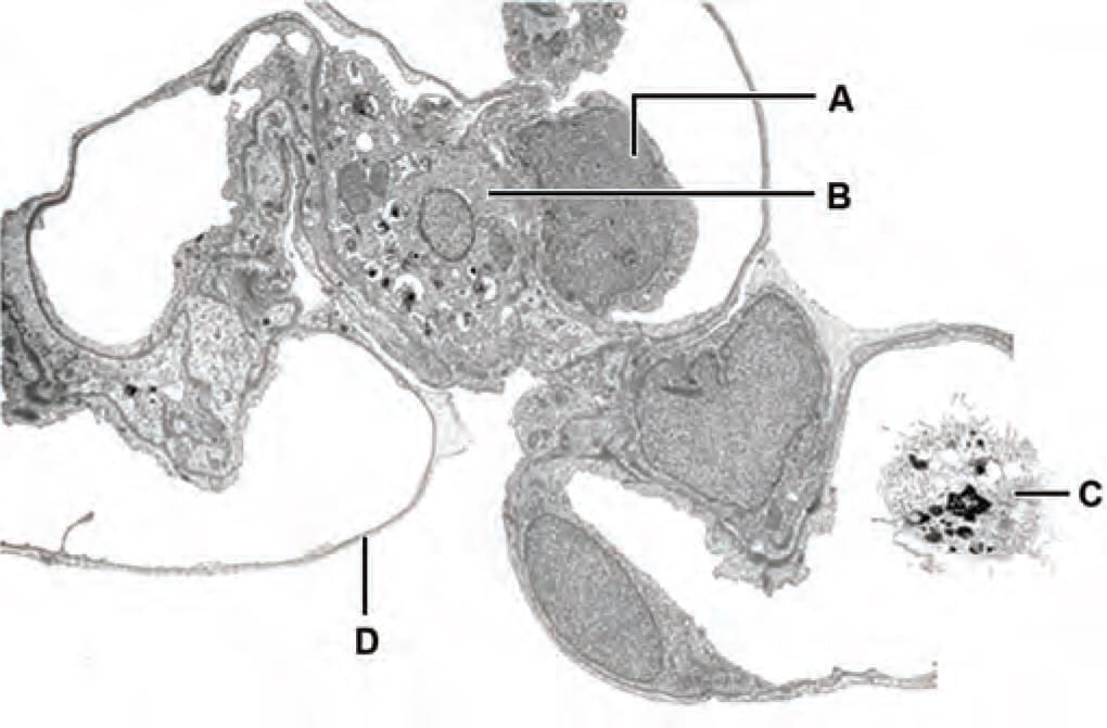
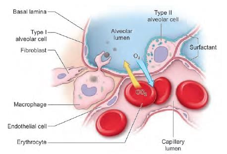
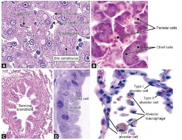
Which of the marked cells in the following picture is involved in respiratory distress syndrome: (INI-CET Nov 2021)

Detailed Solution for Test: Anatomy - 1 - Question 15

The image depicts an electron microscopic view of different cells in relation to lung alveoli. A lack of alveolar surfactant, stemming from the immaturity of alveolar type II epithelial cells/pneumocytes (Marker B), leads to respiratory distress syndrome (RDS). The severe acute respiratory syndrome coronavirus-2 (SARS-CoV-2) enters the lungs via ACE-2 receptors and replicates in AT-II cells, contributing to the development of Coronavirus disease – 2019 (COVID-19). The SARS-CoV-2 virus damages the AT-II cells, resulting in a reduced production of pulmonary surfactant (PS).

  • The clinical signs of acute respiratory distress syndrome (ARDS) in COVID-19 patients resemble those of neonatal respiratory distress syndrome (NRDS).
Test: Anatomy - 1 - Question 16

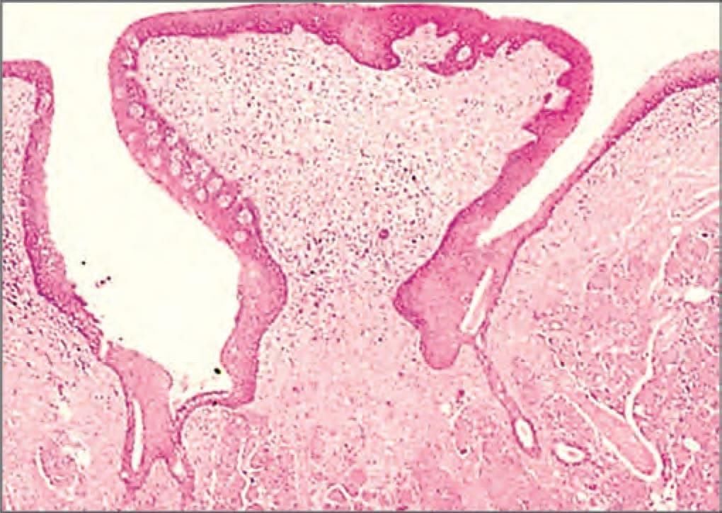
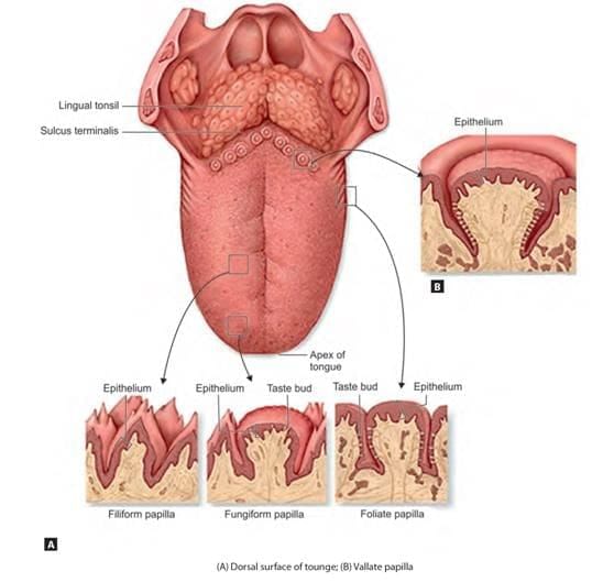
Identify the type of papillae shown in the microscopic picture of tongue: (NEET-PG 2021)

Detailed Solution for Test: Anatomy - 1 - Question 16

Circumvallate papillae are recognised as the largest of all tongue papillae, characterised by a circular trench surrounding them, which is bordered by a circular (circum) wall (valate). The trench tapers, resulting in the base of the papillae being narrower in comparison to the apex. The lateral walls are populated with numerous taste buds (up to 250 per papilla); serous glands (Von Ebner) release their secretions at the base of the trench.

  • Circumvallate (circular vallum/wall) papillae: These are non-keratinised mucosal projections found on the dorsum of the tongue, positioned anterior to the sulcus terminalis.
  • They number between 8 and 12.
  • The taste buds (which appear pale in histological sections) contain gustatory (taste) cells.

Tongue papillae are projections of mucosa, featuring a core of connective tissue and lined with stratified squamous epithelium. They are primarily found on the anterior two-thirds of the tongue (on the dorsal surface) and contain taste buds, with the exception of the filiform papilla, which does not have taste buds.

  • Filiform (pointed) papillae: These are highly keratinised, giving them a white appearance. They create friction between the tongue and food, aiding in its movement.
  • Fungiform (mushroom-shaped) papillae: These are non-keratinised, appear red due to the highly vascular connective tissue, and are dispersed among the filiform papillae.
  • Foliate (leaf-like) papillae: This type is rudimentary in adults and is best observed in young children. They are situated on the sides of the tongue, arranged in multiple lines, separated by vertical trenches.


Test: Anatomy - 1 - Question 17

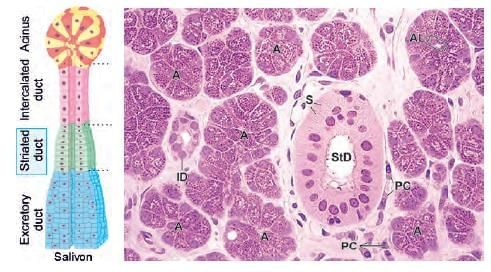
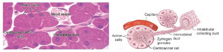
Exocrine pancreas differs from parotid gland histologically by: (AIIMS June 2020)

Detailed Solution for Test: Anatomy - 1 - Question 17

The exocrine pancreas acts as a serous gland and closely resembles the parotid gland, often leading to confusion between the two. A key distinguishing characteristic is the presence of striated ducts in the parotid gland, which are absent in the pancreas.

Parotid gland: It contains the Salivon, the fundamental secretory unit of any salivary gland, comprising the acinus, intercalated duct, striated duct, and excretory duct.

  • Secretion from the acinus is drained by the intercalated duct, which is lined with simple cuboidal epithelium.
  • This duct merges into the striated duct, characterised by simple columnar epithelium with distinct basal striations, and ultimately leads to the excretory duct, which features either stratified cuboidal or columnar epithelium and is surrounded by connective tissue.
  • The cells of striated ducts possess numerous infoldings in the basal plasma membrane that house mitochondria.

  • These infoldings are specialised for the reabsorption of electrolytes from the secretion.
  • Striated ducts play a significant role in modifying the ion composition of primary saliva after it has been produced in the acini.

Pancreas: The secretory units are acinar or tubulo-acinar in shape, formed by a simple epithelium of pyramidal serous cells.

  • The secretory (zymogen) granules are located in the apical cytoplasm, while Golgi complexes, rough endoplasmic reticulum, and a large nucleus are found at the basal domain of the cell.
  • Pancreatic acini are unique in that their intercalated ducts commence within the acinus; thus, the nuclei of duct cells situated inside the acinus are known as centro-acinar cells, a distinctive characteristic of the pancreas.
  • This arrangement is not observed in salivary glands, including the parotid gland.

Note: The intercalated ducts in the pancreas drain acini into larger excretory ducts, with striated ducts being absent.

Test: Anatomy - 1 - Question 18

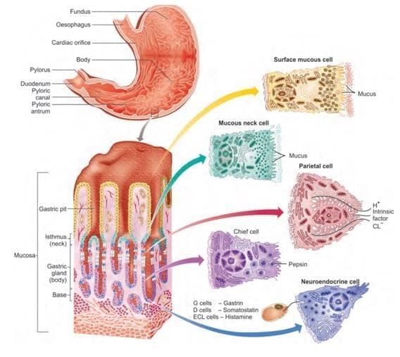
Predominant cells present at isthmus of gastric pit: (AIIMS June 2020)

Detailed Solution for Test: Anatomy - 1 - Question 18

The main cells found at the isthmus of the gastric pit are stem cells. Chief cells and neuroendocrine cells are situated in the lower section of the gland, closer to the base (fundus) of the gastric gland.

  • Parietal cells and mucus neck cells are mainly located in the upper half of the gland.
Test: Anatomy - 1 - Question 19

Ito cells are located in: (AIIMS June 2020)

Detailed Solution for Test: Anatomy - 1 - Question 19

Hepatic stellate cells of Ito are located in the perisinusoidal space of Disse, which is a narrow region between the sinusoids and hepatocytes. These Ito cells are responsible for storing vitamin A and are regarded as the primary profibrogenic cells involved in both acute and chronic liver diseases.

The periportal space of Mall is situated between the stroma of the portal canal and the outermost hepatocytes within the hepatic lobule. It is believed to be one of the origins of lymph in the liver.

Test: Anatomy - 1 - Question 20

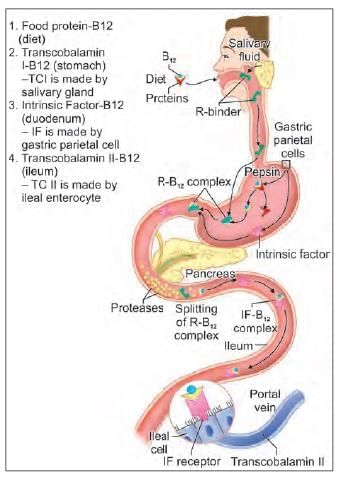
A young male met with a motor bike accident and had severe injury to ileum and jejunum. Therefore, the entire ileum and partial jejunum was resected. Which of the following would the patient suffer from? (NEET-PG 2020)

Detailed Solution for Test: Anatomy - 1 - Question 20

Surgical resection of the terminal ileum can lead to a deficiency in vitamin B12 (cobalamin). Vitamin B12 is present in animal-derived foods. R binder, also referred to as TC-1 (transcobalamin-1), is produced by the salivary glands and forms a complex with vitamin B12. The primary role of R binder is to safeguard the acid-sensitive vitamin B12 as it passes through the stomach.

Once it has bound with intrinsic factor (IF) secreted in the stomach, vitamin B12 becomes resistant to further digestion. In the duodenum, digestive enzymes, specifically pancreatic peptidases, release vitamin B12 from R binder. The liberated vitamin B12 then associates with intrinsic factor, which is a transport and delivery binding protein produced by the parietal cells of the stomach.

  • The complex proceeds to the distal ileum.
  • It attaches to a specific mucosal brush border receptor called cublin.
  • This interaction promotes the uptake of the cobalamin-IF complex through an energy-dependent mechanism.

Once inside the cells, intrinsic factor is removed, and cobalamin is transferred to other transport proteins, such as transcobalamin-2, facilitating its movement into the portal circulation. Vitamin B12 is exclusively found in animal products and is synthesised solely by microorganisms. A significant reserve (lasting several years) is predominantly stored in the liver.

Deficiency can arise from various factors, including:

  • Inadequate intake (e.g., through vegan diets)
  • Achlorhydria
  • Bacterial overgrowth
  • Excessive alcohol consumption
  • Lack of intrinsic factor, such as in pernicious anaemia
  • Removal of the terminal ileum due to surgical procedures, for instance, in Crohn's disease.

Test: Anatomy - 1 - Question 21

Arrange the following cells in basal to luminal side order: (INI-CET May 2022)
1. Spermatogonium
2. Spermatozoa
3. Primary spermatocyte
4. Spermatid
5. Myofibroblast

Detailed Solution for Test: Anatomy - 1 - Question 21

The cells mentioned are located in the seminiferous tubules (STs) of the testis.

  • The Sertoli cells extend from the basal to the luminal side, forming the blood-testis barrier through tight junctions with neighbouring Sertoli cells.
  • Spermatogonia are situated towards the basal side of the STs, undergoing mitotic cell division to transform into primary (1°) spermatocytes.
  • Primary spermatocytes, the largest among germ cells with notably clumped chromatin, undergo meiosis-I to produce secondary (2°) spermatocytes, which are only temporarily present.
  • These secondary spermatocytes proceed through meiosis-II, resulting in spermatids.
  • Subsequently, spermatids undergo spermiogenesis, aided by Sertoli cells, to develop into mature spermatozoa, characterised by a small condensed nucleus and a tail, located in the lumen of the STs.
  • Myofibroblasts, which are contractile, are present at the peripheral margin of the STs. They assist in the further movement of spermatozoa within the lumen.
  • Leydig cells, which secrete testosterone, are large polygonal cells found in the interstitial space between the STs.

Test: Anatomy - 1 - Question 22

Macula densa is located at junction of: (INI-CET Nov 2022)

Detailed Solution for Test: Anatomy - 1 - Question 22

Macula densa is situated at the junction of the thick ascending loop of Henle (TAL) and the distal convoluted tubule (DCT). It is a part of the Juxta-Glomerular Apparatus (JGA), located near the renal glomerulus and the afferent arteriole, with the function of regulating the Glomerular Filtration Rate (GFR).

The JGA develops at the site where a nephron's distal tubule (D) meets the vascular pole of its glomerulus (G). At this juncture, the cells of the distal tubule become columnar, forming a thickened area known as the macula densa (MD). - Smooth muscle cells in the tunica media of the afferent arteriole (AA) transition from a contractile state to a secretory form, known as juxtaglomerular granule cells (JG). - Additionally, lacis cells (L) are present, which are extraglomerular mesangial cells located next to the macula densa, the afferent arteriole, and the efferent arteriole (EA). In this specimen, the lumens of the proximal tubules (P) appear to be filled, and the urinary space (US) is somewhat enlarged.

Test: Anatomy - 1 - Question 23

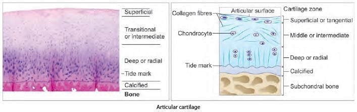
Identify the arrow marked structure in the microscopic picture: (NEET-PG 2023)

Detailed Solution for Test: Anatomy - 1 - Question 23

The structures indicated by arrows represent renal glomeruli in a histological slide of the kidney, characterised by clusters of mesangial cells surrounding a network of capillaries within an apparently empty capsular or renal space.

  • Numerous sections of proximal convoluted tubules (PCTs) and a few distal convoluted tubules (DCTs) are present.
  • PCT and DCT are lined with cuboidal cells around their lumen, with the PCT lumen obscured by microvilli (brush border).

Islets of Langerhans are recognised by groups of various endocrine cells (α, β, δ) located near blood capillaries, encircled by pancreatic acini (which contain centroacinar cells) and ducts. Lymphatic nodules are identified by the presence of lymphoid tissue, such as lymph nodes, tonsils, and the spleen. Note that the thymus does not contain lymphatic nodules.

  • Lymphoid tissue is evident from the large number of lymphocytes, which appear as multiple dark blue cells due to their prominent basophilic nuclei.
  • The lymph node displays a peripheral cortex (housing B cells in lymphatic nodules), a paracortex (containing T cells), and a central medulla (featuring lymphatic sinuses and cords).
  • The tonsil is characterised by lymphatic nodules and crypts lined with stratified squamous epithelium.
  • The spleen consists of red pulp (splenic sinusoids) and white pulp (lymphatic nodules with eccentrically placed central arterioles).
  • The thymus features multiple lobules, including a cortex and medulla with Hassall’s corpuscles.

Interstitial cells of Leydig are identifiable as a cluster of darkly eosinophilic large cells situated between several seminiferous tubules. The seminiferous tubules exhibit multiple layers of cells engaged in gametogenesis, with sperm present in the lumen.

(A) Kidney: Renal glomeruli (indicated by arrows), distal convoluted tubules (DCTs), proximal convoluted tubules (PCTs), and renal space (RS).
(B) Pancreas: Islet of Langerhans (indicated by arrows), pancreatic acini (PAs), duct (D), and blood capillaries (BCs).
(C) Lymph node: Lymphatic nodules (indicated by arrows), cortex (Cx) containing B lymphocytes in lymphatic nodules, and paracortex (PCx) containing T lymphocytes.
(D) Testis: Leydig cells (indicated by arrows), sperms in lumen (Sp), and seminiferous tubules (STs).

Test: Anatomy - 1 - Question 24

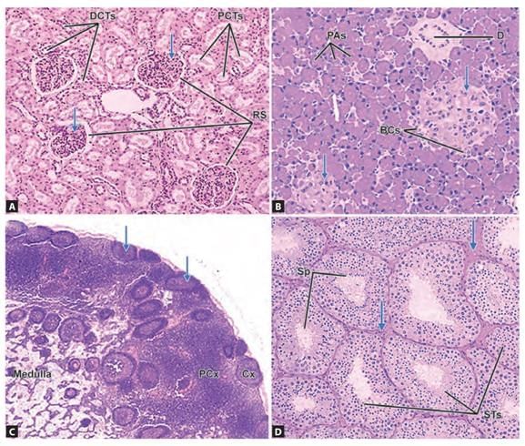
Identify the arrow marked structure in the given microscopic picture: (NEET-PG 2022)

Detailed Solution for Test: Anatomy - 1 - Question 24

The arrow in the histological slide indicates the islet of Langerhans, which is a cluster of endocrine cells, surrounded by serous acini (the exocrine component of the pancreas), along with various ducts and blood vessels present in the section. The islets of Langerhans are characterised by a grouping of multiple endocrine cells (α, β, δ) near blood capillaries. Numerous capillaries close to the islets transport endocrine secretions (hormones) that ultimately drain into larger vessels (veins), as can be observed in the provided histological specimen. Blood vessels are filled with blood, exhibiting numerous RBCs, with veins possessing a wider lumen compared to arteries.

  • This slide displays multiple islets of Langerhans, which are encircled by pancreatic acini (serous acini containing centroacinar cells) and collecting ducts, where the exocrine secretions (digestive enzymes rich in alkaline fluid) are directed.
  • It is noteworthy that pancreatic ducts are lined by simple cuboidal or columnar cells, unlike salivary glands, which may have multilayered cells.

A glomerulus is visible in a histology slide of the kidney, identified by a cluster of mesangial cells and a network of capillaries surrounded by an empty-looking capsular or renal space. It is encircled by various sections of proximal convoluted tubules (PCTs) and a few distal convoluted tubules (DCTs).

Lymphatic nodules are recognised by the presence of lymphoid tissue, such as lymph nodes, tonsils, and the spleen. Lymphoid tissue is evident due to the abundance of lymphocytes, which appear as multiple dark blue cells because of their large basophilic nuclei. A lymph node displays an outer cortex, a middle paracortex, and an inner medulla.

Serous demilunes are observed in mixed-type glands (sero-mucinous), such as the submandibular and sublingual salivary glands. A serous demilune appears as a cap of serous acinus (cuboidal, darkly stained cells) covering the mucinous acinus (columnar, lightly stained cells).

  • In salivary glands, ducts include striated ducts (which concentrate saliva) and multilayered collecting ducts.
  • It is important to note that striated ducts are absent in the pancreas, where the ducts are only a single cell thick (not multilayered).

(A) Pancreas: Islet of Langerhans (indicated by arrows) with nearby blood capillaries (BCs), pancreatic acini (PAs), and duct (D).
(B) Kidney: Renal glomeruli (indicated by arrows), renal space (RS), distal convoluted tubules (DCTs), and proximal convoluted tubules (PCTs).
(C) Lymph node: Lymphatic nodules (indicated by arrows), cortex (Cx) containing B lymphocytes in lymphatic nodules, and paracortex (PCx) containing T lymphocytes.
(D) Submandibular salivary gland: Serous demilunes (indicated by arrows), serous acinus (Sa), mucinous acinus (Ma), and striated duct (Sd).

Test: Anatomy - 1 - Question 25

Correctly match the following pairs: (INI-CET Nov 2022)

Detailed Solution for Test: Anatomy - 1 - Question 25
  • Kupffer cells are the phagocytic cells located in liver sinusoids;
  • Parietal cells are responsible for secreting acid in gastric glands (stomach);
  • Clara (Club) cells are surfactant-secreting cells found in terminal (and respiratory) bronchioles;
  • Dust cells (alveolar macrophages) are present in the lungs.

(A) Kupffer cells: Located in liver sinusoids, functioning as macrophages.
(B) Parietal cells: Found in gastric glands, responsible for secreting hydrochloric acid.
(C) Clara (Club) cells: Present in terminal bronchioles, involved in protection and repair of the respiratory epithelium.
(D) Dust cells (alveolar macrophages): Located in lung alveoli, responsible for phagocytosing inhaled particles.

Information about Test: Anatomy - 1 Page
In this test you can find the Exam questions for Test: Anatomy - 1 solved & explained in the simplest way possible. Besides giving Questions and answers for Test: Anatomy - 1, EduRev gives you an ample number of Online tests for practice
Download as PDF