NEET PG Exam  >  NEET PG Tests  >  Test: Anatomy - 5 - NEET PG MCQ

Test: Anatomy - 5 - NEET PG MCQ


Test Description

25 Questions MCQ Test - Test: Anatomy - 5

Test: Anatomy - 5 for NEET PG 2025 is part of NEET PG preparation. The Test: Anatomy - 5 questions and answers have been prepared according to the NEET PG exam syllabus.The Test: Anatomy - 5 MCQs are made for NEET PG 2025 Exam. Find important definitions, questions, notes, meanings, examples, exercises, MCQs and online tests for Test: Anatomy - 5 below.
Solutions of Test: Anatomy - 5 questions in English are available as part of our course for NEET PG & Test: Anatomy - 5 solutions in Hindi for NEET PG course. Download more important topics, notes, lectures and mock test series for NEET PG Exam by signing up for free. Attempt Test: Anatomy - 5 | 25 questions in 25 minutes | Mock test for NEET PG preparation | Free important questions MCQ to study for NEET PG Exam | Download free PDF with solutions
Test: Anatomy - 5 - Question 1

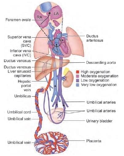
Highest oxygen concentration of fetal circulation is at which site? (NEET-PG 2020p)

Detailed Solution for Test: Anatomy - 5 - Question 1

The inferior vena cava (IVC), situated between the liver and the heart, transports blood with moderate levels of oxygen. It carries the deoxygenated blood from the body below the diaphragm, while also receiving oxygenated blood from the umbilical vein via the ductus venosus.

  • The IVC transfers this moderately oxygenated blood into the right atrium.
  • From the right atrium, blood moves to the left atrium through the foramen ovale.
  • The left atrium then pushes the blood into the left ventricle, which subsequently sends it into the aorta.
  • This moderately oxygenated blood is directed towards the developing brain via the carotid arteries.

The superior vena cava carries deoxygenated blood from the body above the diaphragm into the right ventricle.

Test: Anatomy - 5 - Question 2

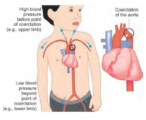
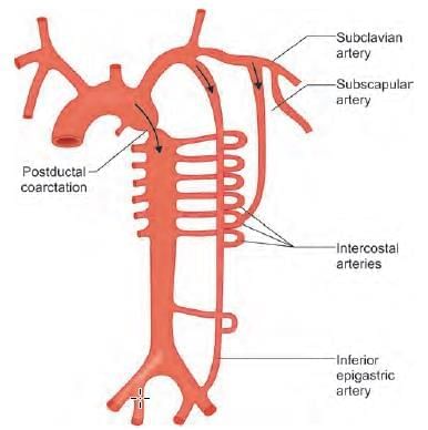
A 10-year-oId child presents with upper limb hypertension, and feeble lower limb pulses. Chest X-ray showed notching of ribs. What is the likely diagnosis? (NEET-PG 2021)

Detailed Solution for Test: Anatomy - 5 - Question 2

Coarctation of the aorta, which is a narrowing of the vessel, leads to elevated blood pressure prior to the constriction (e.g., hypertension in the upper limbs) and reduced blood pressure beyond the constriction (e.g., in the lower limbs, resulting in weak pulses). To compensate, collateral circulation develops to ensure blood supply to the areas affected by ischaemia. - One notable anastomosis occurs between the anterior and posterior intercostal arteries, which transport a significant volume of blood at high pressure. - The strong pulsations of the posterior intercostal arteries, located within the costal groove, can cause erosion or notching of the inferior ribs. Blood flow to the lower limb is preserved through the anastomosis of the superior epigastric artery and the inferior epigastric artery within the rectus sheath. The pathway for blood flow is as follows:

  • Arch of the aorta
  • Subclavian artery
  • Internal thoracic artery
  • Superior epigastric artery
  • Inferior epigastric artery
  • External iliac artery
  • Femoral artery

Test: Anatomy - 5 - Question 3

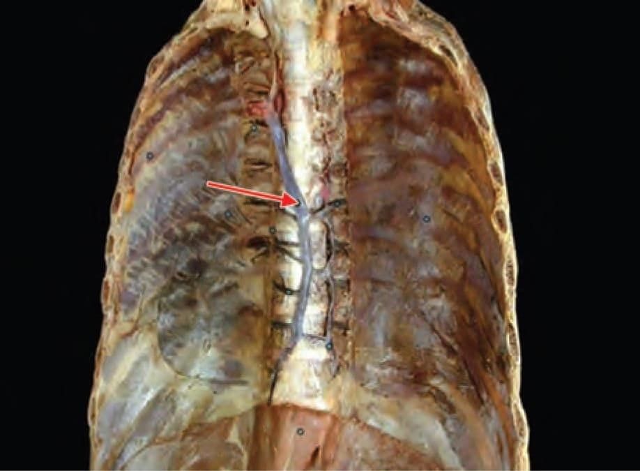
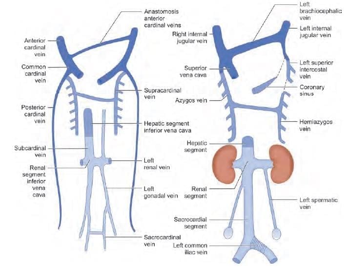
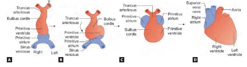
Identify the vein shown below and choose its embryological origin: (INI-CET Nov 2021)

Detailed Solution for Test: Anatomy - 5 - Question 3

The highlighted vein is the azygous vein, which originates from the supracardinal veins during embryonic development. The image depicts a coronal section of the thoracic cavity and the posterior thoracic wall, emphasising the azygous venous drainage. During the 5th to 7th weeks, several veins develop:

  • The subcardinal veins, primarily responsible for draining the kidneys;
  • The sacrocardinal veins, which drain the lower limbs;
  • The supracardinal veins, draining the body wall through the intercostal veins, assuming the roles of the posterior cardinal veins.

Test: Anatomy - 5 - Question 4
Type of joint at site marked by the arrow: (NEET-PG 2020p)
Detailed Solution for Test: Anatomy - 5 - Question 4
The joint located at the indicated area is known as the costo-transverse joint, which is classified as a plane synovial joint.
  • Ribs (costa) connect with the transverse process of the vertebra to create the costo-transverse joint.
  • They also articulate with the body of the vertebra to establish the costo-vertebral joint.
Both are considered plane synovial joints, allowing for free movement.
Test: Anatomy - 5 - Question 5

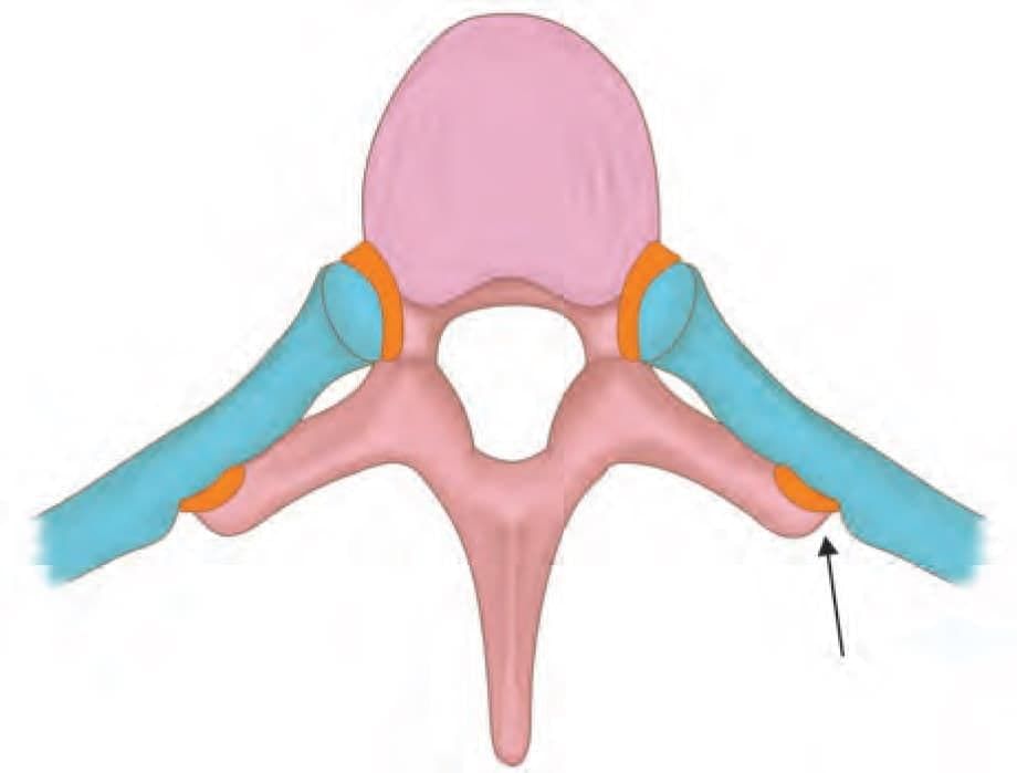
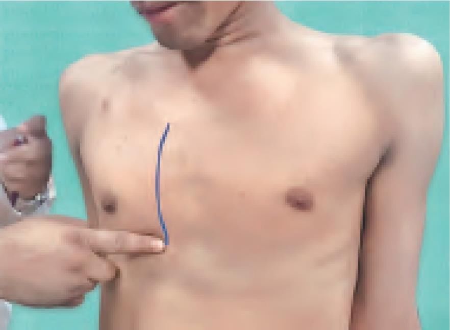
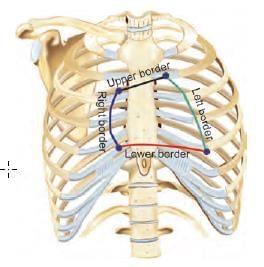
Surface marking of which of the following is demonstrated in the video. A convex line (towards right) is being drawn, starting at right 3rd costal cartilage to the right 6th costal cartilage, as shown in the diagram below: (AIIMS May 2019)

Detailed Solution for Test: Anatomy - 5 - Question 5

The right border of the heart can be delineated on the surface by drawing a convex line (towards the right) that begins at the right 3rd costal cartilage and extends to the right 6th costal cartilage. The surface marking of the heart can be achieved by connecting four points in a specific order:

  • Point A: Lower border of left CC2
  • Point B: Upper border of right CC3
  • Point C: Right CC6
  • Point D: Apex – 95 cm from the midline in ICS 5, medial to MCL, at the level of the diaphragm

Procedure: The upper border (from Point A to B) of the heart can be marked by drawing a line connecting a point on the lower border of the 2nd left costal cartilage, 1.5 inches from the median plane, to a point on the upper border of the 3rd right costal cartilage, 1 inch away from the median plane. The right border (from Point B to C) is a smoothly curved line, convex to the right, extending from the third to the sixth right costal cartilages, typically 1–2 cm lateral to the sternal edge. The inferior border (from Point C to D) reaches the cardiac apex, which is generally located from the level of the fifth to the sixth rib, averaging 8.7 + 1 cm from the midline. The left border (from Point D to A) curves towards the left and extends superomedially from the cardiac apex to meet the second left costal cartilage, roughly 1 cm from the left sternal edge.

Test: Anatomy - 5 - Question 6

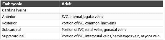
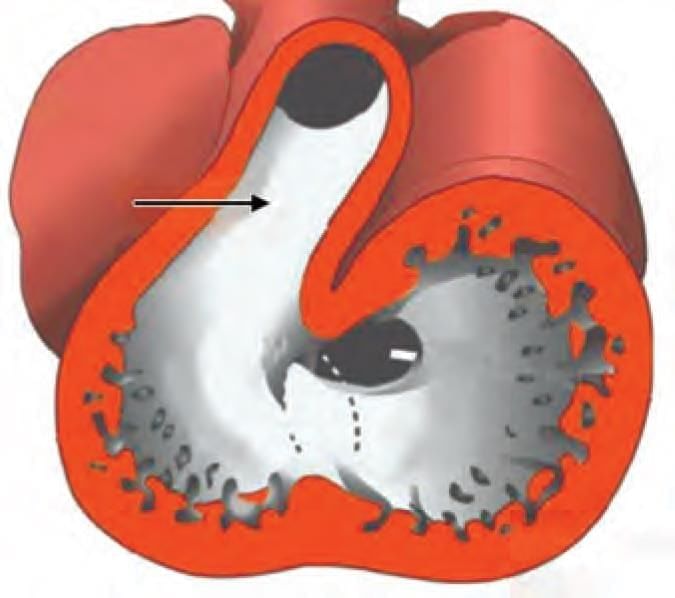
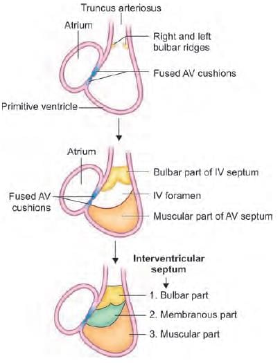
Which of the following structure develop from the arrow marked area? (AIIMS June 2020)

Detailed Solution for Test: Anatomy - 5 - Question 6
The area indicated by the arrow represents the bulbus cordis section of the heart tube, which plays a role in creating the smooth outflow tract of the ventricles, incorporating the conus arteriosus (infundibulum) of the right ventricle.
  • The infundibulum, also referred to as the conus arteriosus, is a conical pouch that originates from the upper and left angle of the right ventricle in an adult heart.
  • From this structure, the pulmonary trunk emerges.

Embryologically, it arises from the bulbus cordis.

Test: Anatomy - 5 - Question 7

Embryological origin of the marked structure: (AIIMS Nov 2019)

Detailed Solution for Test: Anatomy - 5 - Question 7

The arrow symbol indicates the membranous section of the interventricular septum, which originates from the endocardial cushions, themselves derived from neural crest cells. The bulbo-ventricular flange plays a role in forming the lower muscular part of the interventricular septum.

Test: Anatomy - 5 - Question 8

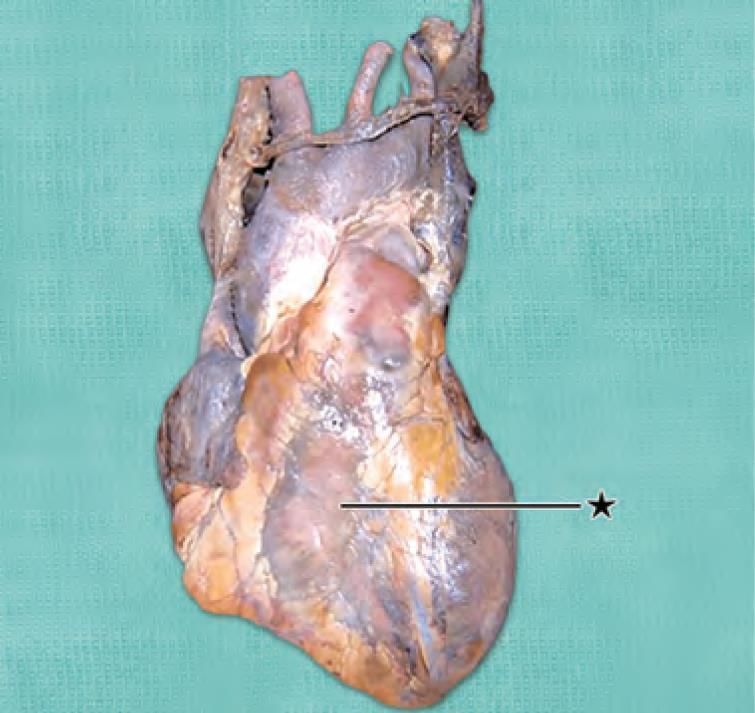
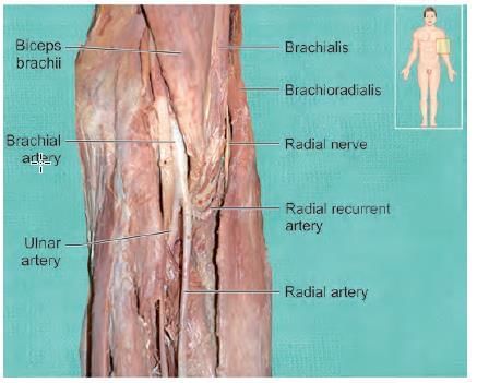
Marked structure is supplied by all the following arteries, except: (AIIMS Nov 2019)

Detailed Solution for Test: Anatomy - 5 - Question 8

The arrow indicates the right ventricle, which does not receive its blood supply from the diagonal artery. Instead, it obtains arterial nourishment from:

  • the right coronary artery,
  • the acute marginal artery,
  • the anterior interventricular artery.

Test: Anatomy - 5 - Question 9

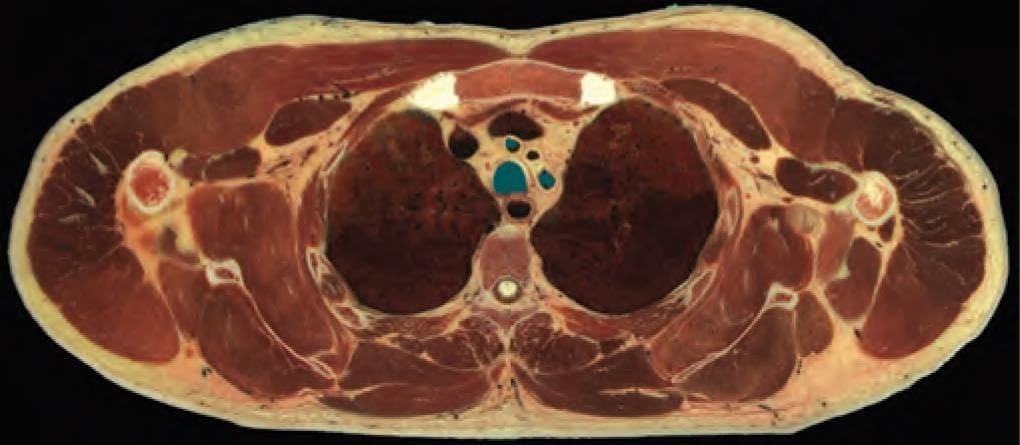
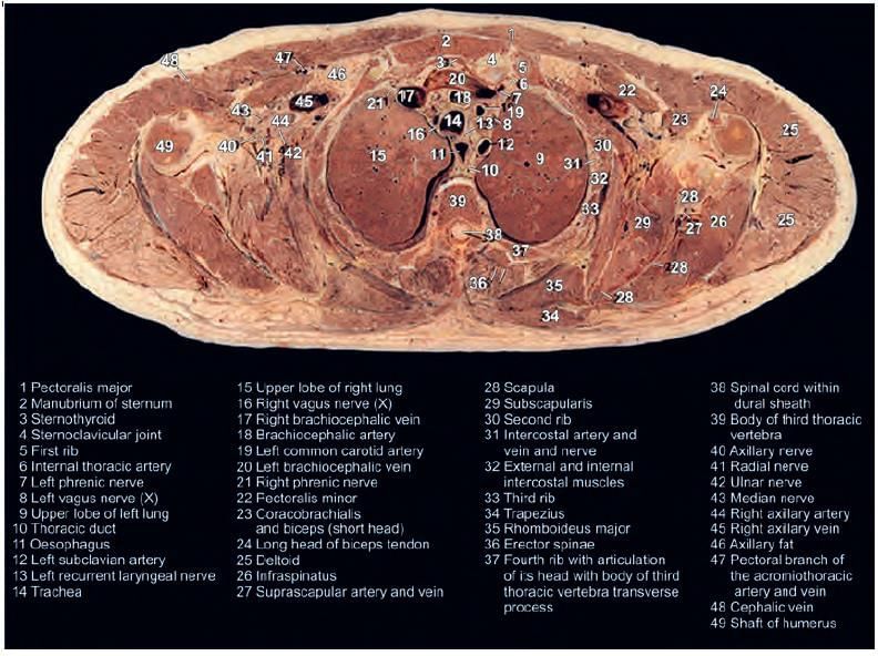
The present transverse section has been taken at which vertebra level? (INI-CET Nov 2020)

Detailed Solution for Test: Anatomy - 5 - Question 9

The current diagram represents a transverse section at the T3-4 vertebral level (inferior view), characterised by the arrangement of five vessels, along with the presence of the trachea and oesophagus.

  • The oesophagus is seen positioned slightly to the left of the midline; it is located between the T3 vertebra posteriorly and the trachea anteriorly.
  • Five vessels include:
    • Right brachiocephalic vein
    • Left brachiocephalic vein
    • Three branches of the aorta:
      • Right brachiocephalic artery
      • Left common carotid artery
      • Left subclavian artery
  • Other notable structures:
    • Manubrium sterni bone
    • Upper lobes of the lungs
    • TS (Humerus bone)

Test: Anatomy - 5 - Question 10

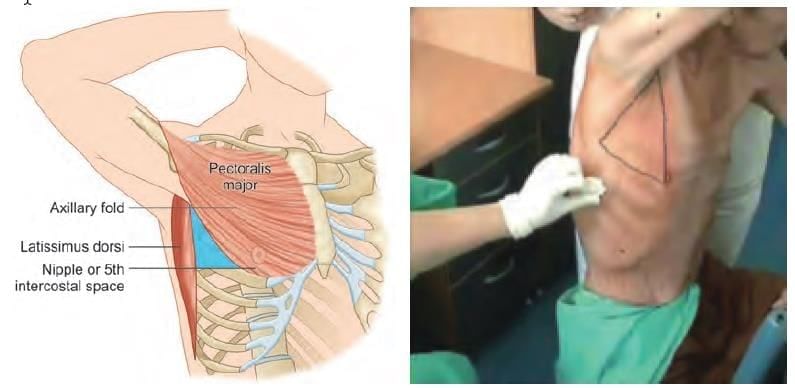
Which of the following is NOT a boundary for triangle of safety? (INI-CET Nov 2022)

Detailed Solution for Test: Anatomy - 5 - Question 10

The mid-clavicular line does not define any boundary for the triangle of safety. This triangle, located in the axilla, serves as a reference for the secure placement of an intercostal drainage tube.

  • In an abducted arm, the apex is situated in the axilla.
  • The triangle is defined by:
    • Anteromedially: the lateral border of the pectoralis major
    • Posteriorly: the lateral border of the latissimus dorsi
    • Inferiorly: a horizontal line at the level of the nipples/xiphoid process (typically at the 5th intercostal space)

Although this triangle is regarded as safe, it is recommended to utilise ultrasound guidance when inserting an intercostal drain. The surgeon should make a horizontal incision (approximately 3 cm) anterior to the mid-axillary line, within the 5th intercostal space at the level of the xiphoid.

Note: As with all intercostal approaches, placing the catheter closer to the superior border of the rib below in the intercostal space should help prevent injury to the intercostal neurovascular bundle located beneath the costal groove of the rib above.

The triangle of safety for the insertion of an intercostal drainage tube is defined as follows:

  • Bounded anteromedially by the lateral border of the pectoralis major
  • Bounded posteriorly by the lateral border of the latissimus dorsi
  • Bounded inferiorly by a horizontal line at the level of the nipples/xiphoid process (typically at the 5th intercostal space)

Test: Anatomy - 5 - Question 11

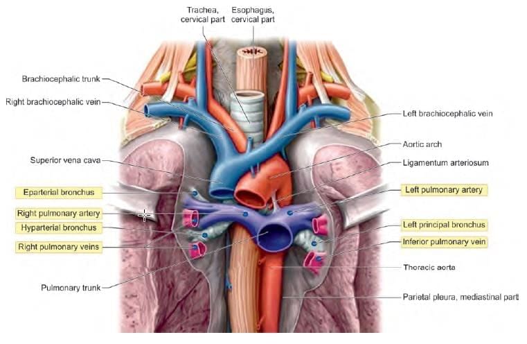
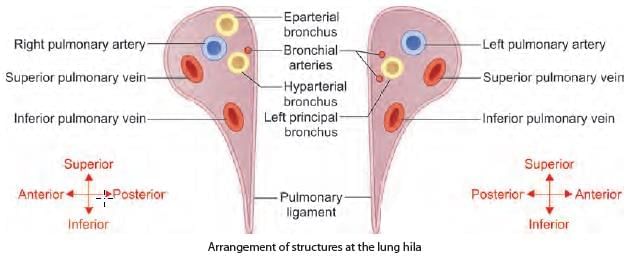
Arrangement of the structures in anterior to posterior relations at lung hilum: (INI-CET May 2022)

Detailed Solution for Test: Anatomy - 5 - Question 11

At both lung hila, the arrangement of structures from anterior to posterior follows the sequence VAB: Vein – Artery – Bronchus.

  • The most anterior structure is the Vein (superior pulmonary vein).
  • The Artery (pulmonary) is positioned in the middle.
  • The Bronchus, which has the thickest wall, is the most posterior.

Note: Bronchial arteries form a plexus around the bronchus, which is more pronounced posteriorly. There are two veins, referred to as superior and inferior based on their location at the hilum.
Note: The inferior pulmonary vein is located significantly more inferiorly and does not align with the anterior to posterior reference line. In the superior to inferior direction, the arrangement of structures in the hilum of the left lung can be recalled using the mnemonic ABV (Atal Bihari Vajpayee):

  • Artery (pulmonary)
  • Bronchus (principal)
  • Vein (inferior pulmonary)

Note: The superior pulmonary vein is positioned more anteriorly and does not fit into this reference line. The same sequence applies to the right lung, but with an additional bronchus located above the artery (eparterial bronchus).

Note: The right principal bronchus is relatively short and divides into two lobar bronchi before entering the lung at the hilum.

  • The upper lobar bronchus passes above the pulmonary artery (eparterial bronchus).
  • The lower lobar bronchus passes below the pulmonary artery (hyparterial bronchus).

This explains the differences in the arrangement of structures from above downward on each side.

*Multiple options can be correct
Test: Anatomy - 5 - Question 12

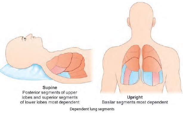
In a bed ridden patient, aspiration pneumonitis is most likely to involve which of the following bronchopulmonary segment? (INI-CET Nov 2022)

Detailed Solution for Test: Anatomy - 5 - Question 12

Aspiration in a supine (bedridden) position typically affects the right lung, specifically the superior (apical) bronchopulmonary segment (BPS) of the right lower lobe. It can also be located in the posterior BPS of the right upper lobe. - Most foreign objects tend to move towards the right lung because the right principal bronchus is shorter, wider, and more vertical compared to the left principal bronchus. - Aspiration in a supine position generally involves the right lower lobar bronchus, with the aspirate settling in the superior (apical) bronchopulmonary segment of the right lower lobe. - In an upright position (sitting or standing), aspirated substances usually enter the right lower lobar bronchus and become lodged in the posterior basal bronchopulmonary segment of the right lower lobe.

Test: Anatomy - 5 - Question 13

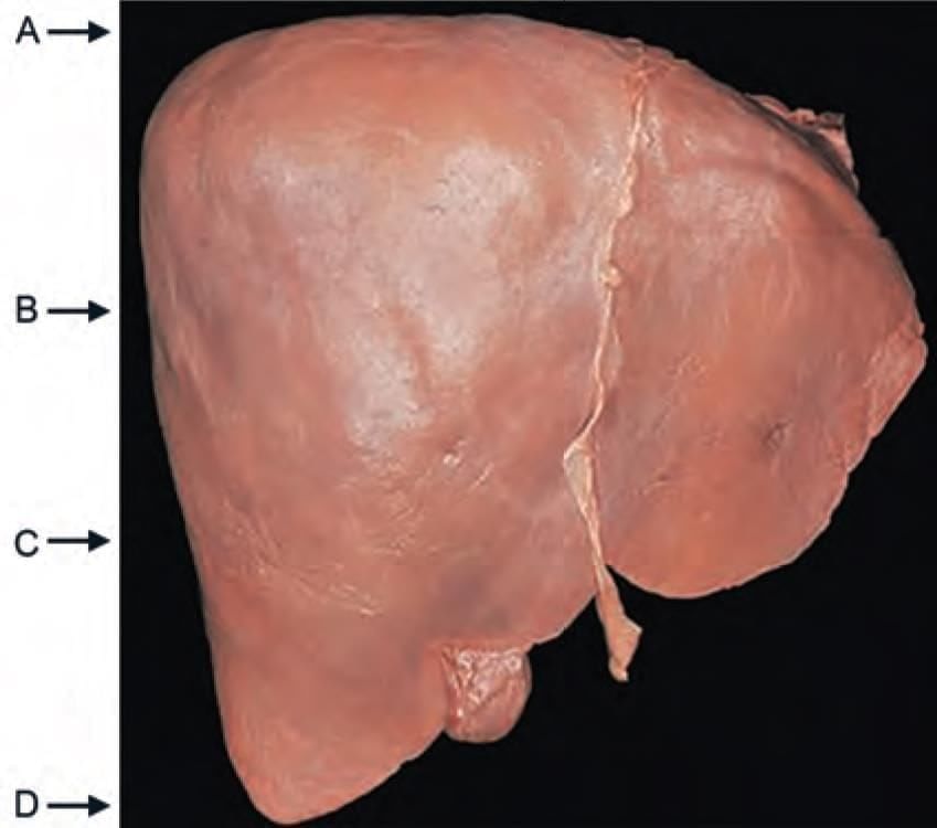
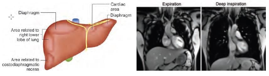
The lung (with visceral pleura) extend upto which level of liver during deep inspiration? (INI-CET Nov 2020)

Detailed Solution for Test: Anatomy - 5 - Question 13

The lung (with visceral pleura) extends up to the upper third of the liver (Marker B) during deep inspiration (refer to the Real Time MRI image shown below).

  • The right lung reaches rib 10 (in the mid-axillary line) during deep inspiration by extending into the costodiaphragmatic recess of the pleura.
  • However, as the diaphragm descends, it pushes the liver down, causing the lung (and the attached visceral pleura) to only reach the upper third of the liver.

Typically, the right surface of the liver is covered by peritoneum and is positioned next to the superior part of the respiratory diaphragm, which separates it from the right lung and pleura, as well as the seventh to the eleventh ribs. The liver is associated with:

  • the diaphragm
  • the right lung and pleura in the upper third (ribs 7 and 8)
  • the right costodiaphragmatic recess in the middle third (ribs 9 and 10)

Lateral to the lower third of the liver, the diaphragm and thoracic wall are in direct contact. It is important to note that although the right costodiaphragmatic recess (parietal pleura) reaches the middle third of the liver, during deep inspiration, the diaphragm's descent pushes the liver down, resulting in the right lung occupying the costodiaphragmatic recess and only reaching the upper third of the liver.

Clinical significance: Liver biopsies are performed during expiration, with the liver accessed at the midaxillary line in the ninth or tenth intercostal space.

Test: Anatomy - 5 - Question 14

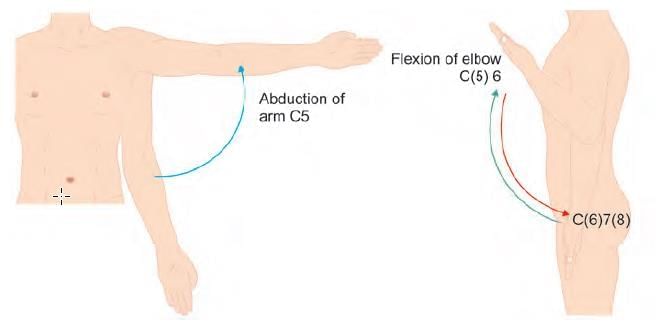
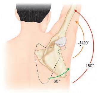
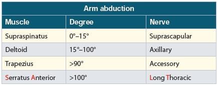
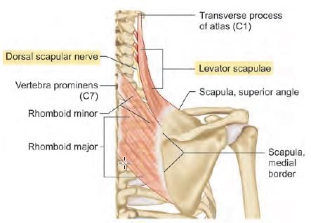
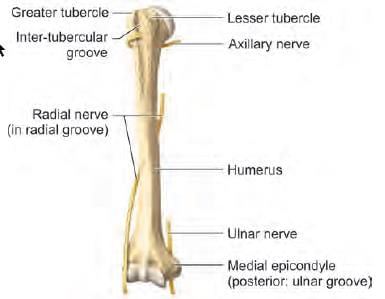
After cervical surgery shoulder abduction NOT possible due to lesioned: (INI-CET Nov 2020)

Detailed Solution for Test: Anatomy - 5 - Question 14

A cervical operation resulting in damage to the C5 root value may affect shoulder abduction. Embryologically, the myotomes (muscles) associated with C5 root values play a role in both shoulder abduction and elbow flexion.

A lesion at C5 (commonly observed in Erb’s palsy) results in weakness in shoulder abduction and elbow flexion.

  • The resulting deformity, known as the "Policeman's tip hand," occurs due to the unopposed action of shoulder adductors and elbow extensors.

Test: Anatomy - 5 - Question 15

Muscles involved in overhead abduction all, except:

Detailed Solution for Test: Anatomy - 5 - Question 15

Pectoralis major performs adduction (not abduction).

  • Overhead abduction (90° to 180°) is primarily executed by the serratus anterior and trapezius.
  • It is important to recognise that all muscles involved in shoulder abduction maintain contraction, regardless of the degree of movement.
  • However, each muscle becomes increasingly active during specific ranges of motion.

For instance, the deltoid muscle is engaged in shoulder abduction throughout the range (0°–180°) but is most active between 15° and 90°.

  • The total range of abduction extends to 180°.
  • Abduction up to 90° occurs at the glenohumeral joint.
  • Abduction from 90° to 120° can only take place if the humerus is laterally rotated.
  • Abduction from 120° to 180° requires the scapula to rotate forwards on the chest wall.

The articular surface of the head of the humerus allows arm elevation only up to 90°; beyond this point, the arm rotates laterally, facilitating abduction up to 120°.

  • Abduction exceeding 120° is only possible if the scapula rotates.
  • Thus, the scapula must rotate forwards (protraction) on the chest wall.

Note: The movement of the humerus and scapula during abduction occurs in a 2:1 ratio; for every 15° of elevation, the humerus moves 10° and the scapula moves 5°.

Test: Anatomy - 5 - Question 16

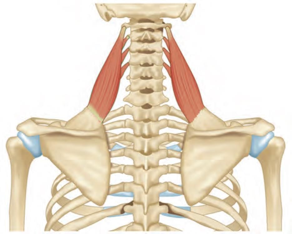
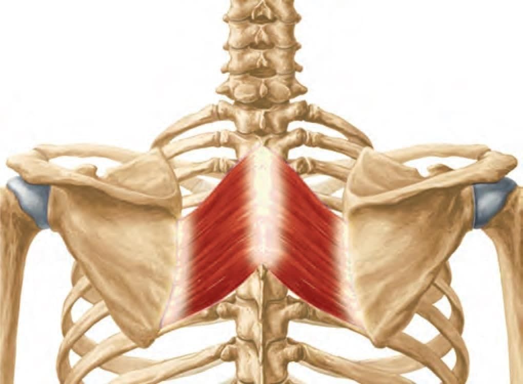
Nerve supply of the shown muscle is: (INI-CET Nov 2020) (NEET-PG 2020p)

Detailed Solution for Test: Anatomy - 5 - Question 16

The displayed muscle is the levator scapulae, which receives its innervation from the dorsal scapular nerve.

*Multiple options can be correct
Test: Anatomy - 5 - Question 17

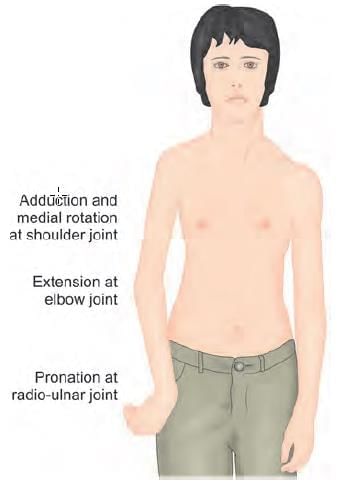
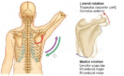
The mechanism of action of the marked muscle is: (INI-CET May 2023)

Detailed Solution for Test: Anatomy - 5 - Question 17

The highlighted muscle is the rhomboid major, which draws the scapula supero-medially, facilitating the retraction and elevation of the scapula. It also plays a role in the medial rotation of the scapula and the inferior rotation of the glenoid cavity.

  • During shoulder abduction, the trapezius and serratus anterior muscles cause the lateral rotation of the scapula and a superior tilt of the glenoid cavity.
  • Conversely, in shoulder adduction, the opposite movements occur: medial rotation of the scapula and inferior tilting of the glenoid cavity (typically due to gravity).
  • However, the rhomboideus major and minor, along with the levator scapulae, assist in this process.

Details: The rhomboid major is a rhombus-shaped muscle, situated beneath the trapezius in the upper thoracic region of the back.

  • Origin: Spinous processes of the T2 to T5 vertebrae, supraspinous ligament.
  • Insertion: Medial border of the scapula.
  • Nerve supply: Dorsal scapular nerve.
  • Action: Retraction and elevation of the scapula (pulls the scapula supero-medially) and rotates the glenoid cavity inferiorly (medial rotation of the scapula).

Applied Anatomy: Paralysis of the rhomboids can lead to winging of the medial border of the scapula and rotation of the inferior scapular angle. The entrapment of the dorsal scapular nerve is the most common cause; direct injury to the dorsal scapular nerve (such as from anterior shoulder dislocation) or overuse in overhead sports (like baseball and volleyball) may also be observed.

Test: Anatomy - 5 - Question 18

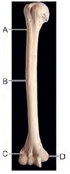
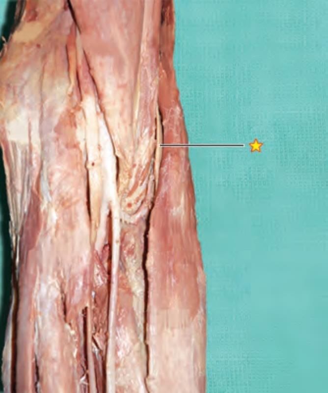
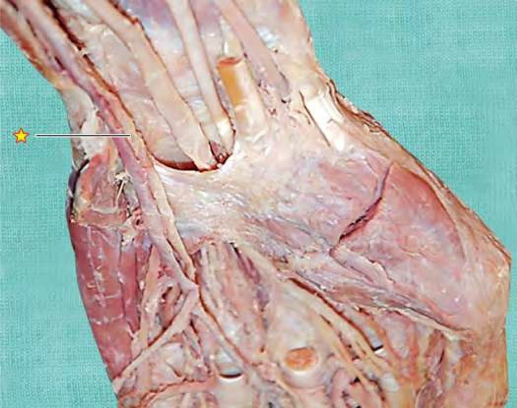
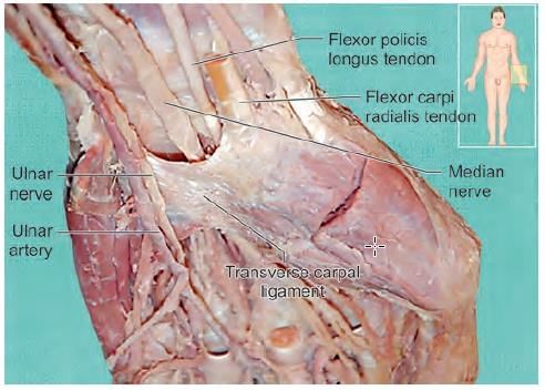
A patient has presented with inability to flex the distal interphalangeal joint of fourth and fifth fingers in right hand. Patient is also unable to hold a paper between fingers. At which of the following marked site, the nerve is most likely injured? (INI-CET July 2021, May 2022, 23)

Detailed Solution for Test: Anatomy - 5 - Question 18

This patient exhibits clinical signs of ulnar nerve damage, likely located at the medial epicondyle of the humerus (marker ‘D’). An injury to the ulnar nerve at this site (e.g., a fracture of the medial epicondyle) results in:

  • Loss of sensation in the medial half of the fingers: the little finger and part of the ring finger (both dorsal and ventral surfaces).
  • Paralysis affecting the medial half of the flexor digitorum profundus, leading to an inability to flex at the distal interphalangeal joint of the fourth and fifth fingers.
  • Paralysis of all interossei muscles.
  • Inability to hold paper between the fingers due to non-functional palmar interossei, which are responsible for adduction at the metacarpophalangeal joint.

Marker A: Axillary nerve (at the surgical neck of the humerus). Marker B: Radial nerve (in the radial groove). Marker C: Radial nerve (anterior to the lateral epicondyle). A lesion of the axillary nerve (at the surgical neck of the humerus) results in:

  • Paralysis of the deltoid muscle (affecting shoulder abduction) and the teres minor (responsible for lateral rotation at the shoulder joint).
  • Loss of sensation over the skin covering the lower half of the deltoid.

A lesion of the radial nerve (in the radial groove) causes:

  • Wrist drop (paralysis of the extensors) and sensory loss on the dorsum of the forearm and hand.

A lesion of the radial nerve (anterior to the lateral epicondyle) leads to:

  • Finger drop at the metacarpophalangeal joint (paralysis of extensors) and sensory loss over the dorsum of the forearm and hand.

Test: Anatomy - 5 - Question 19
A patient presented with multiple humeral shaft fracture, following which he had difficulty in elbow flexion and supination of forearm. He also complains of loss of sensation over lateral side of forearm. Which of the following nerve is most likely involved? (NEET-PG 2021)
Detailed Solution for Test: Anatomy - 5 - Question 19

Damage to the musculocutaneous nerve results in challenges with elbow flexion and forearm supination, primarily because of the paralysis of the biceps brachii. Additionally, there is a corresponding loss of sensation on the lateral side of the forearm.

The musculocutaneous nerve (C5, 6, 7) originates from the lateral cord of the brachial plexus. It innervates three muscles in the anterior arm:

  • coraco-brachialis,
  • brachialis,
  • biceps brachii.

It extends as the lateral cutaneous nerve of the forearm, providing sensation to the lateral aspect of the forearm.

Test: Anatomy - 5 - Question 20

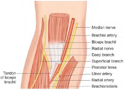
Arrangement of the structures in medial to lateral relations at cubital fossa: (INI-CET May 2022)
1. Biceps tendon
2. Brachial artery
3. Median nerve
4. Radial nerve

Detailed Solution for Test: Anatomy - 5 - Question 20

The contents of the cubital fossa, arranged from medial to lateral, include: the median nerve, brachial artery, biceps tendon, and radial nerve. Details include:

  • Boundaries: The brachioradialis muscle is located laterally, while the pronator teres is found medially; the superior boundary is marked by an imaginary horizontal line that connects the two epicondyles of the humerus.
  • Floor: Composed of the brachialis and supinator muscles.
  • The ulnar nerve runs behind the medial epicondyle and is not considered a part of the cubital fossa.
  • At its distal end, the brachial artery bifurcates into the radial and ulnar arteries.

From medial to lateral, the basilic, median cubital, and cephalic veins are situated within the superficial fascia (at the roof). The fascial roof is reinforced by the bicipital aponeurosis, across which the antecubital vein drains from the cephalic vein into the basilic vein.

Test: Anatomy - 5 - Question 21

Lesion of the marked nerve leads to: (AIIMS Nov 2019)

Detailed Solution for Test: Anatomy - 5 - Question 21

The indicated structure is the radial nerve, which is located at the outermost edge of the cubital fossa. An injury to this nerve leads to symptoms associated with wrist drop (impaired wrist extension). Details include:

  • The cubital fossa comprises four primary vertical components, denoted as R-TAM (from lateral to medial):
  • R – Radial nerve
  • T – Tendon of biceps brachii
  • A – Artery (Brachial)
  • M – Median nerve
Test: Anatomy - 5 - Question 22

Identify the marked structure: (AIIMS Nov 2019)

Detailed Solution for Test: Anatomy - 5 - Question 22

The ulnar nerve runs above the flexor retinaculum at the anterior aspect of the wrist.

Test: Anatomy - 5 - Question 23

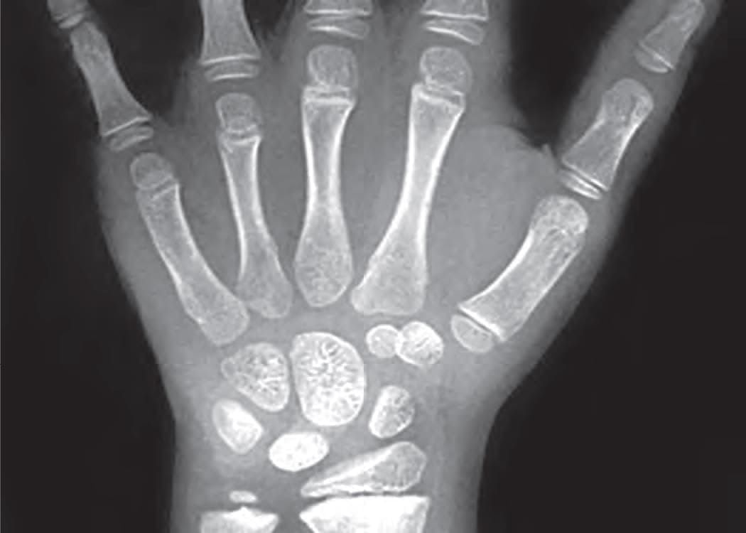
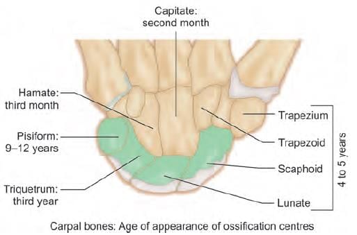
What is the most likely age of this child, a victim of rape being examined under POCSO act? (NEET-PG 2020p)

Detailed Solution for Test: Anatomy - 5 - Question 23

The X-ray of the carpal bones reveals a lack of the pisiform bone, which typically ossifies between the ages of 9 and 12 years after birth.

  • As the trapezoid is visible, it indicates that the individual is older than 5 years.

Test: Anatomy - 5 - Question 24

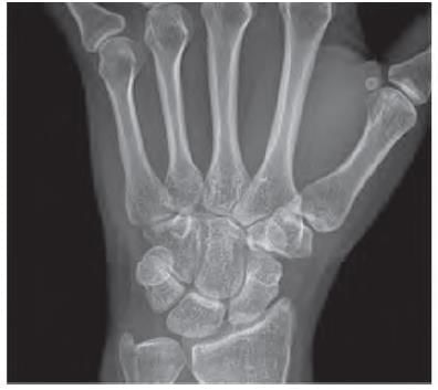
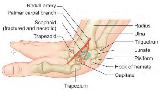
Assertion: In Scaphoid fracture, distal portion gets necrosed

Reason: Scaphoid has arterial supply in retrograde fashion (AIIMS May 2019)

Detailed Solution for Test: Anatomy - 5 - Question 24

In scaphoid fractures, the proximal part of the bone, rather than the distal portion, undergoes necrosis. This occurs due to the retrograde nature of the scaphoid's arterial supply.

Post-traumatic AVN (Avascular Necrosis) of the scaphoid arises from a disruption of the blood supply to the affected bone fragment.

  • There is a notably inadequate blood supply to the proximal pole.
  • This is especially evident when compared to the plentiful supply to the distal two-thirds of the scaphoid.

Test: Anatomy - 5 - Question 25

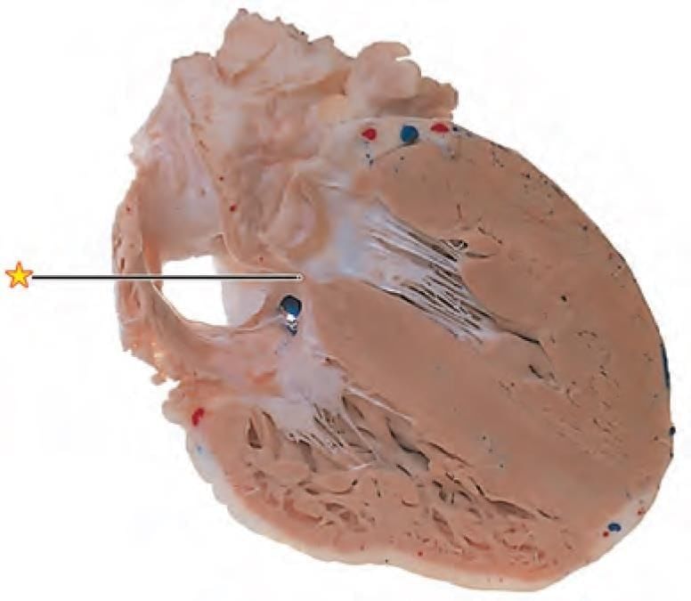
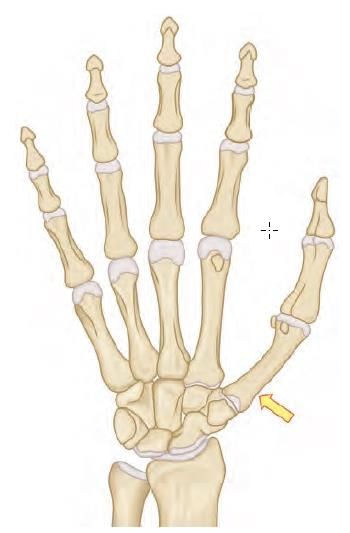
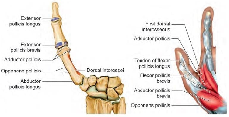
Name the muscle attached to the arrow marked area: (INI-CET Nov 2020)

Detailed Solution for Test: Anatomy - 5 - Question 25

Abductor pollicis longus tendon attaches to the lateral/radial side of the base of the first metacarpal bone (indicated by the arrow).

  • Option a: Palmar interossei (1st) arises from the ulnar/medial side of the base of the first metacarpal bone.
  • Option b: Opponens pollicis connects to the radial/lateral side of the shaft of the first metacarpal bone.
  • Option d: Flexor pollicis brevis (along with abductor pollicis brevis) inserts into the lateral aspect of the base of the proximal phalanx of the thumb.

First dorsal interossei originates from the adjacent sides of the shafts of the first and second metacarpals. Adductor pollicis attaches to the medial/ulnar side of the base of the proximal phalanx of the thumb.

Information about Test: Anatomy - 5 Page
In this test you can find the Exam questions for Test: Anatomy - 5 solved & explained in the simplest way possible. Besides giving Questions and answers for Test: Anatomy - 5, EduRev gives you an ample number of Online tests for practice
Download as PDF