NEET PG Exam  >  NEET PG Tests  >  Test: Anatomy - 6 - NEET PG MCQ

Test: Anatomy - 6 - NEET PG MCQ


Test Description

25 Questions MCQ Test - Test: Anatomy - 6

Test: Anatomy - 6 for NEET PG 2025 is part of NEET PG preparation. The Test: Anatomy - 6 questions and answers have been prepared according to the NEET PG exam syllabus.The Test: Anatomy - 6 MCQs are made for NEET PG 2025 Exam. Find important definitions, questions, notes, meanings, examples, exercises, MCQs and online tests for Test: Anatomy - 6 below.
Solutions of Test: Anatomy - 6 questions in English are available as part of our course for NEET PG & Test: Anatomy - 6 solutions in Hindi for NEET PG course. Download more important topics, notes, lectures and mock test series for NEET PG Exam by signing up for free. Attempt Test: Anatomy - 6 | 25 questions in 25 minutes | Mock test for NEET PG preparation | Free important questions MCQ to study for NEET PG Exam | Download free PDF with solutions
Test: Anatomy - 6 - Question 1

Nerve supply of arrow marked structure is: (INI-CET Nov 2022)

Detailed Solution for Test: Anatomy - 6 - Question 1
The arrow symbol indicates the transverse head of the adductor pollicis muscle, which is innervated by the deep branch of the ulnar nerve. The adductor pollicis consists of two heads (both receiving innervation from the ulnar nerve):
  • Transverse head – Origin: Anterior surface of the third metacarpal.
  • Oblique head – Origin: Bases of the second and third metacarpals, along with the adjoining trapezoid and capitate bones.

The muscle attaches to the medial aspect of the base of the proximal phalanx of the thumb and the ulnar sesamoid bone. Its action is to facilitate the adduction of the thumb at the carpometacarpal joint.

Test: Anatomy - 6 - Question 2

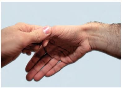
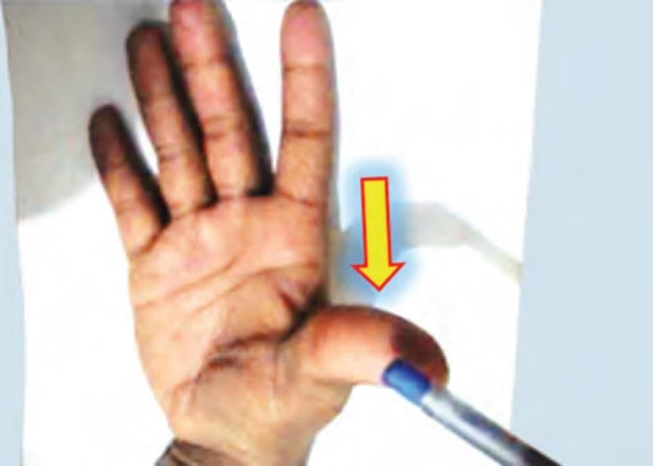
A 30 years female patient is presenting with wrist pain on lateral side. On examination the following test was positive. Tendon sheaths of which two muscle have been involved? (NEET-PG 2020p)

Detailed Solution for Test: Anatomy - 6 - Question 2

The Finkelstein test is employed to identify de Quervain’s tenosynovitis in individuals experiencing wrist discomfort. The examiner holds the thumb and then sharply deviates the hand towards the ulnar side. If a sharp pain is felt along the distal radius, it indicates tenosynovitis of the tendons that create the antero-lateral boundary of the anatomical snuff box, specifically the abductor pollicis longus and extensor pollicis brevis.

Test: Anatomy - 6 - Question 3

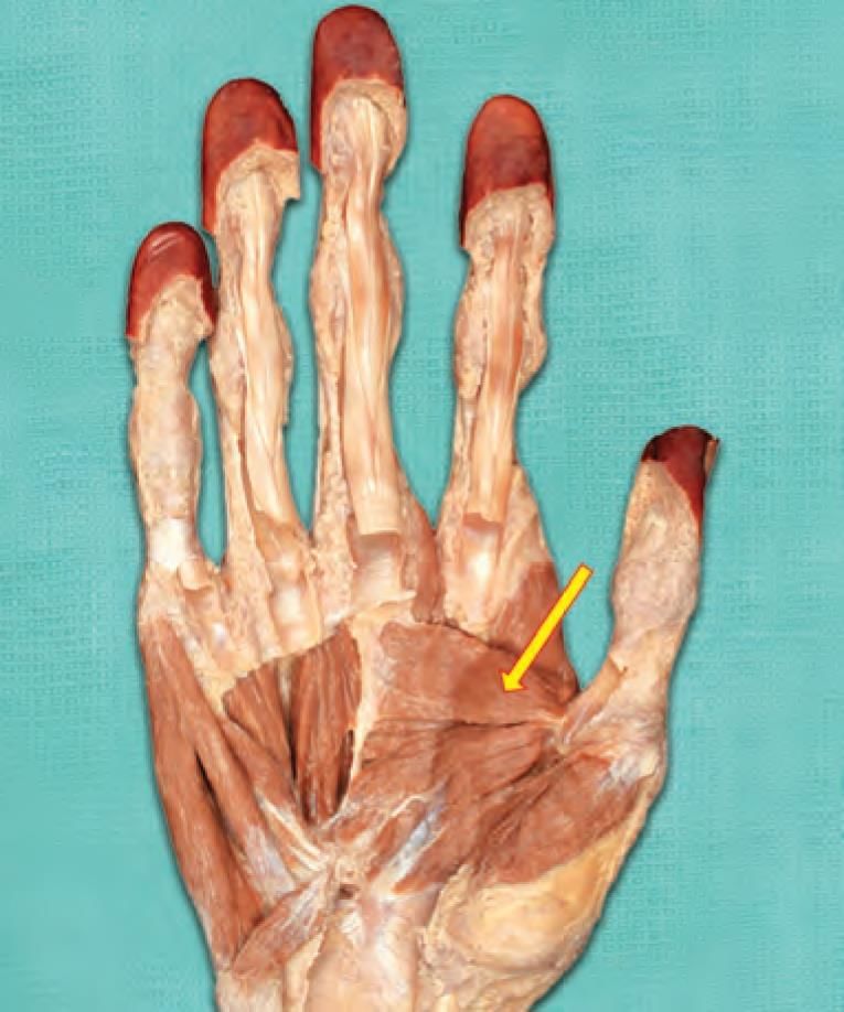
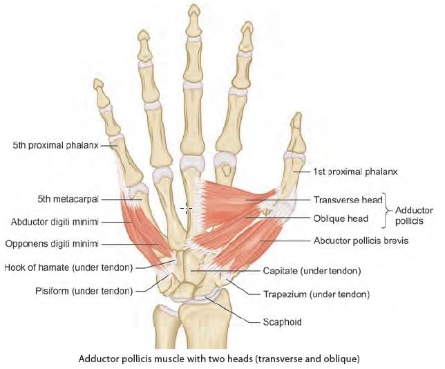
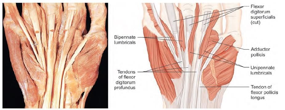
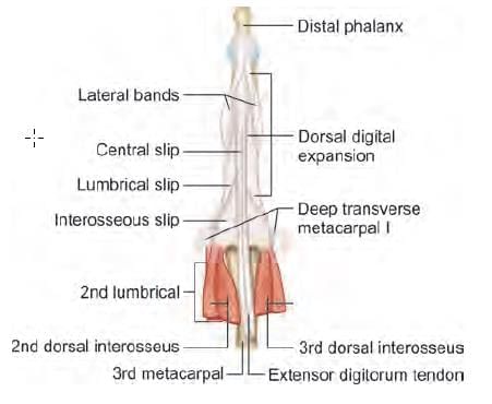
Mention the nerve supply of the marked muscle: (INI-CET May 2022)

Detailed Solution for Test: Anatomy - 6 - Question 3

The muscle indicated by the arrow is the 2nd lumbrical, which is innervated by the median nerve. Lumbricals originate from the tendons of the flexor digitorum profundus (as shown in the photograph), and therefore receive their nerve supply accordingly.

  • Lumbricals 1 & 2 originate from the lateral half of the flexor digitorum profundus, hence they are innervated by the median nerve.
  • Lumbricals 3 & 4 originate from the medial half of the flexor digitorum profundus, thus they are supplied by the ulnar nerve.

Lumbricals 1 & 2 are unipennate, while lumbricals 3 & 4 are bipennate. All four lumbricals traverse the MCP (metacarpo-phalangeal) joint anteriorly (causing MCP flexion), travel laterally to each corresponding finger, and insert into the dorsal digital expansion (resulting in interphalangeal extension).

Test: Anatomy - 6 - Question 4

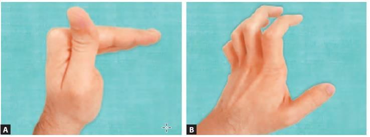
Hand deformity presenting as hyper-extension at the metacarpo-phalangeal joint and flexion at inter-phalangeal joint, occurs due to paralyzed: (NEET-PG 2020p)

Detailed Solution for Test: Anatomy - 6 - Question 4

Hand deformity characterised by hyper-extension at the metacarpo-phalangeal joint and flexion at the inter-phalangeal joint is known as claw hand deformity. This condition may arise from the paralysis of the lumbricals and interossei.

  • (A) The combined action of the lumbricals and interossei results in metacarpo-phalangeal (MCP) flexion and interphalangeal (IP) extension.
  • (B) Their paralysis causes unopposed activity of the posterior forearm muscles, leading to MCP hyperextension, and of the anterior forearm muscles, resulting in IP flexion.

Claw hand deformity.

Test: Anatomy - 6 - Question 5

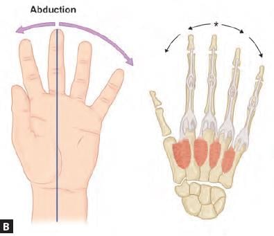
Which of the following finger has two dorsal interossei? (AIIMS May 2019)

Detailed Solution for Test: Anatomy - 6 - Question 5

The middle finger is connected to two dorsal interossei muscles, which enable abduction to either side. The dorsal interossei comprise four muscles in the hand that function to abduct (spread) the index, middle, and ring fingers away from the hand's midline (the middle finger). They also aid in flexion at the metacarpophalangeal joints and extension at the interphalangeal joints of the index, middle, and ring fingers.

  • (A) PAD: The palmar interossei facilitate ADduction, moving the fingers towards the axis (middle finger). It is important to note that the middle finger has no palmar interossei, as it serves as the axis itself.
  • (B) DAB: The dorsal interossei enable ABduction, moving the fingers away from the axis (middle finger). It is noteworthy that the middle finger has two dorsal interossei, and any movement away from the axis is considered abduction.

Test: Anatomy - 6 - Question 6

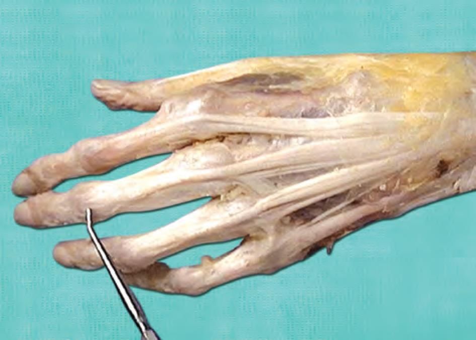
Which of the structure does not contribute to the pointed structure? (AIIMS Nov 2019)

Detailed Solution for Test: Anatomy - 6 - Question 6

The structure identified is the dorsal digital expansion located on the back of the middle finger. This is a modification of the extensor digitorum tendon, which does not receive any insertion from the palmar interossei. It does, however, receive insertions from two dorsal interossei (2 and 3) as well as the second lumbrical, originating from the hand muscles.

Test: Anatomy - 6 - Question 7
Mention the nerve involved in this test: (NEET-PG 2021)
Detailed Solution for Test: Anatomy - 6 - Question 7
The assessment being conducted is referred to as the ‘Pen test’, which evaluates the anterior abduction of the thumb executed by the abductor pollicis brevis, innervated by the median nerve.
  • The Pen test yields a positive result in cases of median nerve lesions, where the thumb is unable to achieve anterior abduction to reach the pen held by the examiner.
In the case of a posterior interosseous nerve (PIN) lesion:
  • The lateral (radial) abduction of the thumb is affected due to the paralysis of the abductor pollicis longus.
The PIN is a branch of the radial nerve that innervates the muscles of the posterior forearm. For an ulnar nerve lesion:
  • Both the Card test (palmar interossei) and the Froment test (adductor pollicis) return positive results.
With a musculocutaneous nerve lesion:
  • There is a noticeable weakness in elbow flexion and supination, among other functions.
Test: Anatomy - 6 - Question 8

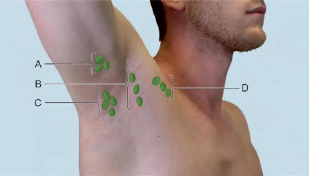
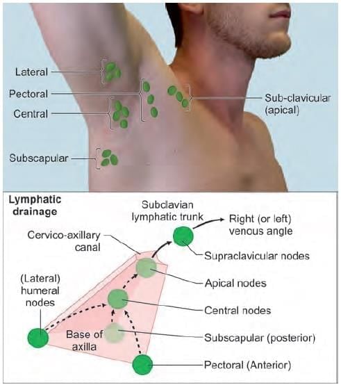
Choose the CORRECT matching pair for axillary lymph nodes in the given diagram: (INI-CET Nov 2022)

Detailed Solution for Test: Anatomy - 6 - Question 8

Anterior (pectoral), posterior (subscapular), and lateral (humeral) lymph nodes direct drainage towards the central axillary lymph nodes. The lymphatics then progress to the apical axillary (infraclavicular) lymph nodes, which represent the final axillary lymph nodes. Ultimately, the lymphatics advance to the supraclavicular lymph node, followed by the subclavian lymphatic trunk, leading to the right lymphatic duct and the right jugulo-subclavian venous angle. The lymphatic drainage is symmetrical on both the right and left sides, with slight variations. On the left side, the left subclavian lymphatic trunk empties into the thoracic duct, which ultimately drains into the left jugulo-subclavian venous angle.

Test: Anatomy - 6 - Question 9
Clinical diagnosis for the patient shown in the diagram is: (NEET-PG 2020p)
Detailed Solution for Test: Anatomy - 6 - Question 9

Gastroschisis is a defect in the anterior abdominal wall, where the intestines extend outward. This occurs to the right of the umbilical ring, while the umbilical cord remains intact on the left side. The protruding contents lack an amniotic covering.

  • Meckel’s diverticulum appears as a minor bulge outside the umbilicus, which includes a small section of the distal ileum.
  • Omphalocele is a midline defect (not positioned on the right).
  • The umbilical cord is abnormal.
  • Urachal diverticulum does not exhibit intestinal herniation.
Test: Anatomy - 6 - Question 10

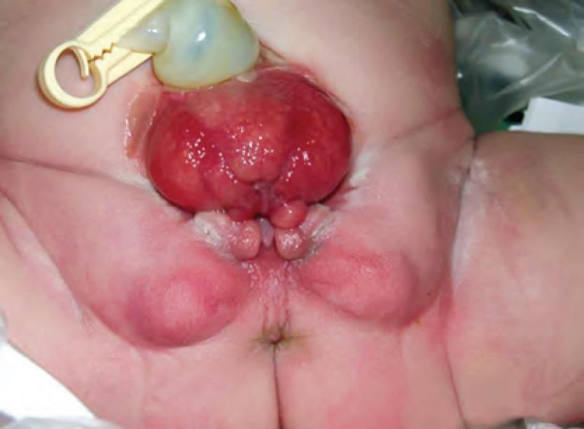
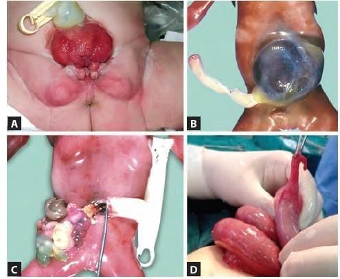
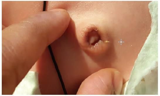
Identify the congenital anomaly shown in the newborn baby: (NEET-PG 2022)

Detailed Solution for Test: Anatomy - 6 - Question 10

The image illustrates a pathology concerning the anterior abdominal wall, situated below the umbilicus (which has a normal umbilical cord). It shows exposed mucosa (pinkish) on the posterior wall of the urinary bladder, as seen in bladder exstrophy (ectopia vesicae). Bladder exstrophy arises from abnormal lateral folding of the embryo, resulting in the failure of the ventral body wall to close. The endodermal wall of the urinary bladder is exposed to the outside (not covered by mesoderm or ectoderm). It is worth noting that some authors suggest this occurs because the mesoderm does not cover the cephalad extension of the cloacal membrane. The umbilicus is positioned lower, and the anus is shifted anteriorly.

  • Associated clinical features include:
  • Urinary incontinence
  • Epispadias (in both genders)
  • Split in the dorsum of the penis or separation of the clitoris into two halves
  • Short penis
  • Dorsal chordee
  • Wide separation of pubic bones
  • Broad-based gait

Other anterior abdominal wall defects such as omphalocele, gastroschisis, and Meckel’s anomaly (persistent vitello-intestinal duct) also present distinctly.

Test: Anatomy - 6 - Question 11

Adult derivative of the arrow marked structure in the following diagram is: (NEET-PG 2023)

Detailed Solution for Test: Anatomy - 6 - Question 11

The structure indicated by the arrow is the allantois (as seen in a side view of a developing fetus), which subsequently transforms into the median umbilical ligament (as noted in adults). Details include:

  • The allantois is a diverticulum of the hindgut that connects to the apex of the urinary bladder and traverses through the umbilicus, becoming part of the umbilical cord.
  • Eventually, it is obliterated to form a fibrous band known as the urachus.
  • The adult remnant of the urachus is referred to as the median umbilical ligament.
  • This ligament is covered by a peritoneal fold, termed the median umbilical fold.
  • The median umbilical ligament (which is covered by the median umbilical fold) is located in the anterior abdominal wall, extending from the apex of the urinary bladder to the umbilicus.

Congenital anomalies of the urachus include:

  • Urachal fistula: The urachus maintains a persistent lumen, allowing urine to flow from the apex of the urinary bladder to the umbilicus, resulting in urine leakage at the umbilicus.
  • Urachal sinus: The upper segment of the urachus (near the umbilicus) retains a persistent lumen, with an opening at the umbilicus.
  • Urachal cyst: A small portion of the urachus remains patent.

Test: Anatomy - 6 - Question 12

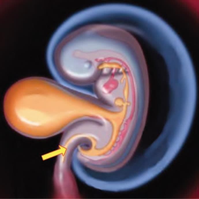
Which of the following ligament develop in the arrow marked structure? (NEET-PG 2022)

Detailed Solution for Test: Anatomy - 6 - Question 12

This is a line diagram illustrating the development of the foregut (for instance, stomach – gastro) and its mesentery (meso-gastrium). The arrow indicates the ventral section of the ventral mesentery, which evolves into the falciform ligament of the liver.

  • The gastro-phrenic ligament, which connects the greater curvature of the stomach (gastro) to the diaphragm (phrenic), arises from the dorsal region of the dorsal mesentery (mesogastrium).
  • The gastro-splenic ligament, linking the greater curvature of the stomach (gastro) to the spleen (splenic), develops in the ventral area of the dorsal mesentery.
  • The lieno-renal ligament, which connects the spleen (lieno) to the kidney (splenic), forms in the dorsal section of the dorsal mesentery.


Test: Anatomy - 6 - Question 13

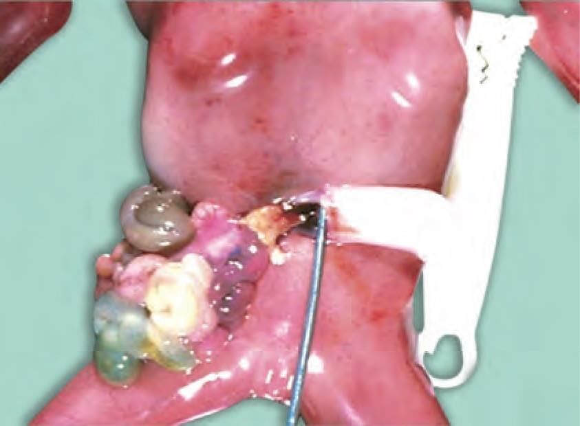
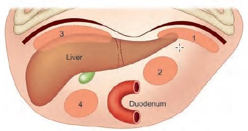
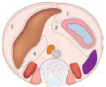
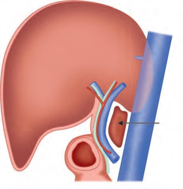
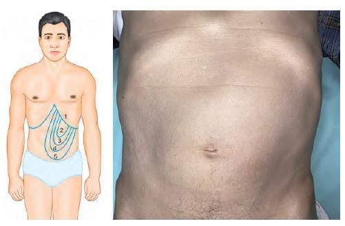
Which of the following is most common site for intraperitoneal abscess in relation to liver? (NEET-PG 2022)

Detailed Solution for Test: Anatomy - 6 - Question 13

Intraperitoneal abscess (a collection of pus) is typically found in the most dependent area – the Hepatorenal pouch of Morrison (located in the right sub-hepatic space). For instance, a posterior perforation of a gastric ulcer can result in the accumulation of gastric contents in the lesser sac or omental bursa (the left sub-hepatic space).

  • Patients are generally positioned supine, causing the pus to gravitate towards the most dependent area – the right subhepatic space (hepato-renal pouch of Morrison), passing through the epiploic foramen of Winslow.
  • As time progresses, if the patient shifts to a semi-recumbent position or stands upright, the pus may migrate further down to the recto-uterine pouch of Douglas (or the recto-vesical pouch in males), which is the lowest point when upright.

A liver-related pathology might result in a right sub-phrenic abscess, while a left sub-phrenic abscess could arise from spleen-related issues.

Locations of intraperitoneal abscesses. A – Frontal view, B – Transverse section. 1: Left sub-phrenic space, 2: Left sub-hepatic space (lesser sac),
3: Right sub-phrenic space, 4: Right sub-hepatic space (hepato-renal pouch of Morrison).

Test: Anatomy - 6 - Question 14

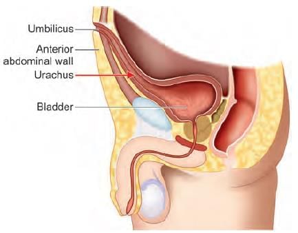
The anatomical structure at the arrow marked location is a defect in: (NEET-PG 2023)

Detailed Solution for Test: Anatomy - 6 - Question 14

The anatomical landmark indicated by the arrow is the deep inguinal ring, which represents a defect in the fascia transversalis. The deep inguinal ring is an oval aperture in the fascia transversalis, located 1.2 cm above the mid-inguinal point and just lateral to the origin of the inferior epigastric artery.

  • The spermatic cord (or round ligament of the uterus) traverses this ring to enter the inguinal canal.
  • An indirect inguinal hernia may occur when a segment of intestine (or omentum) passes through the deep inguinal ring into the inguinal canal.

Additional information:

  • The superficial inguinal ring is an opening in the aponeurosis of the external oblique muscle.
  • The roof of the inguinal canal is formed by the arching fibres of the internal oblique and transversus abdominis muscles.
  • These two muscles together create the conjoint tendon and contribute to the posterior wall of the inguinal canal.

Test: Anatomy - 6 - Question 15

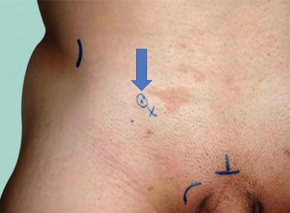
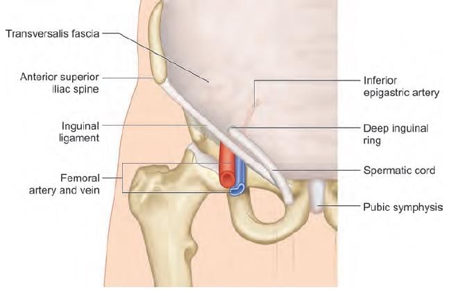
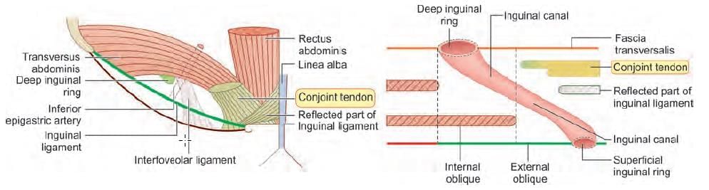
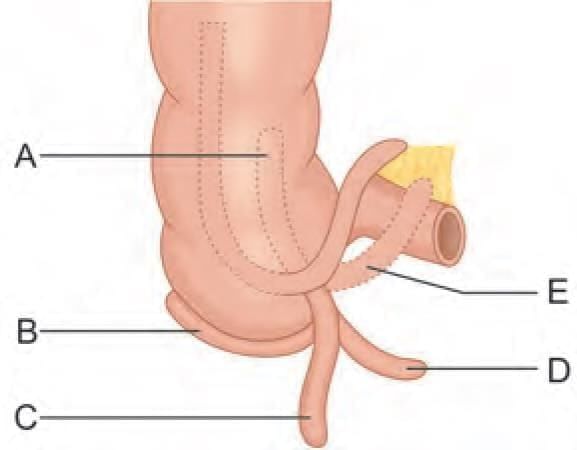
Hernia lying medial to inferior epigastric artery and superior to pubic tubercle, occurs due to weakness in: (NEET-PG 2023)

Detailed Solution for Test: Anatomy - 6 - Question 15

Hernia located medial to the inferior epigastric artery and superior to the pubic tubercle is classified as a direct inguinal hernia. This type of hernia can arise from a weakness in the conjoint tendon.

  • The conjoint tendon originates from a shared aponeurosis of the internal oblique and transverse abdominis muscles.
  • It attaches to the crest of the pubis and the pectineal line, situated deep to the superficial inguinal ring.
  • The conjoint tendon is positioned in the posterior wall of the inguinal canal, providing structural support.

A deficiency in the conjoint tendon may lead to the development of a direct inguinal hernia.

Test: Anatomy - 6 - Question 16

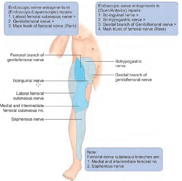
A patient has presented with pain in the right leg region for past 3 months. There is a surgical history of undergoing laparoscopic bilateral inguinal hernia repair. The nerve involved is: (NEET-PG 2023)

Detailed Solution for Test: Anatomy - 6 - Question 16

The patient has a history of having undergone laparoscopic bilateral inguinal hernia repair. They are experiencing pain in the right leg (not the thigh) area for the past three months, which suggests a potential involvement of the femoral nerve. Although it is uncommon for the femoral nerve to be affected in such procedures, if it is involved, it would impact the cutaneous territories, including the medial part of the leg (innervated by the saphenous nerve branch).

Note: If the complaint had been pain in the thigh (rather than the leg) region, the likely diagnosis would have been the lateral cutaneous nerve of the thigh.

  • Details: Nerve entrapments in hernia repairs:
  • Open (anterior) repairs:
    • Ilio-inguinal nerve
    • Ilio-hypogastric nerve
    • Genital branch of genitofemoral nerve
    • Main trunk of femoral nerve (rare)
  • Endoscopic (Laparoscopic) repairs:
    • Lateral femoral cutaneous nerve
    • Genitofemoral nerve
    • Main trunk of femoral nerve (rare)

Test: Anatomy - 6 - Question 17
A postoperative patient of laparoscopic hernia surgery presents with altered sensation at the root of penis. Nerve involved is: (INI-CET May 2022)
Detailed Solution for Test: Anatomy - 6 - Question 17
In laparoscopic hernia surgery, changed sensation at the base of the penis may arise due to the involvement of the genitofemoral nerve.
  • It is important to note that the base of the penis (dermatome L1) is innervated by the ilioinguinal nerve and the genital branch of the genitofemoral nerve.
Nerve entrapments during hernia repairs include:
  • Endoscopic (Laparoscopic) repairs:
    • Lateral femoral cutaneous nerve
    • Genitofemoral nerve
    • Main trunk of femoral nerve (rare)
  • Open (anterior) repairs:
    • Ilio-inguinal nerve
    • Ilio-hypogastric nerve
    • Genital branch of genitofemoral nerve
    • Main trunk of femoral nerve (rare)
Test: Anatomy - 6 - Question 18

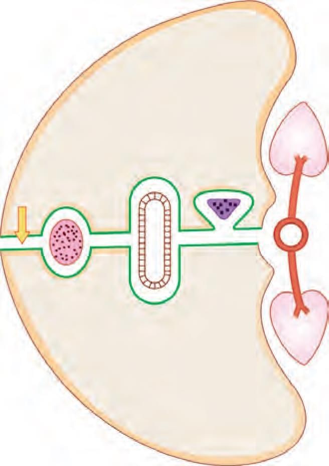
Superior boundary of the arrow marked structure is: (INI-CET July 2021)

Detailed Solution for Test: Anatomy - 6 - Question 18

The caudate lobe (segment 1) of the liver is positioned at the upper boundary of the epiploic foramen of Winslow (the foramen is indicated by an arrow in the diagram). The epiploic foramen of Winslow links the greater sac in the peritoneal cavity to the lesser sac. The boundaries are as follows:

  • Superior: 1st segment of the liver (caudate lobe)
  • Inferior: 1st part of the duodenum
  • Anterior: DAV (Bile Duct, Hepatic Artery, Portal Vein) structures located in the free margin of the lesser omentum
  • Posterior: Inferior vena cava and right adrenal gland on the T-12 vertebra.

Applied anatomy: In instances of posterior perforation of a gastric ulcer, the gastric contents accumulate in the lesser sac (a small peritoneal space located behind the stomach). Surgical exploration and aspiration of the contents in the lesser sac necessitate the passage of instruments from the greater sac to the lesser sac through the epiploic foramen.

Test: Anatomy - 6 - Question 19

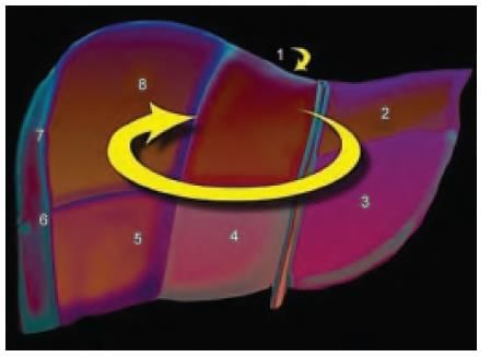
Liver is divided into eight segments according to Couinaud’s classification based upon. (AIIMS May 2019)

Detailed Solution for Test: Anatomy - 6 - Question 19
Liver is categorised into eight segments based on Couinaud’s classification, which relies primarily on the branches of the portal vein, hepatic artery, and bile duct. The Couinaud classification of liver anatomy segments the liver into eight functionally autonomous sections. Each segment possesses its own vascular inflow, outflow, and biliary drainage.
  • At the centre of each segment, there exists a branch of the portal vein, hepatic artery, and bile duct.

Test: Anatomy - 6 - Question 20

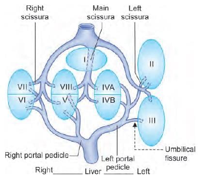
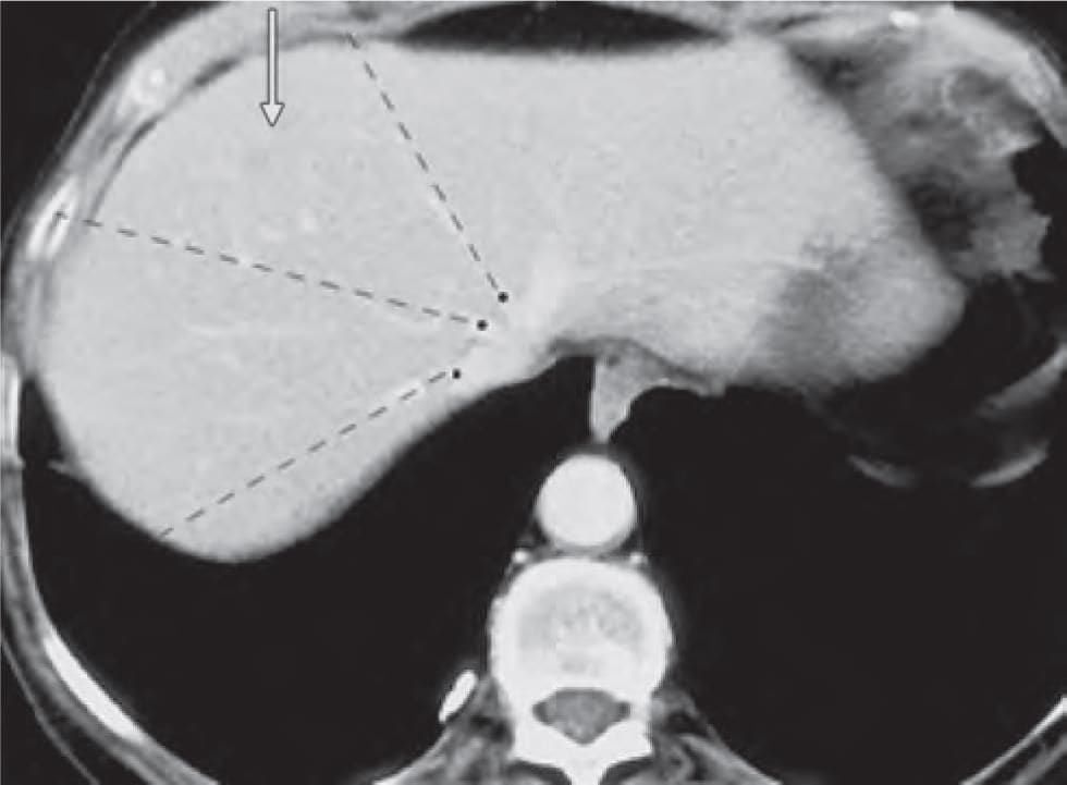
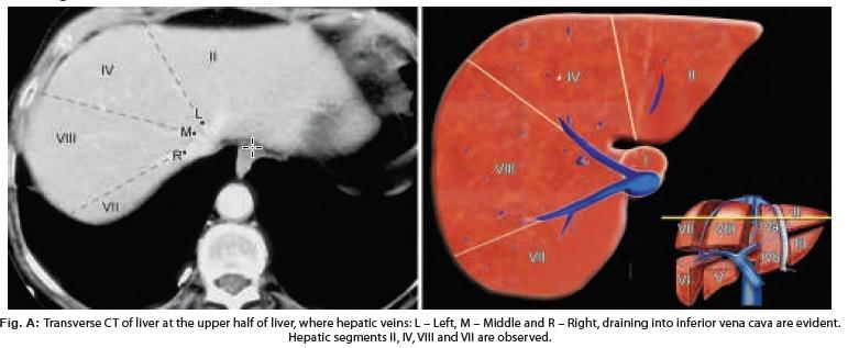
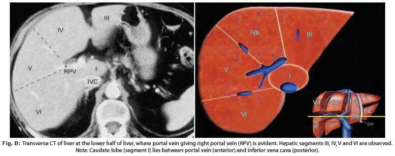
In CT scan of liver, identify the arrow marked segment: (INI-CET Nov 2022)

Detailed Solution for Test: Anatomy - 6 - Question 20

The arrow indicating the hepatic segment represents segment IV, bordered by the left and middle hepatic veins (shown as dotted lines). This transverse CT scan was performed on the upper portion of the liver, where the hepatic veins are clearly visible as they transport blood from the liver to the inferior vena cava. At this level, the upper hepatic segments II, IV, VIII, and VII can be seen (Fig. A).

  • It is important to note that the portal vein is not visible at this level, and its branches are not very distinct.
  • The liver comprises four sectors, defined by three intersectoral hepatic veins, and is further divided into eight segments by the intersegmental branches of the portal vein.
  • The liver is segmented into four sectors by three hepatic veins (right, middle, and left), which are both intersectoral and intersegmental.
  • Eight segments are identified, numbered in an anticlockwise manner from I to VIII.

Note: Segment I (the caudate lobe) is small and not visible from the anterior view. The tributaries of the hepatic veins are more prominent in the upper half of the liver, while the branches of the portal vein are more pronounced in the lower half.

  • In the upper half, segments II, IV, VIII, and VII are observed from left to right.
  • In the lower half, segments III, IV, V, and VI are visible from left to right.

Fig. A: This transverse CT scan of the upper half of the liver shows the hepatic veins: L – Left, M – Middle, and R – Right, all draining into the inferior vena cava. Hepatic segments II, IV, VIII, and VII are noted. The subsequent transverse CT (Fig. B) was conducted on the lower half of the liver, where the portal vein, branching into right and left sections, is evident. At this level, the lower hepatic segments III, IV, V, and VI are visible. Note: The hepatic veins are not particularly prominent in this CT scan. Fig. B: This transverse CT of the lower half of the liver illustrates the portal vein, with the right portal vein (RPV) evident. Hepatic segments III, IV, V, and VI are observed. Note: The caudate lobe (segment I) is located between the portal vein (anteriorly) and the inferior vena cava (posteriorly).

Test: Anatomy - 6 - Question 21

Marked area is related to: (AIIMS Nov 2019)

Detailed Solution for Test: Anatomy - 6 - Question 21

The illustration presented depicts the frontal view of the left kidney, with a marker indicating the gastric region, which is positioned anteriorly to the stomach.

Test: Anatomy - 6 - Question 22

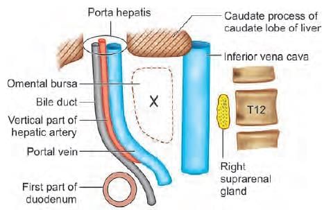
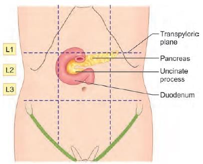
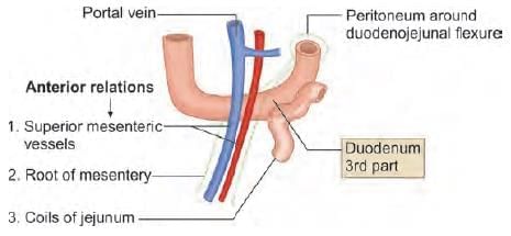
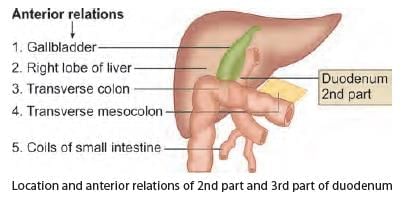
Anterior relations of third part of duodenum are all, except: (INI-CET May 2023)

Detailed Solution for Test: Anatomy - 6 - Question 22
The fundus of the gallbladder is positioned anteriorly to the second (not the third) segment of the duodenum. The anterior connections of the second part of the duodenum include:
  • Superior mesenteric vessels forming the contents of the root of the mesentery.
  • A few loops of the jejunum.

Test: Anatomy - 6 - Question 23

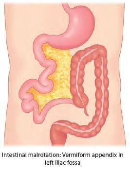
Match the location of vermiform appendix with the labelling given in the diagram: (INI-CET May 2023)

Detailed Solution for Test: Anatomy - 6 - Question 23

The most prevalent position of the vermiform appendix is located behind the caecum, known as retrocaecal (12 o’clock), which is observed in 65–75% of the population. The second most frequent position is directed towards the pelvic cavity infero-medially, termed pelvic (4 o’clock), found in 20–30% of individuals. It can also be situated on the side of the caecum, referred to as paracaecal (11 o’clock), or directly beneath the caecum, known as subcaecal/mid-inguinal (6 o’clock). Occasionally, it may lie anterior or posterior to the terminal ileum, termed preileal or postileal (3 o’clock). Morphologically, the vermiform appendix signifies the underdeveloped distal end of the large caecum. At birth, the appendix is short and broad at its junction with the caecum; however, differential growth of the caecum results in the characteristic tubular form by around the age of 2 years. As the caecum continues to grow, it typically rotates the appendix into a retrocaecal but intraperitoneal position. In roughly 25% of cases, this rotation does not occur, leading to a pelvic, subcaecal, or paracaecal position. Rarely, the caecum fails to migrate during development to its standard location in the right lower quadrant of the abdomen. In such instances, the appendix may be found near the gallbladder or, in cases of intestinal malrotation, in the left iliac fossa, complicating the diagnosis of appendicitis. The base of the appendix maintains a constant position, located at the junction of the three taeniae coli of the caecum, which merge to form the external longitudinal muscle layer of the appendix. During surgical procedures, gentle traction on the taeniae coli, particularly the anterior taenia, guides the surgeon to the base of the appendix.

Test: Anatomy - 6 - Question 24

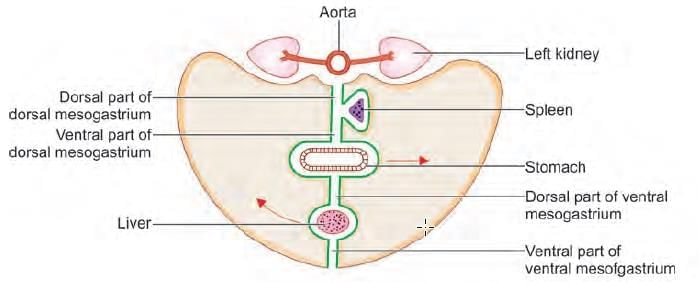
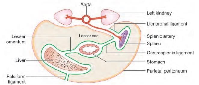
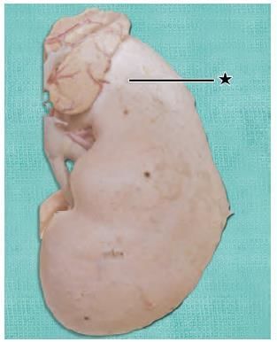
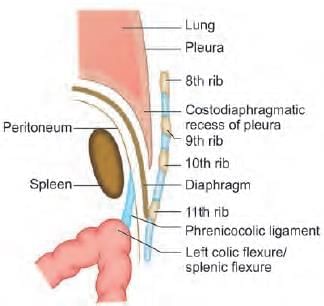
A 25-year-old male presents to emergency with left upper quadrant pain, upon examination spleen was measured to 20 cm and further extending to the mid umbilicus. The extension of the spleen to left lower quadrant was prevented by: (NEET-PG 2021)

Detailed Solution for Test: Anatomy - 6 - Question 24

The spleen enlarges downwards and at an angle towards the right iliac fossa. It is kept from moving to the left side by the phrenico-colic ligament.

The phrenico-colic ligament, also known as the sustentaculum lienis, is a horizontal fold of peritoneum that extends from the splenic flexure of the colon to the diaphragm, which is situated opposite the 11th rib in the midaxillary line.

  • It does not attach to the spleen but provides support to its anterior end.
  • This ligament restricts the upper part of the left paracolic gutter.

Test: Anatomy - 6 - Question 25

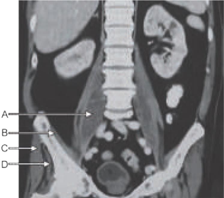
A child was brought to the hospital by his father with complaint of fever, low backache and persistent flexion of the hip joint. He had a history of Spine TB in the past. On examination child has an inguinal swelling. Identify the marked muscle responsible to be involved in this pathology: (NEET-PG 2021)

Detailed Solution for Test: Anatomy - 6 - Question 25

This seems to be a case of a psoas abscess, which can develop as a result of a tubercular infection in the lumbar vertebrae. It may extend between the psoas major muscle (Marker A) and its fascia, progressing behind the inguinal ligament into the femoral triangle. This condition can be confused with swollen lymph nodes.

  • Psoas major muscle (together with the iliacus) serves as the primary flexor at the hip joint and remains contracted in this condition, which accounts for the child's flexed posture at the hip joint.
  • Marker B: Iliacus muscle originates from the medial surface of the iliac part of the hip bone.
  • Marker C: Gluteus maximus originates from the lateral surface of the iliac part of the hip bone.
  • Marker D: Gluteus medius originates from the medial surface of the iliac part of the hip bone.

It is positioned deep and anterior to the gluteus maximus.

Information about Test: Anatomy - 6 Page
In this test you can find the Exam questions for Test: Anatomy - 6 solved & explained in the simplest way possible. Besides giving Questions and answers for Test: Anatomy - 6, EduRev gives you an ample number of Online tests for practice
Download as PDF