NEET PG Exam  >  NEET PG Tests  >  Test: Anatomy - 7 - NEET PG MCQ

Test: Anatomy - 7 - NEET PG MCQ


Test Description

25 Questions MCQ Test - Test: Anatomy - 7

Test: Anatomy - 7 for NEET PG 2025 is part of NEET PG preparation. The Test: Anatomy - 7 questions and answers have been prepared according to the NEET PG exam syllabus.The Test: Anatomy - 7 MCQs are made for NEET PG 2025 Exam. Find important definitions, questions, notes, meanings, examples, exercises, MCQs and online tests for Test: Anatomy - 7 below.
Solutions of Test: Anatomy - 7 questions in English are available as part of our course for NEET PG & Test: Anatomy - 7 solutions in Hindi for NEET PG course. Download more important topics, notes, lectures and mock test series for NEET PG Exam by signing up for free. Attempt Test: Anatomy - 7 | 25 questions in 25 minutes | Mock test for NEET PG preparation | Free important questions MCQ to study for NEET PG Exam | Download free PDF with solutions
Test: Anatomy - 7 - Question 1

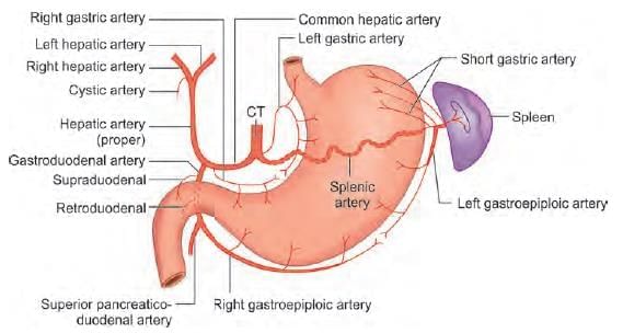
Ligation of the common hepatic artery will compromise blood flow in: (NEET-PG 2020p)

Detailed Solution for Test: Anatomy - 7 - Question 1

Ligation of the common hepatic artery will affect blood circulation in the distal branches, which include:

  • right gastric artery
  • hepatic artery proper
  • gastroduodenal artery

These branches further supply:

  • superior pancreatico-duodenal artery
  • right gastroepiploic artery

Test: Anatomy - 7 - Question 2

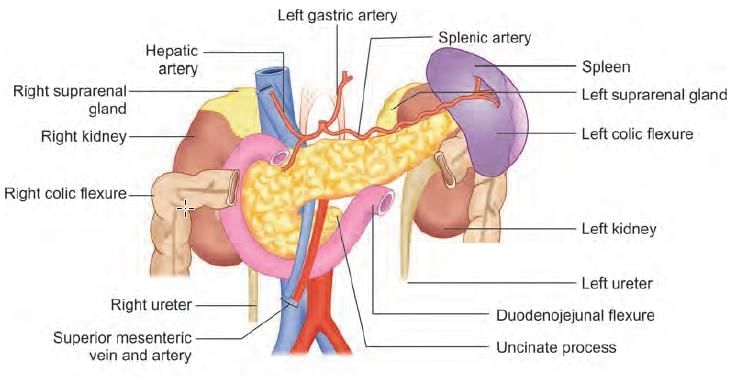
A 54-year-old man comes to a hospital with abdominal pain, jaundice, loss of appetite, and weight loss. On examination of his radiograms and CT scan, a physician finds a slowly growing tumor in the uncinate process of the pancreas. Which of the following structures is most likely compressed by this tumor? (NEET-PG 2020p)

Detailed Solution for Test: Anatomy - 7 - Question 2

The close anatomical connection between the uncinate process and the superior mesenteric vessels leads to early vascular involvement in these types of cancers. The uncinate process of the pancreas extends from the lower section of its head towards the left, positioned posterior to the superior mesenteric vessels.

  • The superior pancreaticoduodenal artery is situated between the duodenum and the head of the pancreas.
  • The splenic artery follows the upper edge of the pancreas.
  • The portal vein is located behind the neck of the pancreas.

Test: Anatomy - 7 - Question 3

Post appendicectomy, on exploration bleeding is noticed. Ligation of which artery is beneficial? (INI-CET Nov 2022)

Detailed Solution for Test: Anatomy - 7 - Question 3

While performing an appendectomy, the inferior branch of the ileocolic artery must be ligated at the base of the appendix. If this is not done, postoperative bleeding may occur. The vermiform appendix receives blood from the appendicular artery, which originates from the ileocolic artery.

  • The appendicular artery travels within the mesoappendix of the vermiform appendix.
  • It runs close to the free margin of the mesoappendix and ends in several small branches that supply the appendix, terminating at its tip.
  • A communicating loop is present between the terminal section of the ileocolic artery and the ileal branch.

The superior mesenteric artery provides blood to the mid-gut derivatives, including the vermiform appendix. The ileocolic artery, which is the lowest branch arising from the concavity of the superior mesenteric artery, supplies the cecum, ileum, and appendix. The middle colic artery, a branch of the superior mesenteric artery, supplies the proximal two-thirds of the transverse colon. It runs intraperitoneally through the root of the transverse mesocolon before dividing into right and left branches. The left colic artery is the first branch of the inferior mesenteric artery (the artery of the hindgut). It supplies blood to the distal third of the transverse colon and the entire descending colon. The appendicular artery arises from the ileocolic artery.

Test: Anatomy - 7 - Question 4

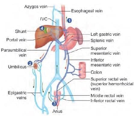
In a patient of liver cirrhosis anastomotic blood flow increases in: (NEET-PG 2023)

Detailed Solution for Test: Anatomy - 7 - Question 4

In liver cirrhosis, fibrosis occurs, leading to a reversal of blood flow in the portal circulation and resulting in portal hypertension. This condition causes:

  • Increased blood flow through porto-systemic anastomosis channels such as the left gastric vein (from portal circulation) and the oesophageal veins (in systemic circulation).
  • This ultimately results in oesophageal varices and haematemesis.

Other possible anastomoses include:

  • Between portal and portal circulation: Middle colic vein & left colic veins, sigmoidal & superior rectal veins.
  • Between systemic and systemic circulation: Superior and inferior phrenic veins.

Common locations of porto-systemic anastomoses in portal hypertension and their associated clinical implications include:

  • Hepatic portal vein tributaries:
    • Left gastric vein → Distal oesophageal veins draining into the azygos and hemiazygos veins → Oesophageal and gastric varices.
    • Superior anorectal veins → Middle and inferior anorectal veins draining into internal iliac and internal pudendal veins → Rectal varices.
    • Persistent tributaries of the left branch of the hepatic portal vein in the round ligament of the liver → Periumbilical branches of epigastric and intercostal veins → ‘Caput medusae’.
    • Tributaries of the right branch of the hepatic portal vein overlying the ‘bare area’ of the liver → Retroperitoneal veins draining into azygos, hemiazygos, lumbar, intercostal, and inferior phrenic veins → Dilated retroperitoneal veins at risk during surgery or interventional procedures.

Test: Anatomy - 7 - Question 5

SRY gene is present on: (INI-CET May 2023)

Detailed Solution for Test: Anatomy - 7 - Question 5

The SRY gene (sex-determining region on the Y chromosome), found at the distal end of the short arm of the ‘acrocentric’ Y chromosome, is crucial for male sex determination in mammals. SRY triggers the series of processes required to develop a testis from an undifferentiated gonad.

  • Klinefelter syndrome: Inherits a normal Y chromosome along with multiple X chromosomes, resulting in a karyotype of XXY. Individuals with this condition are classified as male.
  • Swyer syndrome: If the SRY gene is relocated to the X chromosome instead of remaining on the Y chromosome, testis formation will not occur. This condition is identified by an XY karyotype and a female phenotype. Affected individuals typically possess properly formed uteri and fallopian tubes, but their gonads are non-functional.
  • XX male syndrome: Characterised by a body with female chromosomes, where SRY is translocated to one of them. Individuals have a female genotype but exhibit male physical characteristics.
Test: Anatomy - 7 - Question 6

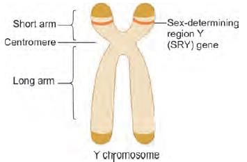
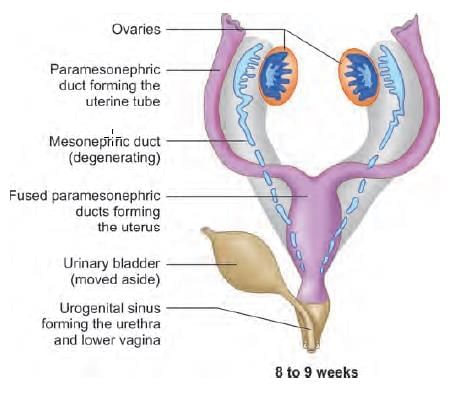
Remnants of Wolffian ducts in a female are found in: (NEET-PG 2020p)

Detailed Solution for Test: Anatomy - 7 - Question 6

Remnants of the Wolffian (mesonephric) ducts in females, such as Gartner’s duct, can be observed in the anterolateral wall of the vagina and the broad ligament.

Test: Anatomy - 7 - Question 7
Which of the following marked structure in the figure given below forms the pelvic diaphragm? (INI-CET Nov 2021)
Detailed Solution for Test: Anatomy - 7 - Question 7

This is a coronal section of the pelvic area illustrating the connections of the ischiorectal fossa. The pelvic diaphragm is primarily formed by the levator ani muscle (Marker B). It constitutes the superomedial limit of the ischiorectal fossa.

  • Marker A: Obturator internus muscle
  • Marker C: Internal anal sphincter
  • Marker D: Perianal fascia
Test: Anatomy - 7 - Question 8

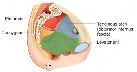
All of the following are true regarding levator ani, except: (AIIMS May 2019)

Detailed Solution for Test: Anatomy - 7 - Question 8

Levator ani muscle is connected to the pubis bone (at the front) and the tendinous arch (of the obturator internus fascia) on the sides. It does not connect to the pelvic brim.

  • Note: The pelvic brim is made up of the outer bony edges of the pelvic inlet.
  • The levator ani muscle is a broad, thin muscle located on the lateral aspect of the pelvis.
  • It attaches to the inner surface of the lateral part of the lesser pelvis and merges with its counterpart on the opposite side.

This forms the major portion of the pelvic cavity floor.

Test: Anatomy - 7 - Question 9

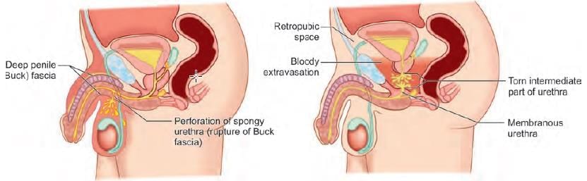
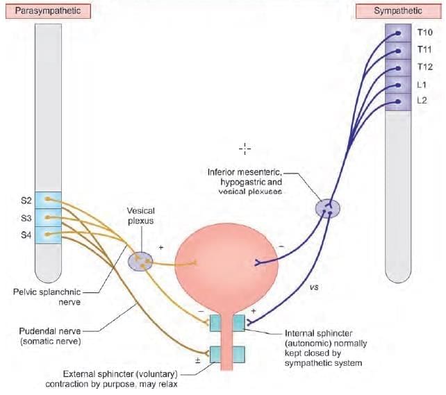
In bulbous rupture of urethra, urine extravasation occurs at all locations, except: (INI-CET Nov 2022)

Detailed Solution for Test: Anatomy - 7 - Question 9

In bulbous rupture of the urethra, urine can extravasate into the superficial perineal pouch, which is situated below the perineal membrane. The urine may then progress into the scrotal layers and the penile shaft, and it might also extend into the anterior abdominal wall, situated deep to Scarpa’s fascia.

  • It is important to note that the deep perineal pouch is located superior to the perineal membrane, which prevents the urine from entering this deep pouch.
  • In the event of a straddle injury, the bulbous part of the spongy urethra can rupture below the urogenital diaphragm (deep perineal pouch).
  • The extravasated urine may flow into the superficial perineal space and spread:
    • Inferiorly into the scrotum,
    • Anteriorly around the penis,
    • Superiorly into the lower section of the anterior abdominal wall.

The urine is unable to spread laterally into the thigh because the inferior fascia of the urogenital diaphragm (the perineal membrane) and the superficial fascia of the perineum are securely attached to the ischiopubic rami and are linked with the deep fascia of the thigh (fascia lata). It is also prevented from moving posteriorly into the anal region (ischiorectal fossa) due to the continuity between the perineal membrane and Colles’ fascia around the superficial transverse perineal muscles. Furthermore, the urine cannot enter the deep perineal pouch because the perineal membrane obstructs this pathway. In cases of straddle injury, the rupture of the bulbous part of the spongy urethra below the perineal membrane allows the extravasated urine to enter the superficial perineal space and spread into the scrotum, around the penis, and into the lower part of the anterior abdominal wall.

Test: Anatomy - 7 - Question 10

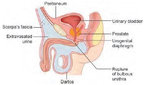
A man was brought to the emergency after meeting with an accident and injured perineum. He feels an urge to micturate but is unable to pass urine. There is blood at the tip of the meatus with extensive swelling of the penis and scrotum. What is the possible site of the injury? (NEET-PG 2020p)

Detailed Solution for Test: Anatomy - 7 - Question 10

Blood at the tip of the meatus, along with significant swelling of the penis and scrotum, indicates a possible bulbar rupture of the urethra.

  • Urethral injuries occur predominantly in men.
  • Presence of blood at the urethral meatus raises suspicion.
  • Urethral catheterisation is generally contraindicated.

Anterior urethral injury and posterior urethral injury involve different parts of the urethra:

  • Bulbar (spongy) urethra
  • Membranous urethra

Mechanisms of injury include:

  • Perineal straddle injury
  • Pelvic fracture

Location of urine leak or blood:

  • Blood accumulates in the scrotum.
  • If Buck fascia is torn, urine may escape into the perineal space.
  • Urine can leak into the retropubic space.

Presentation includes:

  • Blood at the urethral meatus and scrotal haematoma.
  • Blood at the urethral meatus and a high-riding prostate.

The urinary bladder may sustain injuries through both iatrogenic and classic traumatic mechanisms. Indications for bladder imaging encompass:

  • Gross haematuria in the context of injuries correlated with bladder injury.

Test: Anatomy - 7 - Question 11

Episiotomy incision was extended posteriorly beyond perineal body injuring the structure immediately posterior to it. Which structure has been injured? (NEET-PG 2020p)

Detailed Solution for Test: Anatomy - 7 - Question 11

An episiotomy incision was extended backwards past the perineal body, causing damage to the external anal sphincter located directly behind it.

Test: Anatomy - 7 - Question 12

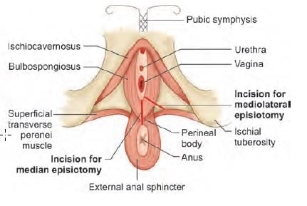
Which of the following structure is NOT cut in mediolateral episiotomy? (AIIMS June 2020)

Detailed Solution for Test: Anatomy - 7 - Question 12

Structures affected in a mediolateral episiotomy include:

  • the posterior vaginal wall
  • superficial and deep transverse perineal muscles
  • bulbospongiosus and a portion of the levator ani
  • transverse perineal branches of pudendal vessels and nerves
  • subcutaneous tissue and skin

Test: Anatomy - 7 - Question 13

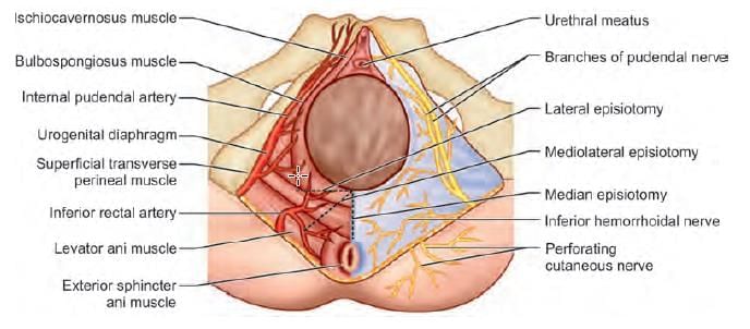
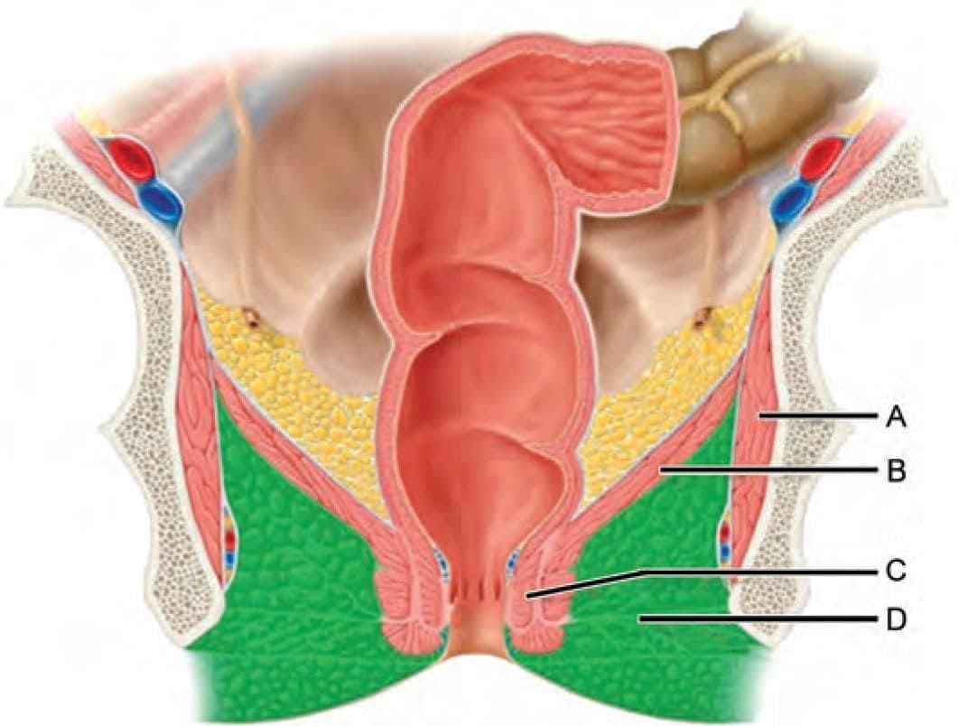
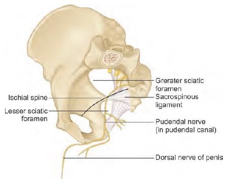
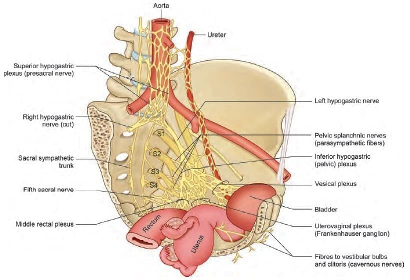
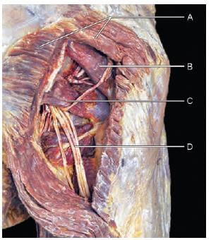
Which of the following nerves are related with the marked structure? (AIIMS Nov 2019)

Detailed Solution for Test: Anatomy - 7 - Question 13

The provided specimen represents the right hip bone (viewed medially), illustrating the ischial spine associated with the pudendal nerve.

Test: Anatomy - 7 - Question 14

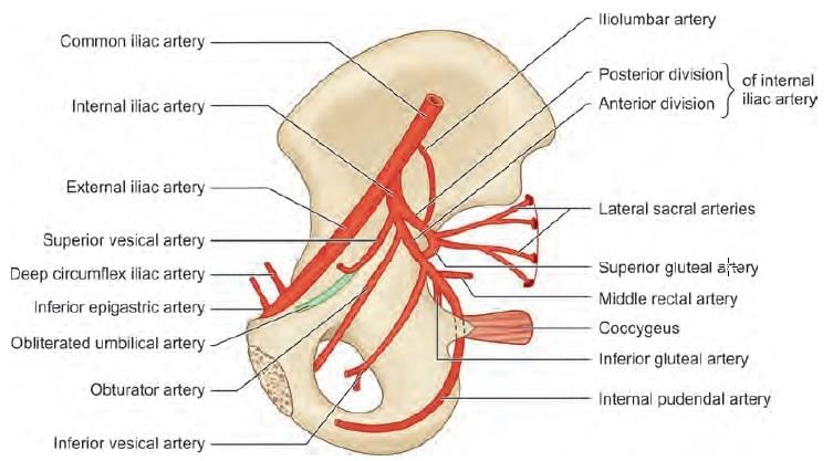
Branches from anterior division of internal iliac artery are: (INI-CET July 2021)

  1. Superior gluteal artery
  2. Inferior gluteal artery
  3. Superior vesical artery
  4. Inferior vesical artery
  5. Vaginal artery
  6. Middle rectal artery
Detailed Solution for Test: Anatomy - 7 - Question 14

Superior gluteal artery (1) is a branch that arises from the posterior (not anterior) division of the internal iliac artery. The posterior division of the internal iliac artery has three branches (SIL):

  • S – Superior gluteal artery
  • Ilio-lumbar artery
  • Lateral sacral artery

Most branches of the internal iliac artery originate from the anterior division. Applied anatomy: In cases of post-partum haemorrhage, for uterine devascularisation, a ligature is applied to the anterior division of the internal iliac artery, beyond the origin of the posterior division. If there is a reduction in blood flow within the posterior division, the patient may experience ischaemia in the gluteal region, particularly due to inadequate blood flow in the superior gluteal artery, which is a branch of the posterior division of the internal iliac artery.

Test: Anatomy - 7 - Question 15

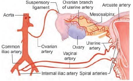
A 25 years old primigravida female after giving birth to a child have continuous bleed from uterus. The patient was taken to OT and planned for uterine devascularization. Which of the following is the correct order of devascularization? (NEET-PG 2021)

Detailed Solution for Test: Anatomy - 7 - Question 15

The sequence for applying ligatures in uterine devascularisation is as follows:

  • Uterine artery
  • Ovarian artery
  • Internal iliac artery

The goal of vascular ligation is to reduce blood supply to the uterus to halt life-threatening post-partum haemorrhage (PPH) when medical or manual interventions prove ineffective. Bilateral uterine artery ligation: Approximately 90% of the uterine blood supply during pregnancy is provided by these vessels. If this intervention does not successfully control the bleeding, the subsequent step involves ovarian artery ligation. Bilateral ovarian artery ligation: This artery emerges from the abdominal aorta and establishes a utero-ovarian vascular anastomosis. A suture is placed around the ovarian artery in a vascular area located within the mesovarium. If this measure also fails to manage the bleeding, the next procedure is internal iliac artery ligation. Internal iliac artery ligation: This procedure can lead to nearly an 85% reduction in pulse pressure in the arteries located distal to the ligature, effectively transforming the arterial pressure system to one that approaches venous circulation levels, facilitating haemostasis through clot formation. Note: The ligature is applied to the anterior division of the internal iliac artery, distal to where the posterior division originates. If blood flow in the posterior division is compromised, the patient may experience ischaemia in the gluteal region, particularly due to reduced blood flow in the superior gluteal artery, a branch of the posterior division of the internal iliac artery.


Test: Anatomy - 7 - Question 16

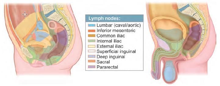
Painless cancerous lesion with inguinal lymph node involvement is due to carcinoma: (NEET-PG 2020p)

Detailed Solution for Test: Anatomy - 7 - Question 16

This case involves penile cancer at an intermediate stage, with the inguinal lymph node affected. It is important to note that most penile cancers do not cause pain. The stages of cancer are as follows:

  • Early disease: The cancer is confined to the foreskin or the surface of the head of the penis.
  • Intermediate disease: The cancer has penetrated beneath the skin into the shaft of the penis, and/or small quantities of cancer cells may be detected in one of the lymph nodes in the groin (microscopic disease).
  • Advanced disease: The cancer is present in one or more lymph nodes in the groin and/or has metastasised to other areas of the body.

Regarding testicular cancer, the lymphatic drainage includes the lumbar (para and pre-aortic) lymph nodes. The lymphatic vessels from the upper regions of the seminal glands and prostate primarily drain into the internal iliac lymph nodes, although some drainage from these may reach the sacral nodes. For the urinary bladder:

  • Lymphatics from the superolateral regions flow to the external iliac lymph nodes;
  • While the fundus and neck drain into the internal iliac lymph nodes.
  • Some vessels from the neck of the bladder also drain into the sacral or common iliac lymph nodes.

Test: Anatomy - 7 - Question 17

Which of the following statement is INCORRECT regarding labour pain? (INI-CET May 2022)

Detailed Solution for Test: Anatomy - 7 - Question 17

The motor innervation of the uterus (smooth muscles) is provided by the autonomic nervous system:

  • Sympathetic – Lumbar splanchnic nerves (T11,12;L1)
  • Parasympathetic – Nervi erigentes (S2,3,4)

These nerves also transmit autonomic sensations and innervate the uterus and vagina through the inferior hypogastric plexus (also known as the Frankenhauser plexus/ganglion). Note: Although the contraction and relaxation of the uterus primarily rely on hormonal influences, the autonomic nervous system additionally contributes to the contraction and relaxation of the uterine smooth muscles. Early labour pain (first stage of labour) results from the contraction of the uterine fundus and is conveyed by the lumbar splanchnic nerves (T11,12;L1). Details: Sympathetic innervation to the pelvic viscera (including the urinary bladder, uterus, rectum, etc.) originates from the superior hypogastric plexus, also referred to as the presacral nerve. This plexus is composed of sympathetic fibres arising from spinal levels T10 through L2. At the level of the sacral promontory, the superior hypogastric plexus bifurcates into a right and a left hypogastric nerve, which descend along their respective lateral pelvic walls. Parasympathetic innervation to the pelvic organs is provided by the nervi erigentes (pelvic splanchnic nerve), which are derived from the anterior primary rami of the S2,3,4 spinal cord segments. The two hypogastric nerves (sympathetic) and the two pelvic splanchnic nerves (parasympathetic) collectively form the inferior hypogastric plexus (Frankenhauser plexus/ganglion), which supplies the pelvic viscera. For the uterus, most sensory fibres (e.g., painful stimuli from contractions) ascend through the inferior hypogastric plexus and enter the spinal cord via the T10,11,12;L1 spinal nerves. These fibres transmit the painful stimuli from contractions to the central nervous system. For the cervix and upper part of the vagina, sensory nerves traverse the nervi erigentes (S2,3,4 spinal segments). Sensations from the lower vagina and perineum are conveyed by the pudendal nerve (somatic nervous system), whose root value matches that of the nervi erigentes (S2,3,4).

Test: Anatomy - 7 - Question 18

The most common complication of major vessel ligation in abdomino-perineal resection of carcinoma rectum is: (NEET-PG 2023)

Detailed Solution for Test: Anatomy - 7 - Question 18

One of the key vessels ligated during an abdomino-perineal resection for rectal carcinoma is the inferior mesenteric artery. - Ligation of this artery proximally can harm the superior hypogastric plexus, which is primarily sympathetic and partially parasympathetic. - This damage can result in: - Bladder dysfunction - Retrograde ejaculation Bladder dysfunction occurs when the sympathetic supply is lost, causing: - Inability to relax the detrusor muscle - Failure to contract the urethral sphincter, leading to impaired urinary storage. Retrograde ejaculation may happen due to the loss of T1 sympathetic supply to the internal urethral sphincter. Details: An abdomino-perineal resection (APR) entails the complete removal of the anal canal and anus, along with the creation of a permanent colostomy from the descending or sigmoid colon. Impotence (erectile dysfunction) can arise from nerve injuries sustained during the APR, which may disrupt the nerve signalling necessary for nitric oxide release, ultimately resulting in erectile dysfunction. Neurogenic bladder dysfunction (inability to urinate post-operatively) might occur due to damage to the inferior hypogastric plexus of nerves.
Note: Loss of perineal sensation may result from injury to the pudendal nerve, although this does not typically occur during this type of surgery.

Test: Anatomy - 7 - Question 19
Action of muscle attached to the area marked on the image shown: (AIIMS May 2019)
Detailed Solution for Test: Anatomy - 7 - Question 19
The illustration depicts the attachment of the gluteus medius muscle to the lateral aspect of the greater trochanter (femur), which facilitates abduction at the hip joint. The gluteus medius arises from the gluteal surface of the ilium and connects to the lateral surface of the greater trochanter of the femur.
Test: Anatomy - 7 - Question 20

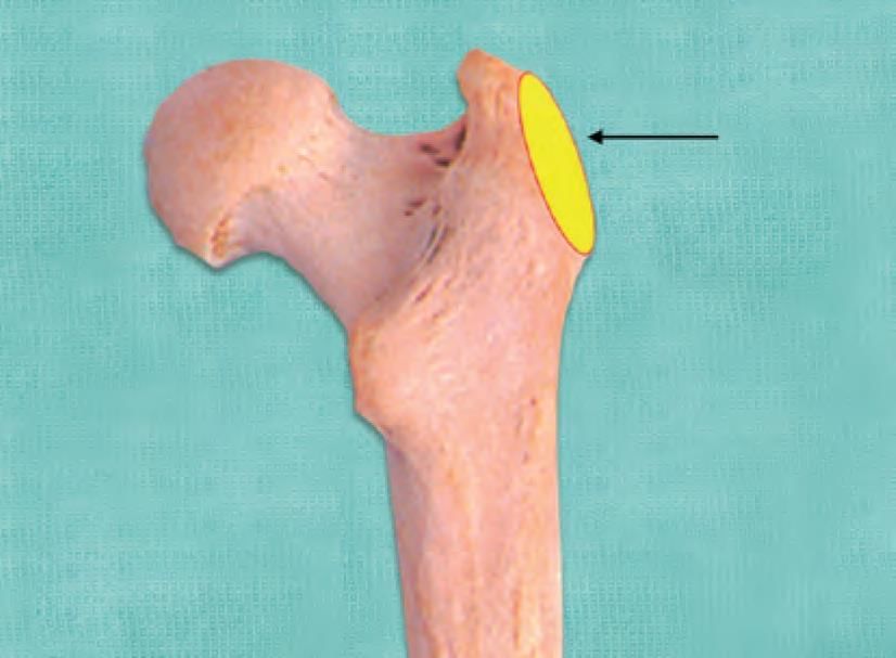
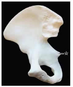
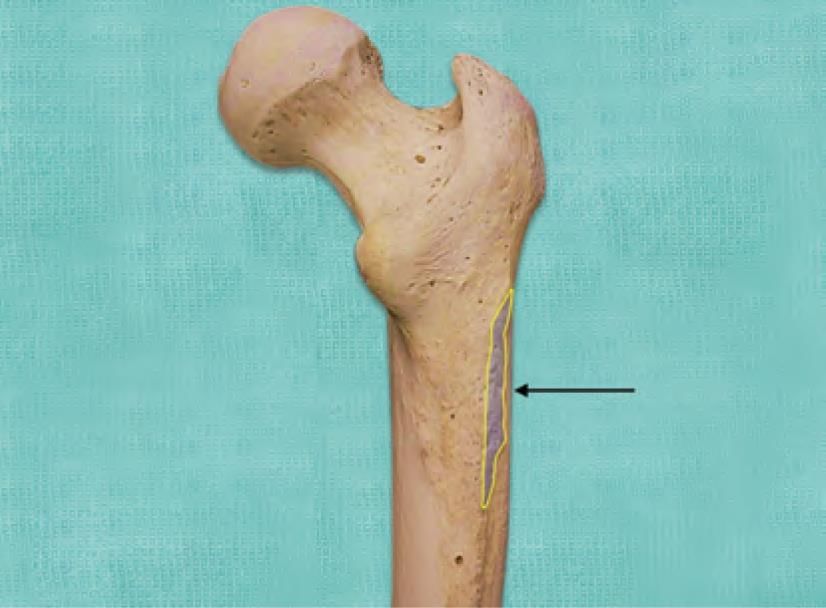
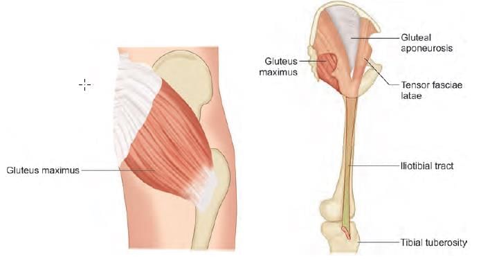
Mention the action of the muscle on hip joint as marked in the diagram below: (AIIMS June 2020)

Detailed Solution for Test: Anatomy - 7 - Question 20

The diagram illustrates the posterior view of the upper femur, highlighting the marking on the gluteal tuberosity, which indicates the partial insertion of the gluteus maximus muscle. This muscle serves as the primary extensor of the hip joint and is the main antigravity muscle when transitioning from a seated to a standing position.

  • Actions of the gluteus maximus (according to its insertion at the gluteal tuberosity):
  • Hip extension
  • Lateral rotation
  • Abduction

The lower portion of the muscle also functions as an external rotator of the limb, while the upper fibres act as abductors of the hip joints. To assess the gluteus maximus, one can test hip extension against resistance, either in a supine or prone position.

It is important to note that 75% of the fibres of the gluteus maximus muscle insert into the tibia via the iliotibial tract, which helps support the extended knee. The gluteus medius muscle attaches to the lateral surface of the greater trochanter, and the gluteus minimus connects to the anterior surface of the greater trochanter.

Test: Anatomy - 7 - Question 21

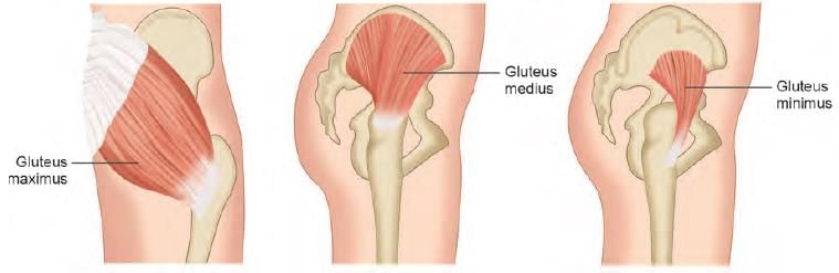
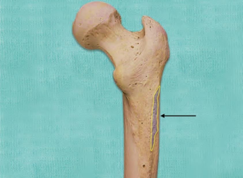
Mention action of the muscle on hip joint, whose attachment is marked in the diagram: (INI-CET Nov 2020)

Detailed Solution for Test: Anatomy - 7 - Question 21

The diagram illustrates the markings on the gluteal tuberosity, which serves as the attachment point for the gluteus maximus muscle. This muscle is the primary extensor of the hip joint and plays a crucial role as the main antigravity muscle when transitioning from sitting to standing.

  • Insertion of gluteus maximus:
    • One quarter of the fibres attach to the gluteal tuberosity.
    • Three quarters of the fibres insert (via the iliotibial tract) into Gerdy’s tubercle, located anterolaterally at the upper end of the tibia.
  • Actions of gluteus maximus:
    • Facilitates hip extension, lateral rotation, and abduction.
    • Supports the extended knee through the iliotibial tract.
    • The lower portion of the muscle functions as an external/lateral rotator of the limb.
    • The upper fibres operate as abductors of the hip joints.

Clinical testing: The functionality of the gluteus maximus can be assessed by performing hip extension against resistance, either while supine or prone.

Test: Anatomy - 7 - Question 22
Muscles supplied by superior gluteal nerve are: (INI-CET May 2022, 23)
Detailed Solution for Test: Anatomy - 7 - Question 22
Muscles innervated by the superior gluteal nerve (L4,5;S1) include the Gluteus medius, Gluteus minimus, and tensor fasciae latae. Some authors also indicate that the Piriformis muscle receives innervation from the superior gluteal nerve. Note: The Gluteus maximus is innervated by the inferior gluteal nerve (L5;S1,2). The superior gluteal nerve originates from the lumbo-sacral plexus. It emerges from the posterior divisions of L4, L5, and S1. It exits the pelvis via the greater sciatic foramen, situated above the piriformis muscle, and is accompanied by the superior gluteal vessels. During normal gait, the gluteus medius and minimus on the stance side help stabilise the pelvis in the coronal plane. A lesion of the superior gluteal nerve leading to paralysis of these muscles results in weakened abduction at the affected hip joint, causing a Trendelenburg lurching gait. In a positive Trendelenburg’s test, the pelvis tilts towards the normal unsupported side (the swing leg). When there is bilateral nerve damage, a waddling gait may occur.
Test: Anatomy - 7 - Question 23

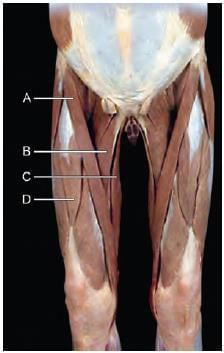
A patient with fall from a height was treated with a cast. After removal of cast he was not able to walk properly, unable to lift the leg and was dragging his right leg while walking. Which of the following marked structure is involved? (INI-CET Nov 2021)

Detailed Solution for Test: Anatomy - 7 - Question 23

The diagram illustrates the muscles located in the gluteal region. This patient exhibits a compromised abductor mechanism at the hip joint, resulting from an injury that affects the gluteus medius (marker B).

In instances of compromised abductor function, which may arise from:

  • Fracture of the head or neck of the femur
  • Superior gluteal nerve damage
  • Paralysis of the gluteus medius or minimus

the patient displays a positive Trendelenburg test. As a result, he struggles to walk correctly, finds it difficult to lift his leg, and tends to drag it while moving. Over time, he adapts by walking with a lurching gait, characterised by tilting of the contralateral spine, to avoid dragging his foot on the ground.

Key: Marker A: Gluteus maximus, Marker C: Piriformis, Marker D: Sciatic nerve.

Test: Anatomy - 7 - Question 24

Which of the following muscle cause flexion, abduction and lateral rotation at hip joint? (INI-CET Nov 2022)

Detailed Solution for Test: Anatomy - 7 - Question 24

The Sartorius muscle (marker A) facilitates flexion, abduction, and lateral rotation at the hip joint.

  • Marker B: Adductor longus (promotes adduction at the hip joint)
  • Marker C: Gracilis (also promotes adduction at the hip joint)
  • Marker D: Rectus femoris (enables hip flexion and knee extension)

The Sartorius is a long, slender, superficial muscle that extends infero-medially in the anterior compartment of the thigh.

  • Origin: Anterior superior iliac spine of the hip bone
  • Insertion: Anteromedial surface of the proximal tibia (within the pes anserinus)
  • Nerve supply: Femoral nerve (anterior division)
  • Action: Flexion at the hip and knee joints, abduction and lateral rotation at the hip joint. (Assists in achieving the sartor/tailor/Palthi posture.)
Test: Anatomy - 7 - Question 25

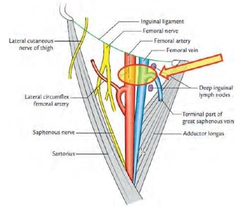
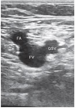
The sign seen on USG while locating the long saphenous vein and common femoral vein lying medial to the common femoral artery is called as: (NEET-PG 2020p)

Detailed Solution for Test: Anatomy - 7 - Question 25

The Mickey Mouse sign is believed to illustrate the typical anatomy of the femoral artery, femoral vein, and greater saphenous vein when viewed via ultrasound just below the inguinal crease. The ultrasound image at the area indicated by the arrow (femoral triangle) displays the lumen of the femoral artery (FA), femoral vein (FV), and great saphenous vein (GSV) as they enter the femoral vein.


Information about Test: Anatomy - 7 Page
In this test you can find the Exam questions for Test: Anatomy - 7 solved & explained in the simplest way possible. Besides giving Questions and answers for Test: Anatomy - 7, EduRev gives you an ample number of Online tests for practice
Download as PDF