NEET PG Exam  >  NEET PG Tests  >  Test: Congenital Heart Disease - NEET PG MCQ

Test: Congenital Heart Disease - NEET PG MCQ


Test Description

25 Questions MCQ Test - Test: Congenital Heart Disease

Test: Congenital Heart Disease for NEET PG 2025 is part of NEET PG preparation. The Test: Congenital Heart Disease questions and answers have been prepared according to the NEET PG exam syllabus.The Test: Congenital Heart Disease MCQs are made for NEET PG 2025 Exam. Find important definitions, questions, notes, meanings, examples, exercises, MCQs and online tests for Test: Congenital Heart Disease below.
Solutions of Test: Congenital Heart Disease questions in English are available as part of our course for NEET PG & Test: Congenital Heart Disease solutions in Hindi for NEET PG course. Download more important topics, notes, lectures and mock test series for NEET PG Exam by signing up for free. Attempt Test: Congenital Heart Disease | 25 questions in 25 minutes | Mock test for NEET PG preparation | Free important questions MCQ to study for NEET PG Exam | Download free PDF with solutions
Test: Congenital Heart Disease - Question 1

In coarctation of aorta, site of rib notching is? (Recent Question 2015-16)

Detailed Solution for Test: Congenital Heart Disease - Question 1

Causes of inferior rib notching include:

  • Arterial: aortic coarctation, aortic thrombosis, pulmonary oligemia/AV malformation, Blalock-Taussig shunt, Tetralogy of Fallot, absent pulmonary artery, and pulmonary stenosis.
  • Venous: AV malformations of the chest wall, superior vena cava, or other central venous obstructions.
  • Neurogenic: intercostal neuroma, neurofibromatosis type 1, poliomyelitis.
  • Osseous: hyperparathyroidism, thalassaemia.

Causes of superior rib notching are:

  • poliomyelitis,
  • osteogenesis imperfecta,
  • neurofibromatosis,
  • Marfan's syndrome,
  • collagen vascular disease,
  • hyperparathyroidism.
Test: Congenital Heart Disease - Question 2

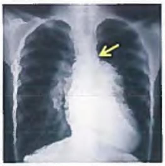
A neonate presents with anoxic spells and single S2. CXR shows all except:

Detailed Solution for Test: Congenital Heart Disease - Question 2

The CXR displays a heart exhibiting a boot-like shape along with decreased vascularity in the lungs. The clinical diagnosis is tetralogy of Fallot, which, as a result of subpulmonic stenosis, manifests with pulmonic oligemia.

Test: Congenital Heart Disease - Question 3

Lithium use in pregnancy leads to which effect on the baby? (AIMS Nov 2018)

Detailed Solution for Test: Congenital Heart Disease - Question 3

Lithium is a drug that can cause teratogenic effects, resulting in Ebstein anomaly. This condition leads to a malformed tricuspid valve, which can cause regurgitation and, in some cases, stenosis. Additionally, certain patients may exhibit Wolff-Parkinson-White (WPW) syndrome.

Test: Congenital Heart Disease - Question 4
Epstein syndrome is characterised by presence of? (Recent Question 2019)
Detailed Solution for Test: Congenital Heart Disease - Question 4

Epstein syndrome (EPTS) is a hereditary condition that follows an autosomal dominant inheritance pattern. It is defined by the following features:

  • Nephritis
  • Mild hearing impairment
  • Thrombocytopenia with enlarged platelets
Test: Congenital Heart Disease - Question 5

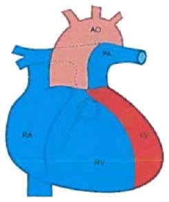
Which of the following is compatible with tet spells in a 4 month old child with TOF? (AIIMS May 2016)

Detailed Solution for Test: Congenital Heart Disease - Question 5

Tetralogy of Fallot is characterised by sub-pulmonic stenosis and right-to-left shunting, resulting in cyanosis. The murmur associated with right ventricular outflow tract obstruction is an ejection systolic murmur. During a tet spell, the right-to-left shunting intensifies.

  • As the right ventricle contains a fixed volume of blood and most of it flows from right to left, the blood flow through the pulmonic valve decreases.
  • This explains the exacerbation of cyanosis during a tet spell.
  • The reduced blood flow across the subpulmonic stenosis may diminish the intensity of the murmur, potentially rendering it inaudible.

Choices C and D represent signs of heart failure, which are excluded. Tet spells frequently occur upon waking or after physical exertion. During these episodes, the child becomes increasingly dyspnoeic and cyanotic, and may eventually lose consciousness, potentially leading to convulsions.

Test: Congenital Heart Disease - Question 6
Congenital heart disease associated with pre-excitation is? (UPSC 2015)
Detailed Solution for Test: Congenital Heart Disease - Question 6
Ebstein's anomaly is the most prevalent congenital defect linked to Wolff Parkinson-White (WPW) syndrome. Typically, Ebstein's anomaly of the tricuspid valve remains asymptomatic unless it is severe, presenting with cyanosis or accompanied by paroxysmal tachycardia due to pre-excitation. This condition can often be inferred from the ECG, which may reveal sinus rhythm with right bundle branch block, frequently showing low voltage secondary R-waves in V1 or right-sided pre-excitation, characterised by a short PR interval and complexes resembling left bundle branch block. Occasionally, the ECG may switch between these two patterns. Patients with Ebstein's anomaly experiencing atrioventricular re-entry tachycardia tend to have more frequent episodes during pregnancy. Congenital conditions associated with pre-excitation include:
  • Ebstein anomaly
  • Tricuspid atresia
  • Dextrocardia
  • Familial cardiomyopathy
  • Fibro-elastosis
Test: Congenital Heart Disease - Question 7
When should a very large ventricular septal defect be operated? (UPSC 2015)
Detailed Solution for Test: Congenital Heart Disease - Question 7
Indications for surgical closure of a VSD encompass patients of any age with considerable defects where:
  • Clinical symptoms and failure to thrive cannot be managed medically.
  • Infants aged between 6 and 12 months with large defects linked to pulmonary hypertension, even if symptoms are managed with medication; and patients older than 24 months with a Qp : Qs ratio exceeding 2 :1.
  • Patients with supra-cristal VSD of any size are typically referred for surgery due to the elevated risk of aortic valve regurgitation.
  • Severe pulmonary vascular disease serves as a contraindication for VSD closure.
The natural progression of a VSD is largely influenced by the size of the defect. A notable proportion (30-50%) of small defects may close spontaneously, predominantly within the first two years of life. Small muscular VSDs have a higher likelihood of closure (up to 80%) compared to membranous VSDs (up to 35%). Most defects that do close occur before the age of 4 years.
Test: Congenital Heart Disease - Question 8

Pulmonary blood flow increases in all except? (PGI Chandigarh May 2015)

Detailed Solution for Test: Congenital Heart Disease - Question 8

ASD, VSD, and PDA are left-to-right shunts that elevate pulmonary blood flow. In transposition of the great arteries, cyanosis arises from the mixing of blood from two circuits. The defect involves the aorta originating from the morphological right ventricle and the pulmonary artery emerging from the morphological left ventricle. As a result, the pulmonary and systemic circulations are linked in parallel instead of the typical in-series connection. In TOF, the pulmonary oligemia results from subpulmonic stenosis.

Test: Congenital Heart Disease - Question 9

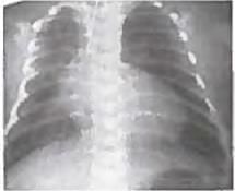
A 5-year old child with episodes of recurrent pneumonia was found to have a mid-diastolic murmur. CXR was performed. All are correct about the image shown except?

Detailed Solution for Test: Congenital Heart Disease - Question 9

The clinical presentation at the age of 5, characterised by recurrent pneumonia, suggests a left to right shunt.
  • The detection of a flow murmur (mid-diastolic murmur) indicates a diagnosis of an atrial septal defect.
  • The large yellow arrow on the right side highlights pulmonary plethora.
  • The large yellow arrow on the left side denotes dilatation of the pulmonary trunk.
  • The small yellow arrow on the left side marks the location of the aortic knuckle.
The red line signifies an enlarged heart.
Test: Congenital Heart Disease - Question 10
Turner's syndrome is associated with? (Recent Pattern 2015-16)
Detailed Solution for Test: Congenital Heart Disease - Question 10

Turner syndrome is linked to isolated non-stenotic bicuspid aortic valves in approximately one third to one half of those affected.

As patients age, the condition of bicuspid aortic valve disease may advance to a dilatation of the aortic root.

  • Less commonly observed defects encompass:
    • aortic coarctation (20%)
    • aortic stenosis
    • mitral valve prolapse
    • anomalous pulmonary venous drainage
Test: Congenital Heart Disease - Question 11

Most common cause of heart block in infants is? (Recent Pattern 2015-16)

Detailed Solution for Test: Congenital Heart Disease - Question 11

In children, the primary reason for permanent acquired complete AV block is surgery for congenital heart conditions.

  • Postsurgical complete atrioventricular block (AVB) is the leading cause of acquired AV block in children, arising from damage to the AV node during surgery (such as from hemorrhage, ischaemia, necrosis, inflammation, or traumatic disruption).
  • The second most prevalent cause is congenital heart disease linked with complete AV block.
  • Other causes of acquired AV block are generally reversible and include intoxications from digitalis and other medications, viral myocarditis, acute rheumatic fever, Lyme disease, and infectious mononucleosis.
Test: Congenital Heart Disease - Question 12

A Preterm baby with PDA will have all except: (AIMS Nov. 2012)

Detailed Solution for Test: Congenital Heart Disease - Question 12

Manifestations of PDA may consist of:

  • Apnea without apparent cause in an infant recovering from RDS.
  • A hyperdynamic precordium, along with bounding peripheral pulses, wide pulse pressure, and a continuous or systolic murmur, which may extend into diastole, or an apical diastolic murmur, accompanied by multiple clicks that resemble the sound of shaking dice.
  • Retention of carbon dioxide.
  • Increased dependence on oxygen.
  • X-ray findings indicating cardiomegaly and heightened pulmonary vascular markings.
  • Hepatomegaly.
  • Pulmonary haemorrhage may arise due to elevated pressure in the pulmonary circulation.

The diagnosis is confirmed through echocardiographic imaging of a PDA with Doppler flow assessment.

Test: Congenital Heart Disease - Question 13
All are causes of sudden death in infant except: (Recent Pattern 2014-15)
Detailed Solution for Test: Congenital Heart Disease - Question 13
Romano Ward syndrome is a variant of long Q-T syndrome, which is part of a group of channelopathies impacting ventricular repolarisation. It is also linked to sudden death and can resemble Sudden Infant Death Syndrome. The mechanism behind sudden death is polymorphic VT (Torsades de pointes).
  • An initial occurrence of sudden cardiac death is observed in 9% of patients.
  • LQT 3 may present with death during sleep in children.
Mortality rates are elevated in patients with severe or critical aortic valve stenosis during the first year of life, particularly in the neonatal period, though this risk has significantly diminished in the last 20 years. If left undetected, severe aortic valve stenosis is a known contributor to sudden death, accounting for about 1% of all sudden death cases among young individuals. Hypoplastic left heart syndrome entails a hypoplastic left ventricle that cannot sustain systemic perfusion and manifests as shock following death. Prostaglandin infusions in these patients can assist in survival. Kawasaki disease is another cause of sudden death in children, but it is considered a better prognosis due to the likelihood of complete recovery without noticeable long-term effects for those who do not develop coronary disease.
  • Recurrent acute illness occurs in merely 1-3% of instances.
  • The outlook for patients with coronary abnormalities is contingent upon the severity of coronary disease.
Overall, 50% of coronary artery aneurysms resolve as observed by echocardiogram 1-2 years post-illness. Giant aneurysms are less likely to resolve and are more prone to lead to thrombosis or stenosis. Amongst coronary artery-related causes of sudden death in children, the anomalous origin of the coronary artery from the Sinus of Valsava is noted to be more prevalent than Kawasaki disease. Coronary artery anomalies are frequently associated with sudden death in children and adolescents. The most common abnormality linked to sudden death is the origin of the left main coronary artery from the right sinus of Valsava. Consequently, the coronary artery travels between the aorta and pulmonary artery. Physical activity causes an increase in pulmonary and aortic pressure, which compresses the left main coronary artery, resulting in ischaemia.
Test: Congenital Heart Disease - Question 14
Neonate with cyanosis, heart failure and systolic murmur is suffering from: (Recent Pattern 2014-15)
Detailed Solution for Test: Congenital Heart Disease - Question 14

TGA is the most prevalent cyanotic heart condition that appears within the first few hours of life, which is a rather startling but true observation. In contrast, TOF typically manifests after the first week of life.

  • Infants diagnosed with transposition of the great arteries (TGA) are generally born at full term, with cyanosis noticeable within hours of their birth.
  • The clinical progression and symptoms are influenced by the degree of inter-circulatory mixing and the existence of associated anatomical abnormalities.

In cases of transposition of the great arteries accompanied by a significant ventricular septal defect:

  • Infants may not show immediate signs of heart disease, although mild cyanosis, especially during crying, is frequently observed.
  • Indicators of congestive heart failure, such as tachypnea, tachycardia, diaphoresis, and inadequate weight gain, may start to appear within the first 3 to 6 weeks as pulmonary blood flow increases.

The presence of heart failure complicates the diagnosis of TOF, even though cyanosis and a murmur are evident. Rheumatic fever typically occurs in individuals aged 5 to 15 years and does not present with cyanosis. VSD becomes apparent at around 6 weeks with signs of CHF.

Test: Congenital Heart Disease - Question 15

Which of the following is not given it of cyanotic spell? (IS Nov. 2013)

Detailed Solution for Test: Congenital Heart Disease - Question 15

Depending on the frequency and severity of hyper-cyanotic attacks, one or more of the following procedures should be implemented in order:

  • Positioning the infant on their abdomen in a knee-chest posture.
  • Providing oxygen (although increasing the inspired oxygen will not reverse cyanosis due to intracardiac shunting).
  • Administering morphine subcutaneously at a dosage not exceeding 0.2 mg/kg. Soothing and holding the infant in a knee-chest position may halt the progression of an early spell.
  • Since metabolic acidosis occurs when arterial PO2 is <40 mm Hg, prompt correction (within minutes) through intravenous sodium bicarbonate is essential if the spell is particularly severe and the child shows no response to the previous treatments.
  • For spells unresponsive to this therapy, medications that enhance systemic vascular resistance, like intravenous phenylephrine, can aid in improving right ventricular outflow, reducing the right-to-left shunt, and alleviating symptoms.
  • β-Adrenergic blockade via intravenous propranolol (0.1 mg/kg administered slowly, up to a maximum of 0.2 mg/kg) is also beneficial.

Clinical presentation of TET SPELL:

  • Typically observed between 1st to 2nd years of age. The infant becomes hyperpneic and agitated, with increasing cyanosis, gasping breaths, and potential syncope.
  • These episodes most often occur in the morning upon waking or following bouts of intense crying. A temporary reduction or disappearance of the systolic murmur is common as blood flow through the right ventricular outflow tract decreases.
  • The spells can last from several minutes to a few hours but are rarely life-threatening. Brief episodes are usually followed by general weakness and sleep. Severe spells may lead to unconsciousness and, in some cases, convulsions or hemiparesis.
Test: Congenital Heart Disease - Question 16
Ductus arteriosus complete closure occurs at how many weeks in a term baby? (Recent Pattern 2014-15)
Detailed Solution for Test: Congenital Heart Disease - Question 16
Functional closure of the Ductus Arteriosus typically occurs within 7 days after birth. O.P. Ghai notes that complete closure is observed by a maximum of 28 days, or 4 weeks.
  • Prostaglandin E2 (PGE2), generated by both the placenta and the Ductus Arteriosus (DA) itself, is the most powerful of the E prostaglandins.
  • Prostaglandin E1 (PGE1) also contributes to maintaining the ductus arteriosus in an open state.
  • PGE1 and PGE2 sustain the ductus open by elevating intracellular cAMP levels, acting through specific receptors like EP2 or EP4, which are responsive to PGE2.
During pregnancy, the DA of the fetus remains patent due to the presence of prostaglandins. However, immediately after delivery, the concentrations of both PGE2 and EP4 receptors drop significantly. This reduction facilitates the closure of the ductus arteriosus, enabling the normal post-natal circulation of the heart to begin.
Test: Congenital Heart Disease - Question 17
Alprostadil is contraindicated In: (Recent Pattern 2014-15)
Detailed Solution for Test: Congenital Heart Disease - Question 17
In TAPVC, the infusion of alprostadil will maintain the patency of the ductus and enhance blood flow into the lungs. This blood will subsequently surge into the abnormal pulmonary veins, which then return this blood to the right atrium rather than the left atrium. As a result, this will cause volume overload on the right side of the heart, as it receives both the normal venous return and the increased blood from the abnormal veins due to our erroneous choice to keep the ductus open.
  • Ductus dependent pulmonary circulation = TOF/TGA/tricuspid atresia.
  • Ductus dependent systemic circulation = mitral atresia/hypoplastic left heart syndrome/aortic atresia/interrupted aortic arch.
  • Ductus independent circulation = TAPVC/ALCAPA/truncus arteriosus.
Test: Congenital Heart Disease - Question 18
Partial anomalous pulmonary venous connection is associated with which of the following defects? (Recent Pattern 2014-15)
Detailed Solution for Test: Congenital Heart Disease - Question 18
PAPVC is analogous to Total Anomalous Pulmonary Venous Connection (TAPVC); however, TAPVC is characterised by all or the majority of pulmonary venous vessels connecting to the right side of the heart. The most prevalent type of ASD linked with PAPVC is the sinus venosus type. The anomalous pulmonary vein, typically the right upper or middle pulmonary vein, can either:
  • Override the intra-atrial septum (anomalous drainage)
  • Drain separately into the superior vena cava (true anomalous connection)
Usually, the connection is unobstructed. It is also associated with the Scimitar sign, where the anomalous venous return on a CXR resembles a curved sword.
Test: Congenital Heart Disease - Question 19
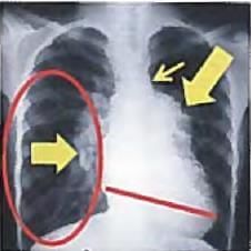
Which of the following conditions causes both superior as well as inferior notching of the ribs? (Recent Pattern 2014 15)
Detailed Solution for Test: Congenital Heart Disease - Question 19

The PTH hormone will lead to bone resorption regardless of the bone surface. In cases of coarctation and interrupted aortic arch:

  • The collateral circulation can result in erosion of the inferior surface of the ribs.

Similarly, in the Blalock-Taussig shunt:

  • The pulsations from the vascular graft may cause similar erosion, affecting only the upper two rib spaces.
Test: Congenital Heart Disease - Question 20
Reversal of shunt is not possible in natural history of: (AIIMS Nov. 2012)
Detailed Solution for Test: Congenital Heart Disease - Question 20
In all left to right shunts, increased pulmonary blood flow leads to pulmonary artery hypertension. This rise in pressure within the pulmonary artery subsequently causes a reversal of the shunt, known as the Eisenmenger complex.
  • As the systolic pressures between the two ventricles are equal, there is minimal or no left to right shunt, making the ventricular septal defect silent.
  • The right to left shunt is also quiet because it occurs with an insignificant pressure difference between the right ventricle and the aorta.
  • The flow from the right ventricle into the pulmonary artery takes place through the pulmonary valve, resulting in an ejection systolic murmur.
  • The severity of pulmonic stenosis correlates with a reduced flow into the pulmonary artery and an increase in the right to left shunt.
In definitive repair, which is performed at 6 months to 1 year of age, the ventricular septal defect (VSD) must be closed. Therefore, a faulty closure can technically lead to shunt reversal.
Test: Congenital Heart Disease - Question 21
A preterm baby with Patent Ductus Arteriosus. All are true except: (AIIMS May 2013)
Detailed Solution for Test: Congenital Heart Disease - Question 21
A significant PDA will lead to noticeable physical signs due to the wide pulse pressure, most notably, bounding peripheral arterial pulses. The apical impulse becomes prominent and, in cases of cardiac enlargement, appears heaving.
  • A thrill, typically strongest in the 2nd left interspace, is frequently observed and may radiate towards the left clavicle, along the left sternal border, or towards the apex.
  • This thrill is generally systolic but can also be felt throughout the cardiac cycle.
The classic continuous murmur is often characterised as having a quality similar to machinery or rolling thunder. The primary risk factor for NEC is prematurity.
  • NEC likely arises from an interplay between the loss of mucosal integrity due to various factors (such as ischaemia, infection, and inflammation) and the host's response to that injury (which includes circulatory, immunologic, and inflammatory reactions), leading to necrosis of the affected region.
Test: Congenital Heart Disease - Question 22

Extremely bad prognosis is seen in which heart disease in pregnancy? (Recent Pattern 2014-15)

Detailed Solution for Test: Congenital Heart Disease - Question 22

Conditions that present a high maternal risk include:

  • Poor functional class prior to pregnancy (NYHA functional classification II or more) or the presence of cyanosis
  • Pulmonary hypertension
  • Eisenmenger syndrome
  • Reduced systemic ventricular function (ejection fraction < 40%)
  • Mitral valve stenosis (area < 2 cm2), aortic valve stenosis (area < 1.5 cm2), left ventricular outflow tract peak pressure gradient greater than 30 mm Hg before pregnancy
  • A preconception history of negative cardiac events, including symptomatic arrhythmia, stroke, transient ischemic attack, and pulmonary edema
  • Marfan syndrome
Test: Congenital Heart Disease - Question 23

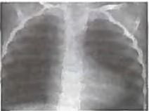
A 6-hour old child with cyanosis. On examination, S2 is loud and single. CXR was done. All arc true about this condition except?

Detailed Solution for Test: Congenital Heart Disease - Question 23

The image shows presence of egg-shaped heart with presence of cyanosis since birth and loud S2.

  • This indicates transposition of great arteries which is a ductal dependent pulmonary circulation.
  • Choice B is correct as PGE1 infusion will keep ductus patent and ensure survival. Balloon atrial septostomy will ensure reduction of cyanosis.
  • Infant of diabetic mother is at risk for development of:
    1. VSD
    2. TGA
    3. Asymmetrical septal hypertrophy
Test: Congenital Heart Disease - Question 24
All of the following are true about ASD except: (AI 2001)
Detailed Solution for Test: Congenital Heart Disease - Question 24

ASD is linked to right atrial (RA) and right ventricular (RV) hypertrophy, but it does not lead to left atrial (LA) hypertrophy.

Test: Congenital Heart Disease - Question 25

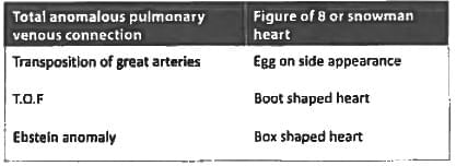
Snow man appearance or figure of 8 appearance is seen in? (Recent Question 2015-16)

Detailed Solution for Test: Congenital Heart Disease - Question 25

Information about Test: Congenital Heart Disease Page
In this test you can find the Exam questions for Test: Congenital Heart Disease solved & explained in the simplest way possible. Besides giving Questions and answers for Test: Congenital Heart Disease, EduRev gives you an ample number of Online tests for practice
Download as PDF