NEET PG Exam  >  NEET PG Tests  >  Test: ECG - NEET PG MCQ

Test: ECG - NEET PG MCQ


Test Description

15 Questions MCQ Test - Test: ECG

Test: ECG for NEET PG 2025 is part of NEET PG preparation. The Test: ECG questions and answers have been prepared according to the NEET PG exam syllabus.The Test: ECG MCQs are made for NEET PG 2025 Exam. Find important definitions, questions, notes, meanings, examples, exercises, MCQs and online tests for Test: ECG below.
Solutions of Test: ECG questions in English are available as part of our course for NEET PG & Test: ECG solutions in Hindi for NEET PG course. Download more important topics, notes, lectures and mock test series for NEET PG Exam by signing up for free. Attempt Test: ECG | 15 questions in 15 minutes | Mock test for NEET PG preparation | Free important questions MCQ to study for NEET PG Exam | Download free PDF with solutions
Test: ECG - Question 1

Inverted T waves are seen in? (Recent Pattern 2014-15)

Detailed Solution for Test: ECG - Question 1

Severe anterior wall ischaemia (with or without infarction) can lead to noticeable T-wave inversions in the precordial leads. This pattern, often called Wellens' T waves, is generally linked to significant stenosis of the left anterior descending coronary artery.

  • Hyperkalemia presents with elevated 'Tented T waves.
  • Coronary syndrome X is defined by the blockage of perforators while the epicardial coronary artery remains normal.
  • In these cases, stenting of the coronaries is ineffective.

Nitrates are the primary treatment option.

Test: ECG - Question 2

The following ECG findings are seen in Hypokalemia: (Recent Pattern 2014-15)

Detailed Solution for Test: ECG - Question 2

The question is cleverly phrased, as choice C may initially seem accurate. However, the QT interval in hypokalaemia is noticeably prolonged because of the merging of T and U waves. This condition also increases the risk of the patient developing torsades de pointes. The ST depression occurs as a result of concordance with the T wave, which becomes inverted in hypokalaemia. Additionally, the PR interval is lengthened due to slow repolarisation.

Test: ECG - Question 3

Which ECG finding is most likely to be seen at the time of cardiac arrest (UPSC 2013)

Detailed Solution for Test: ECG - Question 3
When the onset is sudden or abrupt, the likelihood that the arrest is of cardiac origin is 95%. Continuous electrocardiographic (ECG) recordings, which are sometimes fortuitously captured at the onset of cardiac arrest, typically show alterations in cardiac electrical activity in the minutes or hours leading up to the event.
  • There is a noticeable tendency for the heart rate to rise.
  • Advanced grades of PVCs often develop.
Most cardiac arrests resulting from VF typically start with either a run of nonsustained or sustained VT, which subsequently deteriorates into VF. The chance of achieving successful resuscitation from cardiac arrest is influenced by several factors:
  • the time interval from the onset of circulation loss to the initiation of resuscitative efforts,
  • the context in which the event occurs,
  • the underlying mechanism (VF, VT, PEA, asystole),
  • the clinical condition of the patient prior to the cardiac arrest.
Rates of return of circulation and survival following defibrillation diminish almost linearly from the first minute to 10 minutes. After 5 minutes, survival rates are typically no better than 25-30% in out-of-hospital environments.
Test: ECG - Question 4
Which of the following is cause of RBBB?
Detailed Solution for Test: ECG - Question 4

Factors contributing to RBBB include:

  • Normal physiological conditions.
  • Pulmonary embolism or cor pulmonale.
  • Pulmonary artery hypertension.
  • Atrial septal defect (ASD).
  • Rheumatic heart disease.
Test: ECG - Question 5

Alternating RBBB with Left anterior hemiblock is seen in? (Recent Pattern 2014-15)

Detailed Solution for Test: ECG - Question 5

Bifascicular block refers to the combination of RBBB with either left anterior hemiblock or left posterior hemiblock. Trifascicular block involves RBBB along with either LAHB or LPHB, in addition to first-degree AV block. Complete heart block results from the destruction of the AV node, causing AV dissociation.

Test: ECG - Question 6

Which is the following is the commonest ECG finding in pulmonary embolism? (Recent Pattern 2014-15)

Detailed Solution for Test: ECG - Question 6

Most frequently observed ECG finding in pulmonary embolism is sinus tachycardia. The most distinctive ECG characteristic of pulmonary embolism is S1 Q3 T3. The S1Q3T3 pattern represents the ECG manifestation of acute pressure and volume overload in the right ventricle. An S wave in lead I indicates either a complete or, more commonly, an incomplete RBBB. In lead III, one should look for a Q wave, a slight ST elevation, and an inverted T wave. These observations result from the pressure and volume overload on the right ventricle, leading to repolarisation abnormalities.

Test: ECG - Question 7

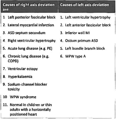
Left axis deviation is seen in all except? (Recent Pattern 2014-15)

Detailed Solution for Test: ECG - Question 7


The ostium primum is linked to a cleft mitral valve, which can result in left atrial enlargement and left axis deviation.

Test: ECG - Question 8

Electrical alternans is seen in? (Recent Pattern 2014-15)

Detailed Solution for Test: ECG - Question 8

Electrical alternans arises from the back-and-forth swinging motion of the heart while it is surrounded by a pericardial sac filled with a substantial amount of fluid.

Test: ECG - Question 9

Osborn wave is seen in? (Recent Pattern 2014-15)

Detailed Solution for Test: ECG - Question 9
  • Characteristically seen in hypothermia (typically T<30°C), but they are not pathognomonic.
  • J waves can be observed in several different conditions:
    • Normal variant.
    • Hypercalcaemia.
    • Medications.
    • Neurological injuries like intracranial hypertension, significant head trauma, and subarachnoid haemorrhage.
Test: ECG - Question 10
All are ECG changes in hypokalemia, except: (Recent Pattern 2014-15)
Detailed Solution for Test: ECG - Question 10

It is important to note that the QT interval exhibits an inverse relationship with the levels of potassium, calcium, and magnesium. A comparison of ECG characteristics in hypokalemia and hyperkalemia includes:

  • Hypokalemia:
    • Increased amplitude and duration of the P wave.
    • Prolongation of the PR interval.
    • Flattening and inversion of the T wave.
    • ST segment depression.
    • Prominent U waves (most visible in the precordial leads).
    • Apparent long QT interval due to the fusion of the T and U waves (equivalent to a long QU interval).
  • Hyperkalemia:
    • Tall, tented T waves.
    • Prolongation of the PR interval.
    • Absence of P waves.
    • Widening of the QRS complex.
    • Sine wave pattern.
Test: ECG - Question 11

Low voltage ECG is seen in? (Recent Pattern 2014-15)

Detailed Solution for Test: ECG - Question 11

The QRS is considered low voltage when:

  • The amplitudes of all QRS complexes in the limb leads are below 5 mm; or
  • The amplitudes of all QRS complexes in the precordial leads are below 10 mm.

There are various causes of low voltage ECG:

  • Obesity.
  • Emphysema.
  • Pericardial effusion and pleural effusion.
  • Severe hypothyroidism (myxedema).
  • Subcutaneous emphysema.
  • Pneumothorax or pneumopericardium.
  • Old large myocardial infarction.
  • End-stage dilated cardiomyopathy.
  • Infiltrative/restrictive diseases such as amyloidosis or hemochromatosis.
Test: ECG - Question 12

PR interval is reduced in? (Recent Pattern 2014-15)

Detailed Solution for Test: ECG - Question 12

The PR interval in an ECG exhibits an inverse relationship with heart rate.

  • Options A and D refer to heart block, indicating that bradycardia occurs, which results in PR interval prolongation.
  • Patients with hypothyroidism also experience bradycardia, contributing to P-R prolongation.
  • In Wolff-Parkinson-White (WPW) syndrome, the bundle of Kent serves as an accessory pathway, facilitating rapid conduction to the ventricles.
  • This, in turn, leads to symptoms such as palpitations and a decreased PR interval.
Test: ECG - Question 13
Low voltage in ECG indicates? (Recent Pattern 2014-15)
Detailed Solution for Test: ECG - Question 13

Low voltage signifies situations in which the electrical signal from the heart is diminished, either due to an inherent heart issue or external factors. Potential causes encompass:

  • Pericardial effusion
  • Constrictive pericarditis
  • Severe ischaemic heart disease
  • Infiltrative diseases, such as amyloidosis
  • Hypothyroidism
  • Pulmonary emphysema
  • Pleural effusion
  • Pneumothorax
  • Generalised oedema
Test: ECG - Question 14

In LVH, SV1 + RV6 is more than ____ mm? (Recent Pattern 2014-15)

Detailed Solution for Test: ECG - Question 14

Left Ventricular hypertrophy criteria is defined as follows:

  • The sum of the S wave in V1 and the R wave in V6 must be greater than 35 mm (SV1 + RV6 > 35).
  • Alternatively, the R wave in aVL should exceed 11 mm.
Test: ECG - Question 15
In left sided massive pneumothorax, ECG shows all, except: (Recent Pattern 2014-15)
Detailed Solution for Test: ECG - Question 15
The ECG indicators of a left tension pneumothorax include:
  • A rightward deviation of the mean frontal QRS axis.
  • A decrease in precordial R-wave voltage.
  • A reduction and/or variation in QRS amplitude (electrical alternans).
  • Inversion of precordial T-waves.
Typically, two or three of these manifestations are observed, while all four are less frequently seen. Non-infarction Q waves can arise from:
  • Normal variant Q waves in V1-V3, III, aVF.
  • Left pneumothorax or dextrocardia.
  • Myocarditis.
  • Left bundle branch block (LBBB).
  • Wolff-Parkinson-White (WPW) syndrome.
Information about Test: ECG Page
In this test you can find the Exam questions for Test: ECG solved & explained in the simplest way possible. Besides giving Questions and answers for Test: ECG, EduRev gives you an ample number of Online tests for practice
Download as PDF