NEET PG Exam  >  NEET PG Tests  >  Test: Embryology and Anatomy of Ear - 2 - NEET PG MCQ

Test: Embryology and Anatomy of Ear - 2 - NEET PG MCQ


Test Description

25 Questions MCQ Test - Test: Embryology and Anatomy of Ear - 2

Test: Embryology and Anatomy of Ear - 2 for NEET PG 2025 is part of NEET PG preparation. The Test: Embryology and Anatomy of Ear - 2 questions and answers have been prepared according to the NEET PG exam syllabus.The Test: Embryology and Anatomy of Ear - 2 MCQs are made for NEET PG 2025 Exam. Find important definitions, questions, notes, meanings, examples, exercises, MCQs and online tests for Test: Embryology and Anatomy of Ear - 2 below.
Solutions of Test: Embryology and Anatomy of Ear - 2 questions in English are available as part of our course for NEET PG & Test: Embryology and Anatomy of Ear - 2 solutions in Hindi for NEET PG course. Download more important topics, notes, lectures and mock test series for NEET PG Exam by signing up for free. Attempt Test: Embryology and Anatomy of Ear - 2 | 25 questions in 25 minutes | Mock test for NEET PG preparation | Free important questions MCQ to study for NEET PG Exam | Download free PDF with solutions
Test: Embryology and Anatomy of Ear - 2 - Question 1

Horizontal semicircular canal responds to: (UP 2005, Exam 2017)

Detailed Solution for Test: Embryology and Anatomy of Ear - 2 - Question 1
  • The horizontal/lateral semicircular canal along with posterior and superior semicircular canal contains cristae which sense angular or rotational acceleration.
  • Horizontal acceleration, i.e. linear acceleration, gravitational movements and head tilt movements are sensed by maculae in the utricle and saccule.
Test: Embryology and Anatomy of Ear - 2 - Question 2

Angular movements are sensed by: (JIPMER 93, Exam 2013)

Test: Embryology and Anatomy of Ear - 2 - Question 3

Stapes foot plate covers: (AIIMS May 2003, Exam 2017)

Detailed Solution for Test: Embryology and Anatomy of Ear - 2 - Question 3
  • The Oval window is covered by the footplate of stapes.
  • The Round window is covered by secondary tympanic membrane.
  • The part of middle ear medial to the descending part of facial nerve is called sinus tympani.
  • Pyramid is the projection on posterior wall from which originates the stapedius muscle.
Test: Embryology and Anatomy of Ear - 2 - Question 4

Movement of stapes causes vibration in: (Exam 2002, Exam 2017)

Test: Embryology and Anatomy of Ear - 2 - Question 5

Where is electrode kept in cochlear implant? (Exam 2013, AIIMS 2008)

Detailed Solution for Test: Embryology and Anatomy of Ear - 2 - Question 5

The electrodes of cochlear implant are placed into the Scala tympani by passing through the round window.

Test: Embryology and Anatomy of Ear - 2 - Question 6

Micro Wick and micro catheter sustained release device are used in (AIIMS 2011)

Test: Embryology and Anatomy of Ear - 2 - Question 7

Perilymph contains: (Exam 2013)

Detailed Solution for Test: Embryology and Anatomy of Ear - 2 - Question 7
  • Perilymph is the fluid which fills the bony labyrinth.
  • Perilymph is actually an extension of CSF which enters from the subarachnoid space into the scala tympani through Cochlear aqueduct.
  • CSF being an extracellular fluid therefore perilymph is rich in Na+.
  • Endolymph filling the membranous labyrinth and secreted from stria vascularis is rich in K+.
Test: Embryology and Anatomy of Ear - 2 - Question 8

Site where endolymph is seen: (Kerala 97, Exam 2013)

Detailed Solution for Test: Embryology and Anatomy of Ear - 2 - Question 8
  • Endolymph is present in the membranous labyrinth, whereas perilymph is present in the bony labyrinth.
  • Hence scala vestibuli, scala tympani and their interconnection helicotrema, which are parts of bony labyrinth, are filled with perilymph.
  • Scala media, utricle, saccule and semicircular canals, which are parts of membranous labyrinth, are filled with endolymph.
Test: Embryology and Anatomy of Ear - 2 - Question 9

Fluid, which has high potassium and low sodium content, is: (JIPMER 2003, Exam 2017)

Detailed Solution for Test: Embryology and Anatomy of Ear - 2 - Question 9

Rest are extracellular hence have more of sodium

Test: Embryology and Anatomy of Ear - 2 - Question 10

Cochlear aqueduct: (Exam 2017)

Detailed Solution for Test: Embryology and Anatomy of Ear - 2 - Question 10
  • The internal ear communicates with the cranium via two openings:
    • Cochlear aqueduct: via this CSF in the subarachnoid space enters scala tympani and becomes perilymph which circulates in the bony labyrinth.
    • Internal acoustic meatus
  • Membranous cochlea is a part of membranous labyrinth which is a closed sac and is not connected to vestibule which is a part of the bony labyrinth.
  • Endolymphatic sac is a blind pouch responsible for absorption of endolymph and is situated in between the endosteal and meningeal layer of the dura mater.
Test: Embryology and Anatomy of Ear - 2 - Question 11

Most potential route for transmission of inner ear infection leading to Meningitis is: (AI 2009, AIIMS 2011)

Detailed Solution for Test: Embryology and Anatomy of Ear - 2 - Question 11
  • • Endolymphatic sac is a closed sac. It does not communicate with CSF.
  • The bony canal around the utriculo-saccular duct and endolymphatic duct is called vestibular aqueduct
  • Hyrtle fissure is a tympanomeningeal fissure which obliterates by 26 weeks period of gestation.
  • If persistent it can lead to a connection between CSF and middle ear.
Test: Embryology and Anatomy of Ear - 2 - Question 12

Infection of CNS spreads in inner ear through: (AIIMS 2010, Exam 2013)

Test: Embryology and Anatomy of Ear - 2 - Question 13

The commonest genetic defect of inner ear causing deafness is: (AIIMS 2010)

Test: Embryology and Anatomy of Ear - 2 - Question 14

Skin over pinna is: (JIPMER 95, Exam 2017)

Detailed Solution for Test: Embryology and Anatomy of Ear - 2 - Question 14

The skin on the lateral side of pinna is firmly attached because of which any inflammatory condition on the lateral side is more painful.

Test: Embryology and Anatomy of Ear - 2 - Question 15

Length of external auditory canal is: (Exam 2013)

Test: Embryology and Anatomy of Ear - 2 - Question 16

Cartilaginous part of external auditory canal is: (Exam 2017)

Test: Embryology and Anatomy of Ear - 2 - Question 17

Ceruminous glands present in the ear are: (AIIMS May 2005, Exam 2017)

Detailed Solution for Test: Embryology and Anatomy of Ear - 2 - Question 17
  • Ceruminous glands are modified sudoriferous glands (sweat glands) located subcutaneously in the external auditory canal. They are apocrine glands, i.e. while discharging the secretions their cell’s apical parts are shed off.
  • Eccrine or merocrine glands secretions are thrown out of the cells by a process of exocytosis, the cell remaining intact, e.g. sweat glands.
  • In some glands the entire cell disintegrates while discharging its secretion. These are said to be holocrine glands, e.g. sebaceous glands.
  • Apocrine, merocrine and holocrine are the descriptions of exocrine glands i.e. the glands which pour their secretions on to an epithelial surface directly or through ducts.
  • Endocrine glands pour their secretions into blood.
Test: Embryology and Anatomy of Ear - 2 - Question 18

Dehiscence in the external auditory canal cause infection in the parotid gland via: (AIIMS 2004, Exam 2017)

Detailed Solution for Test: Embryology and Anatomy of Ear - 2 - Question 18
  • The notch of Rivinus is the upper attachment of pars flaccida.
  • Petro-tympanic fissure is present on the anterior wall of middle ear, on which attaches the anterior malleolar ligament.

Retropharyngeal fissure does not exist.

Test: Embryology and Anatomy of Ear - 2 - Question 19

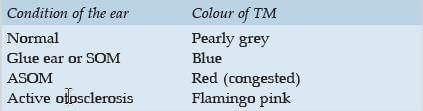
What is the colour of the normal tympanic membrane: (CUPGEE 96, Exam 2017)

Detailed Solution for Test: Embryology and Anatomy of Ear - 2 - Question 19

Test: Embryology and Anatomy of Ear - 2 - Question 20

Pars Flaccida of the tympanic membrane is also called: (MP 2007, Exam 2013)

Detailed Solution for Test: Embryology and Anatomy of Ear - 2 - Question 20
  • Reissner’s membrane separates Scala media from Scala vestibuli in the inner ear.
  • Basilar membrane separates Scala media or cochlear duct from Scala tympani in the inner ear. The organ of Corti rests on the basilar membrane.
  • Secondary TM overlies the round window in the middle ear.
Test: Embryology and Anatomy of Ear - 2 - Question 21

The most mobile part of the tympanic membrane: (TN 98, Exam 2017)

Test: Embryology and Anatomy of Ear - 2 - Question 22

Surface area of tympanic membrane: (Manipal 2006, Exam 2013)

Test: Embryology and Anatomy of Ear - 2 - Question 23

The effective vibratory area of the tympanic membrane: (UP 2005, Exam 2017)

Test: Embryology and Anatomy of Ear - 2 - Question 24

Which nerve supplies the tragus? (AIIMS 96, Exam 2017)

Test: Embryology and Anatomy of Ear - 2 - Question 25

Nerve supply for external ear are all except: (MAHE 2007, Exam 2016)

Information about Test: Embryology and Anatomy of Ear - 2 Page
In this test you can find the Exam questions for Test: Embryology and Anatomy of Ear - 2 solved & explained in the simplest way possible. Besides giving Questions and answers for Test: Embryology and Anatomy of Ear - 2, EduRev gives you an ample number of Online tests for practice
Download as PDF