NEET PG Exam  >  NEET PG Tests  >  Test: General Anatomy - NEET PG MCQ

Test: General Anatomy - NEET PG MCQ


Test Description

20 Questions MCQ Test - Test: General Anatomy

Test: General Anatomy for NEET PG 2025 is part of NEET PG preparation. The Test: General Anatomy questions and answers have been prepared according to the NEET PG exam syllabus.The Test: General Anatomy MCQs are made for NEET PG 2025 Exam. Find important definitions, questions, notes, meanings, examples, exercises, MCQs and online tests for Test: General Anatomy below.
Solutions of Test: General Anatomy questions in English are available as part of our course for NEET PG & Test: General Anatomy solutions in Hindi for NEET PG course. Download more important topics, notes, lectures and mock test series for NEET PG Exam by signing up for free. Attempt Test: General Anatomy | 20 questions in 20 minutes | Mock test for NEET PG preparation | Free important questions MCQ to study for NEET PG Exam | Download free PDF with solutions
*Multiple options can be correct
Test: General Anatomy - Question 1

Endochondral ossification is/are seen in: 

Detailed Solution for Test: General Anatomy - Question 1
  • Long bones and medial end of clavicle bone develop by endochondral ossification.
  • Flat bones of skull; facial skeleton; mandible and lateral end of clavicle develop by intramembranous ossification.
*Multiple options can be correct
Test: General Anatomy - Question 2

Bones ossified at birth: 

Detailed Solution for Test: General Anatomy - Question 2
  • Secondary centers around knee joint (distal femur and proximal tibia) appear during last weeks of intrauterine life (or immediately after birth).
  • Primary center of all tarsal bones (except talus, calcaneum and cuboid) appear after birth.
Test: General Anatomy - Question 3

Nutrient artery runs: 

Detailed Solution for Test: General Anatomy - Question 3

Nutrient artery enters the shaft (diaphysis) of the bone, divides into ascending and descending branches, which run towards and terminate in the adult metaphysis by anastomosing with the epiphyseal, metaphyseal and periosteal arteries.

Test: General Anatomy - Question 4

All of the following statements are true for metaphysis of bone EXCEPT: 

Detailed Solution for Test: General Anatomy - Question 4
  • The strongest part of bone is diaphysis (not metaphysis).
  • During growth of bone maximum activity occurs at growth plate (physis) and the adjacent section of the metaphysis.
  • Metaphysis is richly supplied with arteries forming hairpin bends, hence becomes a common site of osteomyelitis in children, as infectious agents are easily trapped in sluggish blood flow in hairpin bends.
Test: General Anatomy - Question 5

TRUE statement is: 

Detailed Solution for Test: General Anatomy - Question 5
  • Osteoblasts that get trapped in Haversian lamellae become osteocyte and assume the function of bone maintenance. They are no longer involved in bone formation.
  • Growth activity is maximum at the growth plate (physis) and adjacent metaphysis.
  • Metaphysis is present between diaphysis and epiphysis.
  • Interphalangeal joint is a hinge variety of synovial joint.
*Multiple options can be correct
Test: General Anatomy - Question 6

Traction epiphysis is/are: 

Detailed Solution for Test: General Anatomy - Question 6
  • Traction epiphysis are usually present at the ends of bones and develop due to traction by the attached muscles (and are therefore extracapsular), e.g. greater (and lesser) tubercles in humerus and greater (and lesser) trochanter in femur.
  • Pressure epiphysis are involved in weight transmission (and are intracapsular) e.g. head of humerus and femur and condyles of humerus, femur, tibia, etc.
  • Coracoid process in scapula is an example of atavistic epiphysis.
  • Deltoid tuberosity is not an epiphysis (it is present on the shaft/diaphysis).
Test: General Anatomy - Question 7

Which of the following is an aberrant epiphysis? 

Detailed Solution for Test: General Anatomy - Question 7

Aberrant epiphyses are deviations from the normal anatomy and are not always present. For example, the epiphysis at the head of the first metacarpal bone and at the bases of other metacarpals.

*Multiple options can be correct
Test: General Anatomy - Question 8

Bones which is/are pneumatic: 

Detailed Solution for Test: General Anatomy - Question 8
  • Pneumatic bones have air spaces within them and are present around the nasal cavity.
  • Temporal bone is morphologically classified as pneumatic bone because it has an internal air sinus and mastoid air cells.
  • Parietal bone is not a pneumatic bone.
Test: General Anatomy - Question 9

What type of joint is the growth plate? 

Detailed Solution for Test: General Anatomy - Question 9
  • Growing bones have epiphyseal (growth) plate between the epiphysis and diaphysis, this epiphyseo-diaphyseal joint is primary cartilaginous (synchondrosis).
  •  It is found in the growing bone, where the growth plate (hyaline cartilage) connects the epiphysis with the diaphysis, creating a hyaline cartilaginous joint.
  • At this site the fate of synchondrosis is synostosis (bony fusion) after the growth plate gets removed and replaced by the bone.
*Multiple options can be correct
Test: General Anatomy - Question 10

Atlanto-occipital joint is of synovial variety:

Detailed Solution for Test: General Anatomy - Question 10
  • Functionally it is an ellipsoid synovial joint but structurally it is a condylar synovial joint.
  • Head flexion and extension occurs at this joint for the nodding (yes) movement.
Test: General Anatomy - Question 11

Which of the following is a compound condylar joint? 

Detailed Solution for Test: General Anatomy - Question 11
  • Knee joint has more than two bones participating (hence compound joint). It is formed by the lateral and medial femorotibial and the femoropatellar joints.
  • It is a compound synovial joint incorporating two condylar joints between the condyles of the femur and tibia and one saddle joint between the femur and the patella.
  • TM joint is a condylar joint but it involves only 2 bones (not a compound joint).
  • Wrist joint is an ellipsoid synovial joint and elbow is a hinge synovial joint.
*Multiple options can be correct
Test: General Anatomy - Question 12

Metacarpophalangeal joint is:

Detailed Solution for Test: General Anatomy - Question 12
  • Metacarpophalangeal joint has a condyle with elliptical articular surface.
  • Structurally it is condylar but functionally ellipsoid synovial joint.
Test: General Anatomy - Question 13

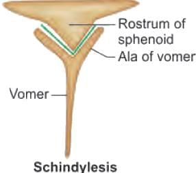
Vomer ala and sphenoidal rostrum junction is: 

Detailed Solution for Test: General Anatomy - Question 13

Spheno-vomerine joint is a schindylesis suture at the roof of the nasal cavity.

*Multiple options can be correct
Test: General Anatomy - Question 14

Synchondrosis is/are seen in:

Detailed Solution for Test: General Anatomy - Question 14
  • Synchondroses are primary cartilaginous unions between bone composed entirely of hyaline cartilage. These joints are immovable and mostly temporary in nature. As the growth ceases they undergo synostosis (i.e. plate of hyaline cartilage is completely replaced by bone).
  • Spheno-occipital joint is a synchondrosis joint between the basisphenoid and basiocciput bones, which together when joined from the clivus. When fused, the synchondrosis is often called the spheno-occipital suture- this is a misnomer-as anatomically it is not a suture.
  • Epiphysio-diaphyseal joint between the epiphyses and diaphysis of a growing long bone, is a synchondrosis, which later undergoes synostosis.
  • Xiphisternal joint is a synchondrosis joint. Some authors mention it as symphysis.
  • Costo-sternal joints are plane synovial joints of the costal cartilages of the true ribs with the sternum, with the exception of the first, which is the so called synchondrosis (a misnomer) since the cartilage is directly united with the sternum.
  • Manubrio-sternal joint is a secondary cartilaginous (symphysis) joint.
Test: General Anatomy - Question 15

Which of the following is a synovial joint of the condylar variety? 

Detailed Solution for Test: General Anatomy - Question 15
  • This a wrong Questions, since both the joints have condyles with ellipsoid articular surface - are structurally condylar but functionally ellipsoid synovial joints.
  • Some authors mention metacarpophalangeal as condylar synovial joint only (hence the answer of first preference).
Test: General Anatomy - Question 16

All are composite muscles EXCEPT: 

Detailed Solution for Test: General Anatomy - Question 16
  • Flexor carpi ulnaris is supplied by a single nerve and is not a composite/hybrid muscle.
  • Composite/hybrid muscles have more than one set of fibers and are supplied usually by different nerves for different set of fibers.
  • Pectineus has anterior set of fibers supplied by the femoral nerve, whereas, posterior set of fibers may be supplied by the obturator nerve.
  • Long head of biceps femoris is supplied by the tibial part of sciatic nerve, and the short head is supplied by the common peroneal nerve. This reflects the composite derivation from the flexor and extensor musculature.
  • Radial half of flexor digitorum profundus is supplied by the median nerve and the ulnar half is supplied by the ulnar nerve.
*Multiple options can be correct
Test: General Anatomy - Question 17

Muscle having double nerve supply: 

Detailed Solution for Test: General Anatomy - Question 17
  • Anterior belly of digastric is supplied by trigeminal nerve and posterior belly by facial nerve.
  • Spinal accessory nerve gives motor fibers to trapezius muscle, whereas ventral rami of C3, C4 are proprioceptive.
  • Adductor magnus ischial part is supplied by tibial part of sciatic nerve and adductor part by obturator nerve.
  • Inferior belly of the omohyoid is innervated by C1–C3 and the superior belly C1 fibers of ansa cervicalis.
  • Thyrohyoid muscle is innervated by C1 fibers traveling with the hypoglossal nerve.
Test: General Anatomy - Question 18

Digastric muscles are the following EXCEPT: 

Detailed Solution for Test: General Anatomy - Question 18
  • Sternocleidomastoid is a muscle with two heads and one belly, like the biceps brachii.
  • Digastric muscles have two bellies.
  • Occipitofrontalis muscle has two bellies: Occipital belly and frontal belly.
  • Omohyoid: Superior and inferior belly.
  • Ligament of Treitz is a digastric muscle with a skeletal muscle belly, which arises from the left crus of diaphragm and a smooth muscle belly which arises from the duodeno-jejunal junction. It has an intermediate tendon attaching to the connective tissue around the celiac trunk of aorta.
Test: General Anatomy - Question 19

Which of the following is multipennate muscle? 

Detailed Solution for Test: General Anatomy - Question 19

Multipennate muscle has the fiber bundles converge to several tendons.

Test: General Anatomy - Question 20

Muscle with parallel fibers are all EXCEPT:

Detailed Solution for Test: General Anatomy - Question 20
  • The individual fibers of a muscle are arranged either parallel or oblique to the long axis of the muscle.
  • Tibialis anterior muscle is a multipennate muscle with oblique fibers.
  • Muscles with parallel fasciculi: These are muscles in which the fasciculi are parallel to the line of pull and have greater degree of movement. Few examples are:
    • Strap-like, e.g. sternohyoid and sartorius
    • Strap-like with tendinous intersections, e.g. rectus abdominis
Information about Test: General Anatomy Page
In this test you can find the Exam questions for Test: General Anatomy solved & explained in the simplest way possible. Besides giving Questions and answers for Test: General Anatomy, EduRev gives you an ample number of Online tests for practice
Download as PDF