NEET PG Exam  >  NEET PG Tests  >  Test: Arrhythmias and Emergency Medicine- 2 - NEET PG MCQ

Test: Arrhythmias and Emergency Medicine- 2 - NEET PG MCQ


Test Description

20 Questions MCQ Test - Test: Arrhythmias and Emergency Medicine- 2

Test: Arrhythmias and Emergency Medicine- 2 for NEET PG 2025 is part of NEET PG preparation. The Test: Arrhythmias and Emergency Medicine- 2 questions and answers have been prepared according to the NEET PG exam syllabus.The Test: Arrhythmias and Emergency Medicine- 2 MCQs are made for NEET PG 2025 Exam. Find important definitions, questions, notes, meanings, examples, exercises, MCQs and online tests for Test: Arrhythmias and Emergency Medicine- 2 below.
Solutions of Test: Arrhythmias and Emergency Medicine- 2 questions in English are available as part of our course for NEET PG & Test: Arrhythmias and Emergency Medicine- 2 solutions in Hindi for NEET PG course. Download more important topics, notes, lectures and mock test series for NEET PG Exam by signing up for free. Attempt Test: Arrhythmias and Emergency Medicine- 2 | 20 questions in 20 minutes | Mock test for NEET PG preparation | Free important questions MCQ to study for NEET PG Exam | Download free PDF with solutions
Test: Arrhythmias and Emergency Medicine- 2 - Question 1

Which of the following is recommended for management of symptomatic bradycardia non responsive to 0.5mg iv atropine? (Recent Pattern Questions)

Detailed Solution for Test: Arrhythmias and Emergency Medicine- 2 - Question 1

For the management of symptomatic bradycardia, the following medications are advised:

  • Atropine 0.5 mg (maximum of 3 mg)
  • Epinephrine 2-10 µg/kg/min
  • Dopamine 2-10 µg/kg/min
Test: Arrhythmias and Emergency Medicine- 2 - Question 2

A 50-year-old patient presents with features of poor perfusion following MI. On examination heart rate is 40/min with BP of 60mmHg systolic. Atropine was given twice over 5 minutes, but the condition of patient is not improving. What is the next best step? (Recent Pattern Questions)

Detailed Solution for Test: Arrhythmias and Emergency Medicine- 2 - Question 2

In cases of symptomatic bradycardia accompanied by indications of inadequate perfusion, transcutaneous pacing is advised according to the ACLS 2015 guidelines.

Test: Arrhythmias and Emergency Medicine- 2 - Question 3

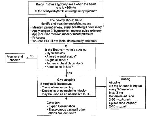
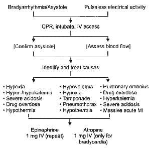
Which drugs are involved in bradycardia algorithm? (Recent Pattern Questions)

Detailed Solution for Test: Arrhythmias and Emergency Medicine- 2 - Question 3
First-line treatment for managing symptomatic bradycardia is atropine, administered up to a maximum dose of 3 mg. If the patient remains unstable, the following actions are suggested:
  • Transcutaneous pacing (TCP) should be considered.
  • If TCP is unavailable, the administration of two medications, dopamine and epinephrine, is recommended.
  • Should the patient still be unstable, transvenous pacing is performed.
Test: Arrhythmias and Emergency Medicine- 2 - Question 4
The primary decision point in bradycardia algorithm is determination of? (Recent Pattern Questions)
Detailed Solution for Test: Arrhythmias and Emergency Medicine- 2 - Question 4
The sufficiency of organ perfusion serves as the main criterion for starting the bradycardia management protocol.
Test: Arrhythmias and Emergency Medicine- 2 - Question 5

After determination of inadequate perfusion of organs in a patient with bradycardia, which of the following is the first step?  (Recent Pattern Questions)

Detailed Solution for Test: Arrhythmias and Emergency Medicine- 2 - Question 5

The initial and most important action in the treatment of symptomatic bradycardia is the provision of atropine.

Test: Arrhythmias and Emergency Medicine- 2 - Question 6
Comment on the diagnosis. (Recent Pattern Questions)
Detailed Solution for Test: Arrhythmias and Emergency Medicine- 2 - Question 6

The ECG indicates a heart rate of 40 beats per minute, featuring a P wave of normal duration preceding each normal qRS complex. The PR interval remains consistent, and there are no dropped beats in the observed rhythm. The patient is experiencing sinus bradycardia.

Test: Arrhythmias and Emergency Medicine- 2 - Question 7

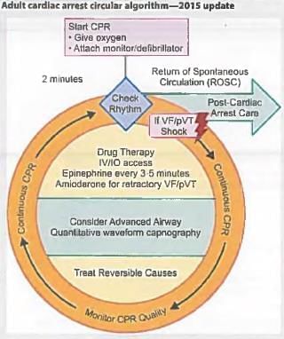
Which of the following is not a cause of PEA? (Recent Pattern Questions)

Detailed Solution for Test: Arrhythmias and Emergency Medicine- 2 - Question 7
  • The five Hs and five Ts associated with pulseless electrical activity (PEA) are outlined below.
  • PEA indicates that while electrical activity is observed on an ECG, there is no detectable pulse.
  • The primary cause of PEA is hypoxia due to respiratory failure, which accounts for approximately 40-50% of cases, followed by hypovolemia.

Test: Arrhythmias and Emergency Medicine- 2 - Question 8

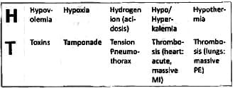
Which of the following drugs is used first line in management of PEA? (Recent Pattern Questions)

Detailed Solution for Test: Arrhythmias and Emergency Medicine- 2 - Question 8

In PEA, the treatment protocol to follow is for asystole. Always administer epinephrine and assess for reversible causes.

CPR Duality

  • Push hard (at least 2 inches (5cm) and fast (100-120/min) and allow complete chest recoil Minimize interruptions in compressions Avoid excessive ventilation
  • Rotate compressor every 2 minutes, or sooner if fatigued
  • If no advanced airway, 30:2 compression-ventilation ratio
  • Quantitative waveform capnography
    • If PETCO, <10 mm Hg, attempt to improve CPR quality
  • Intra-arterial pressure
    • If relaxation phase (diastolic) pressure <20 mm Hg, attempt to improve CPR quality

Shock energy for defibrillation

  • Biphasic: Manufacturer recommendation (e g. initial dose of 120- 200 J); if unknown, use maximum available. Second and subsequent doses should be equivalent, and higher doses may be considered.
  • Monophasic: 360 J

Drug therapy

  • Epinephrine IV/IO dose: 1 mg every 3-5 minutes
  • Amiodarone IV/IO dose: First dose: 300 mg bolus. Second dose: 150 mg

Advanced airway

  • Endotracheal intubation or supraglottic advanced airway
  • Waveform capnography or capnometry to confirm and monitor ET tube placement
  • Once advanced airway in place, give 1 breath every 6 seconds (10 breaths/mln) with continuous chest compressions

Return of spontaneous circulation (ROSC)

  • Pulse and blood pressure
  • Abrupt sustained increase In PETCO2 (typically 240 mm Hg)
  • Spontaneous arterial pressure waves wth intra-arterial monitoring

Reversible causes

  • Hypovolemia - Tension pneumothorax
  • Hypoxia - Tam ponade, cardiac
  • Hydrogen ion (acidosis) - Toxins
  • Hypo-/hyperkaiemia - Thrombosis, pulm onary
  • Hypothermia - Thrombosis coronary
Test: Arrhythmias and Emergency Medicine- 2 - Question 9

What is the cause of death in Commotio cordis? (Recent Pattern Questions)

Detailed Solution for Test: Arrhythmias and Emergency Medicine- 2 - Question 9

Non-penetrating blunt trauma to the chest can induce ventricular fibrillation, potentially resulting in sudden cardiac death. This phenomenon is known as commotio cordis and is frequently observed in sports-related injuries.
When trauma occurs to the chest at the peak of the T wave, a time often referred to as the vulnerable period of the heart, it can trigger ventricular fibrillation.

Test: Arrhythmias and Emergency Medicine- 2 - Question 10

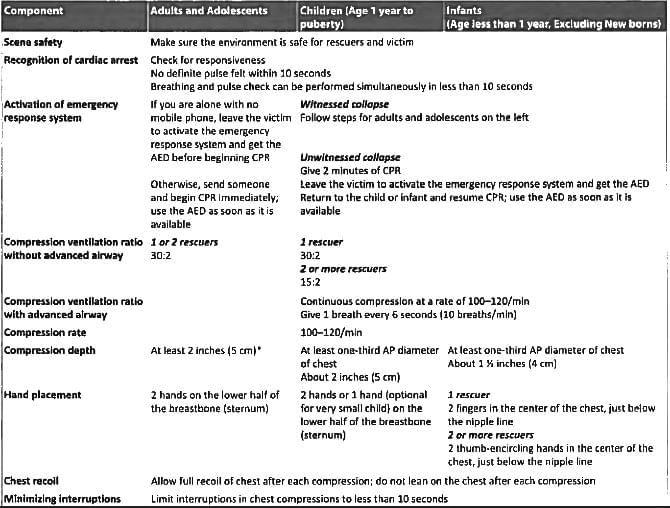
An unresponsive patient is brought to the emergency department with no proper history. What will be your next step? (AIIMS Nov 2016)

Detailed Solution for Test: Arrhythmias and Emergency Medicine- 2 - Question 10

According to the AHA 2015 BLS guidelines, as the patient is already noted as unresponsive, the rescuer should promptly check the carotid pulse and breathing for no longer than a 10-second interval.

  • Call for assistance to activate the emergency response system.
  • If the unresponsive patient is not breathing and has no carotid pulse, the C-A-B sequence is used, and chest compressions commence at a rate of 30:2 for all age groups.
  • If the unresponsive patient is not breathing but has a detectable carotid pulse, administer rescue breaths once every 6 seconds to reach a minimum of 10 breaths per minute.
Test: Arrhythmias and Emergency Medicine- 2 - Question 11

The ratio of chest compression to rescue breath ratio for a lone rescuer in CPR for all ages is? (AIIMS Nov 2016)

Detailed Solution for Test: Arrhythmias and Emergency Medicine- 2 - Question 11

The ratio of chest compressions to ventilations is 30:2 across all age groups, except for neonatal resuscitation, where it is 3:1. This 30:2 ratio also applies to infants and children. For adults, regardless of the number of rescuers, the ratio remains 30:2. In the case of children with one rescuer, the ratio is 30:2, while with two or more rescuers, it adjusts to 15:2.

  • Table: Summary of High-Quality CPR Components for BLS Providers.
Test: Arrhythmias and Emergency Medicine- 2 - Question 12

Which of the following statements is true about atropine and its role in cardiac arrest protocol? (Recent Question 2016-17)

Detailed Solution for Test: Arrhythmias and Emergency Medicine- 2 - Question 12

Atropine is indicated solely for the treatment of bradycardia, and its routine use during resuscitation is discouraged.

Test: Arrhythmias and Emergency Medicine- 2 - Question 13

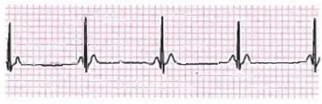
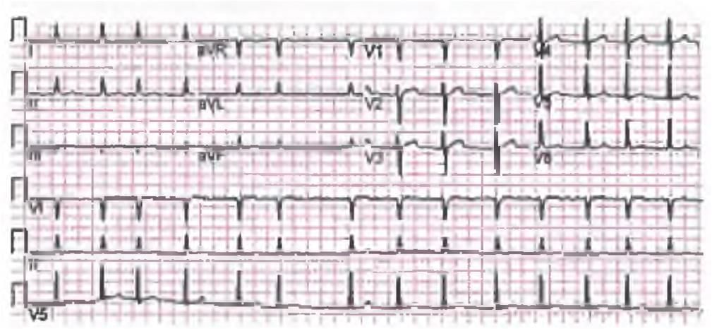
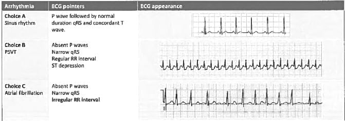
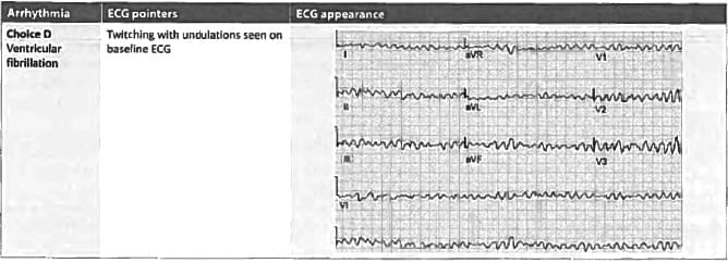
A 30-year-old man presents with recurrent attacks of feeling dizzy. ECG was done. What is the diagnosis? (Recent Question 2016-17)

Detailed Solution for Test: Arrhythmias and Emergency Medicine- 2 - Question 13
  • The heart rate is consistently irregular because of rhythm variations.
  • The estimated heart rate is 60 beats per minute (6 QRS complexes in 6 seconds in lead II [30 large squares]).
  • The axis remains normal.
  • There is a progressive lengthening of the PR interval that is followed by a dropped beat.
  • The PR interval prior to the dropped beat is longer than the PR interval in the subsequent beats.
  • The diagnosis is Mobitz I heart block.
Test: Arrhythmias and Emergency Medicine- 2 - Question 14
A 60-year-old retired banker complains of feeling dizzy with palpitations and breathlessness. His BP becomes un­recordable while ECG is being recorded. What should be done as first step in management of this patient? (Recent Question 2016-17)
Detailed Solution for Test: Arrhythmias and Emergency Medicine- 2 - Question 14

The ECG displays a broad complex QRS tachycardia, which is indicative of ventricular tachycardia. Given that the patient's blood pressure is rapidly declining, a DC shock must be administered without delay.

Test: Arrhythmias and Emergency Medicine- 2 - Question 15
Most common paroxysmal regular SVT is? (Recent Question 2016-17)
Detailed Solution for Test: Arrhythmias and Emergency Medicine- 2 - Question 15
Atrioventricular nodal reentrant tachycardia (AVNRT) represents the most prevalent form of paroxysmal regular supraventricular tachycardia (SVT).
  • AVNRT occurs without the presence of structural heart disease and is typically well tolerated.
  • Neck pulsations are often perceived due to the simultaneous contraction of the atria and ventricles.
  • A 'frog sign' may be observed during a physical examination when the arrhythmia is present.
This condition arises from having two electrophysiologically distinct pathways for conduction within the intricate syncytium of muscle fibres that constitute the AV node.
  • The fast pathway, located in the upper section of the node, has a longer refractory period.
  • Conversely, the pathway situated lower in the AV node area conducts at a slower rate but has a shorter refractory period.
Test: Arrhythmias and Emergency Medicine- 2 - Question 16
A 55-year-old diabetic hypertensive male smoker came with chest pain for last one day, His ECG is shown. Which of the following is the correct diagnosis? (APPG 2016)
Detailed Solution for Test: Arrhythmias and Emergency Medicine- 2 - Question 16

The ECG displays a fluctuating heart rate with the RR interval in lead II (refer to the lowest lead) ranging from 150 to 300 bpm. The axis remains normal.

  • The P wave is absent, accompanied by a shortened RR interval and narrow complex qRS.
  • Hyper-acute T waves are observed in leads V2 to V5, indicating an anterior wall myocardial infarction (MI).
  • Hypertension serves as a risk factor for both subendocardial ischaemia and atrial fibrillation.

Ventricular tachycardia presents with a wide complex qRS, thereby eliminating choices A and B.

Test: Arrhythmias and Emergency Medicine- 2 - Question 17

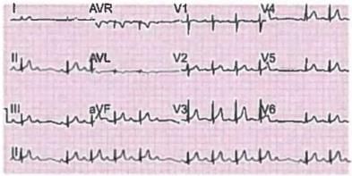
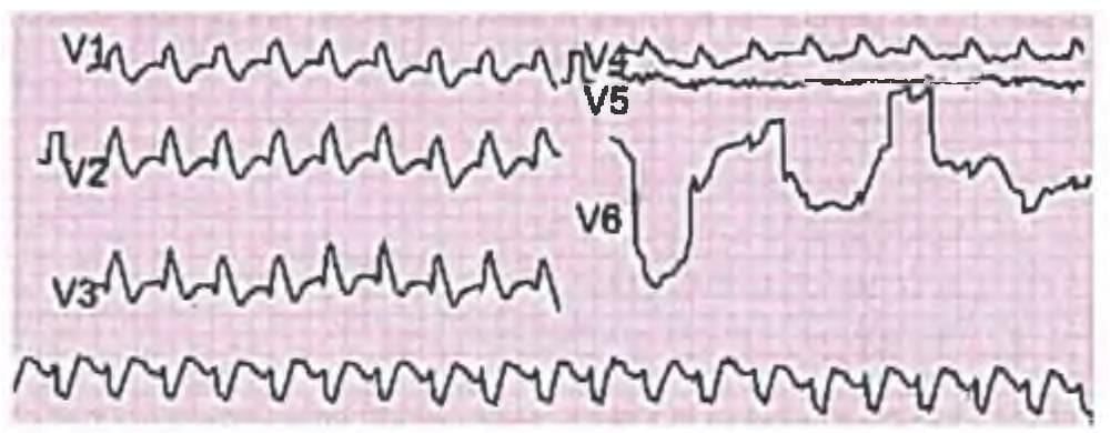
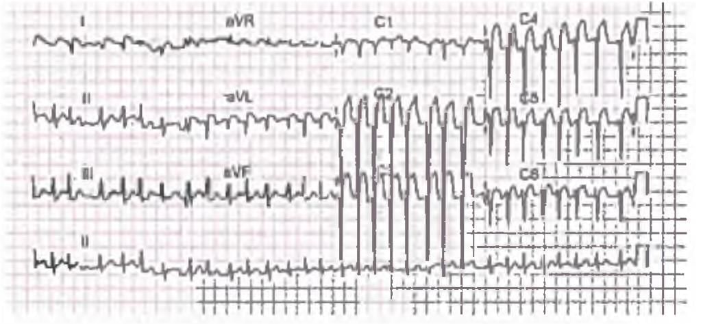
Identify the ECG given in the figure below? (AIIMS Nov 15)

Detailed Solution for Test: Arrhythmias and Emergency Medicine- 2 - Question 17

The ECG indicates a variable heart rate resulting from an irregularly irregular RR interval. The P waves are absent in the ECG, while the QRS complex has a normal duration. These findings suggest the presence of atrial fibrillation.


Atrial Fibrillation (AF)

Test: Arrhythmias and Emergency Medicine- 2 - Question 18

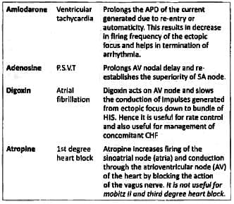
Match List I with List II and select the correct answer using the code given below the lists: (UPSC 2015) 
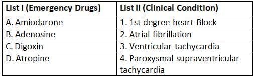
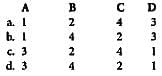
Codes:

Detailed Solution for Test: Arrhythmias and Emergency Medicine- 2 - Question 18

Test: Arrhythmias and Emergency Medicine- 2 - Question 19

Which of the auscultatory sign is absent in mitral stenosis in the presence of atrial fibrillation? (UPSC 2015)

Detailed Solution for Test: Arrhythmias and Emergency Medicine- 2 - Question 19

The presystolic accentuation of the mid-diastolic murmur associated with atrial fibrillation arises from atrial contraction against a closed mitral valve. As atrial fibrillation begins, the atria will twitch, causing the presystolic accentuation to vanish. Other alterations in the findings of mitral stenosis with the initiation of atrial fibrillation include:

  • The first heart sound shows variability in intensity due to the irregularly irregular heart rhythm.
  • The opening snap persists as long as the valves remain pliable and non-calcified.
  • The A2-O snap interval is generally less affected by atrial fibrillation, as it is primarily influenced by the mean left atrial pressure at the start of diastole, which exhibits minimal variation from beat to beat.
  • The duration of the diastolic murmur fluctuates.
Test: Arrhythmias and Emergency Medicine- 2 - Question 20

What is the drug of choice to control sudden onset supraventricular tachycardia? (Bihar PG 15)

Detailed Solution for Test: Arrhythmias and Emergency Medicine- 2 - Question 20

Treatment focuses on modifying conduction within the AV node. Vagal stimulation, such as that produced by the Valsalva manoeuvre or carotid sinus massage, can sufficiently slow conduction in the AV node to successfully terminate AVNRT. In cases where physical maneuvers fail to resolve the tachyarrhythmia:

  • The administration of adenosine, typically 6-12 mg IV, often proves effective.
  • Intravenous beta blockade or calcium channel therapy should be contemplated as a secondary treatment option.

If there is a presence of haemodynamic compromise, R wave synchronous DC cardioversion at 100-200 J can effectively terminate the tachyarrhythmia.

Information about Test: Arrhythmias and Emergency Medicine- 2 Page
In this test you can find the Exam questions for Test: Arrhythmias and Emergency Medicine- 2 solved & explained in the simplest way possible. Besides giving Questions and answers for Test: Arrhythmias and Emergency Medicine- 2, EduRev gives you an ample number of Online tests for practice
Download as PDF