NEET PG Exam  >  NEET PG Tests  >  Test: Anatomy - 2 - NEET PG MCQ

Test: Anatomy - 2 - NEET PG MCQ


Test Description

25 Questions MCQ Test - Test: Anatomy - 2

Test: Anatomy - 2 for NEET PG 2025 is part of NEET PG preparation. The Test: Anatomy - 2 questions and answers have been prepared according to the NEET PG exam syllabus.The Test: Anatomy - 2 MCQs are made for NEET PG 2025 Exam. Find important definitions, questions, notes, meanings, examples, exercises, MCQs and online tests for Test: Anatomy - 2 below.
Solutions of Test: Anatomy - 2 questions in English are available as part of our course for NEET PG & Test: Anatomy - 2 solutions in Hindi for NEET PG course. Download more important topics, notes, lectures and mock test series for NEET PG Exam by signing up for free. Attempt Test: Anatomy - 2 | 25 questions in 25 minutes | Mock test for NEET PG preparation | Free important questions MCQ to study for NEET PG Exam | Download free PDF with solutions
Test: Anatomy - 2 - Question 1

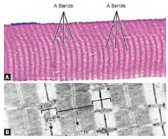
Arrange the parts of sarcomere from periphery to centre: (INI-CET May 2023)
1. Z line
2. A band
3. M line
4. H zone

Detailed Solution for Test: Anatomy - 2 - Question 1

The arrangement of sarcomere components from the outside to the centre is: Z – I – A – H – M. The sarcomere serves as the fundamental structural and functional unit of muscle tissue. It is defined by two Z lines at the edges and an M line in the centre.

The striations observed in muscles, specifically in skeletal and cardiac types, result from the systematic organisation of contractile proteins, namely actin and myosin. These proteins are structured in alternating lighter I bands (Isotropic in polarised light) and darker A bands (Anisotropic).

  • Fine dark lines, referred to as Z lines (Zwischenscheiben), are present, cutting through the I bands.
  • Z lines are the most electron-dense structures and segment each myofibril into many contractile units known as sarcomeres, which are aligned sequentially.
  • The dark A band is divided by the lighter H (Heller) band, which is additionally split by a denser M (Mittelscheibe) line.


(A) Skeletal muscle displays striations due to alternating dark A bands and light I bands.
(B) Sarcomeres, the contractile units of myofibrils, are arranged end to end, separated by dense Z lines. Each sarcomere contains light I bands (isotropic) and dark A bands (anisotropic). Z lines bisect the I bands, while the A band is divided by a lighter H band, which is further split by a denser M line.

Test: Anatomy - 2 - Question 2

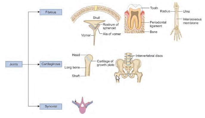
Type of joint present at middle radio-ulnar joint: (INI-CET Nov 2020)

Detailed Solution for Test: Anatomy - 2 - Question 2

The middle radio-ulnar joint is a fibrous (syndesmosis) joint that has limited mobility. Joints can be categorised structurally into three main types:

  • Fibrous
  • Cartilaginous
  • Synovial

Fibrous joints are further divided into:

  • Sutures
  • Gomphosis (tooth and socket)
  • Syndesmosis (for example, the middle radio-ulnar joint and the inferior tibio-fibular joint)

Cartilaginous joints are classified as:

  • Primary cartilaginous (synchondrosis), such as the epiphysio-diaphysial joint
  • Secondary cartilaginous (symphysis), for example, the pubic symphysis and the sacrococcygeal joint

Synovial joints can be subdivided into:

  • Plane joints, for instance, the intercarpal joints in the wrist
  • Uniaxial joints, which include:
    • Pivot joints (e.g., superior and inferior radio-ulnar joints)
    • Hinge joints (e.g., elbow and ankle joints)
  • Bi-axial joints, such as:
    • Condyloid joints (e.g., temporo-mandibular and knee joints)
    • Ellipsoid joints (e.g., wrist (radio-carpal) and metacarpo-phalangeal joints)
  • Multiaxial joints, which comprise:
    • Saddle joints (e.g., sterno-clavicular and 1st carpo-metacarpal joints)
    • Ball and socket joints (e.g., incus-stapedial and talo-calcaneo-navicular joints)

Test: Anatomy - 2 - Question 3

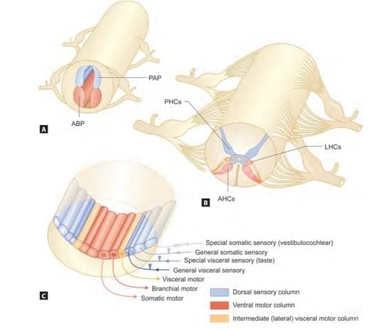
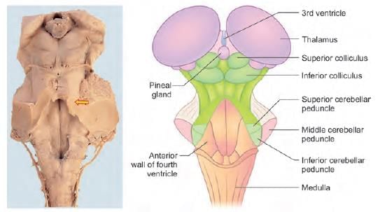
Arrange the following neural columns from medial to lateral at the floor of 4th ventricle:
1. General somatic afferent
2. General visceral afferent
3. Special visceral afferent
4. Special somatic afferent

Detailed Solution for Test: Anatomy - 2 - Question 3

The arrangement of the afferent (sensory) neural columns from medial to lateral at the base of the 4th ventricle is as follows:

  • General visceral afferent
  • Special visceral afferent
  • General somatic afferent
  • Special somatic afferent

During development, the neural tube forms an anterior (ventral) basal plate and a posterior (dorsal) alar plate. The anterior basal plate gives rise to:

  • Anterior motor columns (to regulate skeletal muscles)
  • Intermediate (lateral) column to manage visceral (cardiac & smooth) muscles

The posterior alar plate provides a posterior sensory column responsible for transmitting various sensations. The anterior basal plate results in three neural columns arranged from medial to lateral:

  • GSE (General Somatic Efferent/Somatic motor) – Controls general skeletal muscles in the body
  • SVE (Special Visceral Efferent/Branchial motor) – Regulates pharyngeal/branchial arch muscles
  • GVE (General Visceral Efferent/Visceral motor) – Governs visceral (cardiac & smooth) muscles in the body

The posterior alar plate yields four neural columns arranged from medial to lateral:

  • GVA (General Visceral Afferent/Sensory) – Transmits visceral sensations (such as stretch/ischaemia) from the carotid body/sinus and body viscera
  • SVA (Special Visceral Afferent/Sensory) – Carries special sensations like smell and taste
  • GSA (General Somatic Afferent/Sensory) – Conveys general somatic sensations (like pain/temperature) from the body
  • SSA (Special Somatic Afferent/Sensory) – Relays special sensations of vision, hearing, and balance


(A) The neural tube differentiates into the anterior basal plate (ABP) and posterior alar plate (PAP).
(B) The anterior basal plate forms anterior horn cells (AHCs), which innervate skeletal muscles, and lateral horn cells (LHCs), which regulate visceral muscles (cardiac and smooth). The posterior alar plate develops posterior horn cells (PHCs) responsible for transmitting various sensations.
(C) Neural columns are organized from medial to lateral.

Test: Anatomy - 2 - Question 4

Fornix fibres contain: (AIIMS June 2020)

Detailed Solution for Test: Anatomy - 2 - Question 4

The fornix comprises all three categories of white matter fibres: Commissural, Association, and Projection fibres. It carries fibres from the hippocampus towards the mammillary bodies of the hypothalamus and the anterior nuclei of the thalamus.

  • Commissural fibres of the fornix cross beneath the splenium to the contralateral fornix and terminate in the hippocampus on the opposite side.
  • Association fibres connect the hippocampus with adjacent regions, including the parahippocampal gyrus and septal areas.
  • Projection fibres link the hippocampus to the neurons of the mammillary body within the hypothalamus.

The fornix is a C-shaped bundle of fibrous commissural fibres (axons) that extends from the hippocampus to the mammillary bodies of the hypothalamus and the anterior nuclei of the thalamus, forming an arch over the thalamus. It is situated on the medial surfaces of the cerebral hemispheres. The fornix serves as the primary efferent pathway of the hippocampus and is a crucial component of the limbic system.

The limbic system is involved in motivation, learning, emotion, memory, and various cognitive functions. The fornix plays a role in the transfer of complex cognitive information between the cerebral hemispheres.

Arterial supply primarily comes from the short medial central arteries, which are branches of the proximal anterior cerebral artery.

Applied Anatomy: Cutting through the fornix at its body level can result in memory impairment (amnesia syndromes).

Test: Anatomy - 2 - Question 5

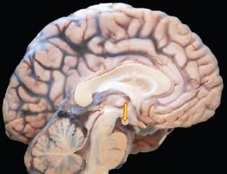
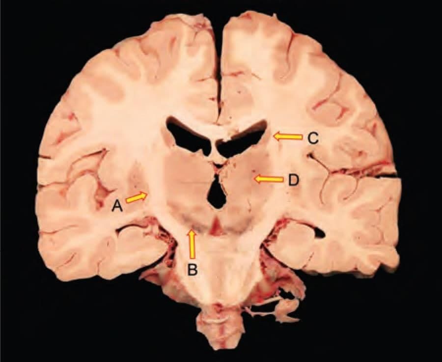
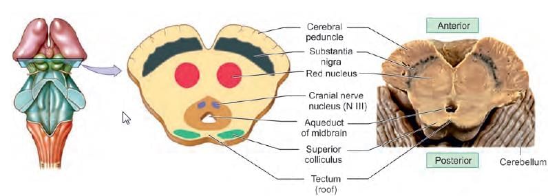
Fibres from the arrow marked structure in the following diagram, reach which nucleus of thalamus? (INI-CET Nov 2021)

Detailed Solution for Test: Anatomy - 2 - Question 5

The anterior nucleus of the thalamus receives input from the hypothalamus, specifically from the mammillary body/nucleus through the mammillothalamic tract. It also obtains input from the hippocampus via the fornix and sends projections to the cingulate gyrus.

Additionally, this nucleus is part of the Papez circuit, which is significant for learning and memory. The Papez circuit forms a neural loop that extends from the hippocampal formation to the mammillary body in the hypothalamus, then to the anterior nucleus of the thalamus, proceeding to the cingulate gyrus (parahippocampal region), and returning to the hippocampal formation.

Test: Anatomy - 2 - Question 6

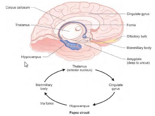
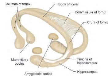
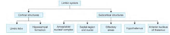
Which of the following are components of limbic system? (INI-CET Nov 2022)
1. Thalamus
2. Subthalamus 
3. Amygdala
4. Hippocampus
5. Hypothalamus

Detailed Solution for Test: Anatomy - 2 - Question 6

The limbic system comprises structures located just below the medial temporal lobe of the cerebrum (telencephalon) and their connections to the diencephalon (thalamus and hypothalamus). It is associated with:

  • Emotion
  • Behaviour
  • Memory consolidation
  • Olfaction

This involves the Papez circuit. It is important to note that although the subthalamic nucleus is a functional nucleus of the basal ganglia, it is typically not classified under the limbic system; however, it plays a crucial role in the 'limbic' functions of the basal ganglia.

Test: Anatomy - 2 - Question 7

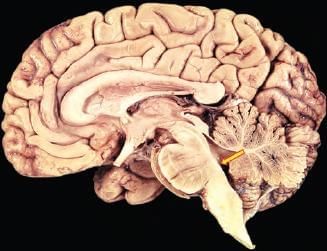
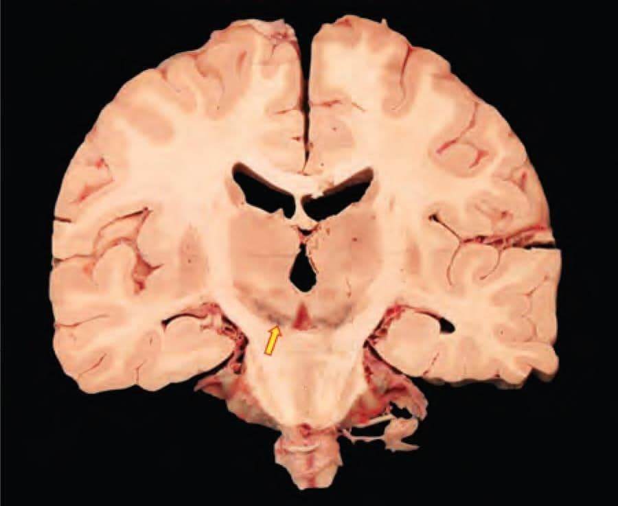
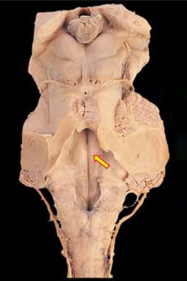
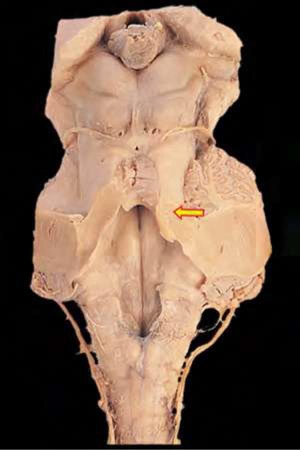
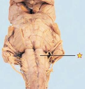
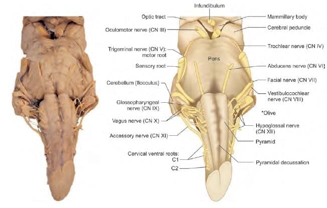
Which cranial nerve fibres are found at point at arrow marked region? (INI-CET Nov 2021)

Detailed Solution for Test: Anatomy - 2 - Question 7

This is a sagittal section of the brain depicting the area of the facial colliculus, located on the dorsum of the lower pons, at the base of the fourth ventricle. The facial colliculus is elevated due to the axonal fibres of the facial nerve (CN VII).

A transverse section of the pons illustrates the facial colliculus, which appears as a rounded prominence created by the axons of the facial nerve as they loop around the abducent nucleus.

The facial colliculus is positioned on the dorsal side of the lower pons and can be observed at the floor of the fourth ventricle.

Test: Anatomy - 2 - Question 8
A 65-year-old lady presents with a cerebrovascular accident involving inferior frontal gyrus. Which functional area would mostly be affected? (NEET-PG 2020)
Detailed Solution for Test: Anatomy - 2 - Question 8
A cerebrovascular accident affecting the inferior frontal gyrus disrupts Broca’s area, leading to Motor (non-fluent) aphasia characterised by speech hesitancy. Note: Damage to the posterior end of the superior temporal gyrus impacts Wernicke’s sensory speech area, which is crucial for language comprehension. This results in fluent yet nonsensical speech, provided Broca’s area remains unaffected.
  • Type: Speech
  • Fluency: Comprehension
  • Comments: Repetition impaired
  • Broca (expressive)
    • Nonfluent
    • Intact
    • Broca area in the inferior frontal gyrus of the frontal lobe.
    • Patient appears frustrated, insight intact.
  • Wernicke (receptive)
    • Fluent
    • Impaired
    • Wordy but nonsensical.
    • Patients lack insight.
    • Wernicke area in the superior temporal gyrus of the temporal lobe.
  • Conduction
    • Fluent
    • Intact
    • Can result from damage to the arcuate fasciculus.
  • Global
    • Nonfluent
    • Impaired
    • Involves the arcuate fasciculus; affects both Broca and Wernicke areas (all regions).
Test: Anatomy - 2 - Question 9
A boy met with a motor bike accident. CT brain shows injury to posterior end of superior temporal gyrus. He is likely to suffer from: (NEET-PG 2020)
Detailed Solution for Test: Anatomy - 2 - Question 9
Injury to the posterior aspect of the superior temporal gyrus affects Wernicke’s sensory speech area, which is crucial for the understanding of language. This results in continuous and nonsensical speech that lacks comprehensibility, while maintaining good fluency, as Broca’s area remains intact.
Test: Anatomy - 2 - Question 10
Neurotransmitter secreted from the arrow marked area is: (AIIMS June 2020)
Detailed Solution for Test: Anatomy - 2 - Question 10
The area indicated by the arrow is the substantia nigra (mid-brain), recognised by the dark hue of neuromelanin. Neuromelanin is synthesised directly from L-DOPA, which is a precursor to dopamine. Neurons that contain neuromelanin within the substantia nigra experience neurodegeneration in Parkinson’s disease. Treatment options for parkinsonism comprise L-DOPA along with centrally acting anticholinergics.
Test: Anatomy - 2 - Question 11

60-year-old patient complains of pain on touching cold water from tap upto shoulder on right side. No motor deficit is observed upon examination. Which of the following structure is involved? (INI-CET July 2021)

Detailed Solution for Test: Anatomy - 2 - Question 11

This patient exhibits typical characteristics of Dejerine Roussy syndrome, which involves the thalamus (Marker D). This syndrome is often a result of the blockage of a posterior thalamo-geniculate artery.

  • Classic signs include:
  • Contralateral hemiparesis;
  • Contralateral hemianesthesia;
  • Increased pain threshold;
  • Spontaneous, agonising, burning pain (hyperpathia);
  • Athetotic posturing of the hand (thalamic hand).

The thresholds for pain, touch, and temperature are diminished on the opposite side of the body (thalamic overreaction). However, once the threshold is reached, the sensations become exaggerated, distorted, and unpleasant; even a light touch can trigger severe pain. In some cases, this spontaneous pain may become unmanageable and resist treatment from potent analgesic medications.

Test: Anatomy - 2 - Question 12

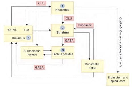
Arrange the following structures in direct pathway of basal ganglia: (INI-CET May 2022)
1. Striaum
2. Substantia nigra
3. Globus pallidus
4. Thalamus
5. Cerebral cortex

Detailed Solution for Test: Anatomy - 2 - Question 12

Dopamine from the Substantia Nigra compactum (SNc), associated with the nigrostriatal pathway, activates the direct pathway by binding to the D1 receptor in the striatum, thereby facilitating movement.

  • The direct (excitatory) pathway involves cortical input via glutamate, which prompts GABA release from the striatum.
  • This GABA release inhibits GABA release from the Globus Pallidus internum (GPi), leading to the disinhibition (activation) of the Thalamus and an increase in motion.

Note: The basal ganglia are responsible for planning and programming voluntary motor activities to ensure smooth movement.

  • The direct (excitatory) pathway operates similarly: cortical input via GLU (glutamate) stimulates GABA release from the striatum.
  • This inhibits GABA release from the Globus Pallidus, resulting in the disinhibition (activation) of the Thalamus and enhanced motion (GLU).

Note: The Substantia nigra plays a crucial role in stimulating the direct pathway and facilitating movement.

Test: Anatomy - 2 - Question 13

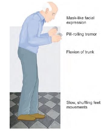
A 75-year-old female has presented with inability to express feelings, bradykinesia, rigidity and resting tremors. The structure most likely involved is: (NEET-PG 2023)

Detailed Solution for Test: Anatomy - 2 - Question 13

An elderly patient exhibiting an inability to convey emotions (mask-like facies), bradykinesia, rigidity (cogwheel or lead-pipe), and resting tremors suggests a diagnosis of Parkinson’s disease, a disorder of the basal ganglia. They also display a stooped posture and a festinating gait.

The hippocampus is associated with short-term memory and is a part of the limbic system, which plays a role in memory consolidation, learning, and spatial navigation. Damage to the hippocampus may manifest as:

  • Short-term memory issues
  • Anterograde amnesia
  • Disorientation and confusion

The cerebellum is responsible for coordinating voluntary motor activities, maintaining posture, and ensuring equilibrium. Clinical signs related to the cerebellum may include:

  • Incoordination of voluntary movements (difficulty walking in a straight line)
  • Intention tremors

The internal capsule contains axons (projection fibres) that transmit sensory and motor information, linking the upper brain centres with the lower ones and vice versa. A lesion in this area can lead to contralateral hemiplegia, hemiparesis, and other related issues.

Test: Anatomy - 2 - Question 14

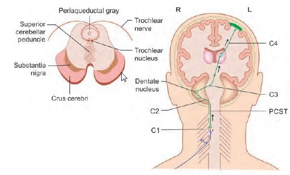
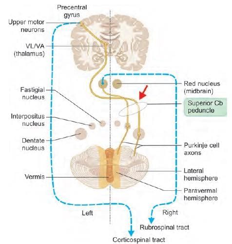
Decussation of superior cerebellar peduncle occurs at: (AIIMS June 2020)

Detailed Solution for Test: Anatomy - 2 - Question 14

The decussation of the superior cerebellar peduncle refers to the crossing of the fibres of the superior cerebellar peduncle through the midline and is situated at the level of the inferior colliculi in the midbrain. It includes:

  • The cerebellothalamic tract, originating from the dentate nucleus (also referred to as the dentatothalamic tract).
  • The cerebello-rubral tract, which arises from the globose and emboliform nuclei, projecting to the contralateral red nucleus and ultimately forming the rubrospinal tract.

This structure serves as a significant anatomical landmark; lesions positioned above it result in contralateral cerebellar signs, whereas lesions located below it lead to ipsilateral cerebellar signs.

Test: Anatomy - 2 - Question 15

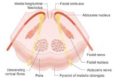
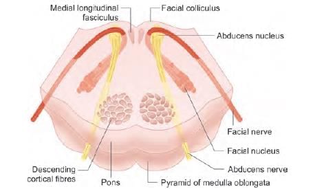
Lesion of the structure producing the elevation marked leads to paralysis of which of the following muscle: (INI-CET May 2022)

Detailed Solution for Test: Anatomy - 2 - Question 15

This dissected specimen reveals the base of the fourth ventricle, with the arrow indicating the facial colliculus (located on the dorsum of the lower pons). The facial colliculus is a rounded projection formed by the axonal fibres of the facial nerve (CN VII). Damage to this structure may result in ipsilateral paralysis of facial expression muscles, such as the risorius.

  • Deep to the facial colliculus lies the abducent nucleus, which may be affected if the injury extends further, potentially causing paralysis of the lateral rectus muscle.
  • Since the facial colliculus comprises both CN VII and CN VI, lesions here can lead to symptoms at multiple levels.
  • Involvement of CN VII can cause ipsilateral Bell's palsy.
  • Lesions of CN VI may lead to difficulties with horizontal eye movements, including lateral gaze palsy and one-and-a-half syndrome.

A transverse section of the pons illustrates the area of the facial colliculus (on the dorsum of the lower pons; at the base of the fourth ventricle). The facial colliculus is elevated by the axonal fibres of the facial nerve (CN VII), which loop around the deeply positioned abducent nerve (CN VI) nucleus.

  • Risorius muscle
  • Origin: (variable) Zygomatic arch, parotid fascia
  • Insertion: Modiolus
  • Action: Pulls the angle of the mouth laterally (grinning/laughing)
  • Innervation: Buccal branch of the facial nerve
  • Arterial supply: Superior labial artery (facial artery)

Note: The modiolus serves as a common insertion point for several facial muscles, held together by fibrous tissue, and is located lateral and slightly superior to each corner of the mouth. It plays a crucial role in mouth movement, facial expression, and is vital for the stability of the lower denture.

Test: Anatomy - 2 - Question 16

Fibres carried by the marked structure terminate in: (INI-CET July 2021)

Detailed Solution for Test: Anatomy - 2 - Question 16

The marker illustrates the superior cerebellar peduncle in a posterior view of the brainstem, following the removal of the cerebellum by severing the peduncles. It contains the dentato-rubro-thalamic tract, with some fibres extending to the opposite red nucleus in the midbrain (hence the term "rubro"). The superior cerebellar peduncle serves as the primary ascending efferent pathway from the cerebellum.

  • It is situated within the lateral wall of the fourth ventricle.
  • A multitude of fibres that enter the midbrain tegmentum originate from the cerebellar nuclei (for example, the dentate nucleus).
  • These fibres travel ventromedially and cross the midline through the decussation of the superior cerebellar peduncle, located at the junction of the pons and midbrain.

Above the decussation, the ascending fibres infiltrate the red nucleus, where some fibres or their collaterals terminate. However, the majority of the superior cerebellar fibres continue to ascend, terminating in the lateral nucleus of the thalamus. The superior cerebellar peduncle carries the efferent fibres of the dentato-rubro-thalamic tract, which decussate at the level of the inferior colliculus (midbrain) and synapse in the contralateral red nucleus and thalamus.

Note: The right cerebellum plays a role in coordinating voluntary motor activities on the right side of the body (lesions exhibit ipsilateral clinical symptoms).

Test: Anatomy - 2 - Question 17
The marked cell inhibits which of the following structure? (INI-CET Nov 2021; NEET-PG 2020)
Detailed Solution for Test: Anatomy - 2 - Question 17

This slide presents the Purkinje cell within the middle layer of the cerebellar cortex. These are a type of GABAergic neuron featuring intricate dendritic branching and are distinguished by a high number of dendritic spines. Purkinje cells provide inhibitory projections to the deep cerebellar nuclei and represent the exclusive output for all motor coordination in the cerebellar cortex.

The slide includes a histological image of the cerebellar cortex, with Purkinje cells indicated by an arrow. These cells are located in the middle layer and are the largest among all cells, having a flask-like shape, with dendrites extending towards the outer molecular layer.

  • The cerebellar cortex comprises five types of cells organised into three layers:
  • Molecular layer: This outer layer, situated beneath the pia mater, contains stellate and basket cells, along with the dendritic arbor of the Purkinje cells. It is characterised by a less dense cell distribution, as indicated by a comparatively lower number of nuclei.
  • Purkinje cell layer: This layer is positioned between the outer molecular layer and the inner granule cell layer.
  • Granule layer: The inner layer that lies above the white matter, containing granule and Golgi cells, as well as cerebellar glomeruli. This layer is densely packed with cells, evident from the large number of nuclei present.

Note: A cerebellar glomerulus is composed of a mossy fibre rosette, granule cell dendrites, and a Golgi cell.

Test: Anatomy - 2 - Question 18

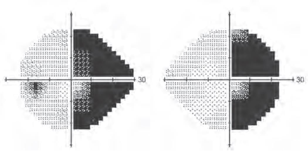
A 23-year-old patient has presented with visual defect as shown in the following diagram. Identify the most likely level of lesion: (INI-CET July 2021, NEET-PG 2021, NEET-PG 2023)

Detailed Solution for Test: Anatomy - 2 - Question 18

The visual anomaly observed in the provided image illustrates right homonymous hemianopia (HHA), accompanied by macular sparing (MS):

  • The right side of each eye is blind while central vision remains intact.
  • This condition may arise from an obstruction in the left posterior cerebral artery.
  • Macular sparing (the preservation of central vision) is a result of collateral circulation from the middle cerebral artery.
  • A lesion affecting the left optic nerve would cause a total loss of vision in the left eye.
  • The same principle applies to the right eye.
  • A lesion in the optic chiasm leads to bitemporal hemianopia (tunnel vision) – a bilateral loss of peripheral visual fields.
  • A lesion in the left optic tract results in right homonymous hemianopia.
  • Lesions posterior to the chiasm cause contralateral homonymous hemianopia, but macular sparing will not occur.
  • A lesion in the right occipital cortex causes left homonymous hemianopia (loss of vision in the left side of each eye).
  • This condition exhibits macular sparing if it results from occlusion of the posterior cerebral artery.


Lesions at different levels of the optic pathway cause distinct visual deficits:
(a) Left optic nerve: Blindness in the left eye.
(b) Optic chiasm: Bitemporal hemianopia (loss of outer visual fields in both eyes).
(c) Left optic tract: Right homonymous hemianopia (loss of right visual field in both eyes).
(d) Left optic radiation: Right homonymous hemianopia, often with macular sparing if caused by posterior cerebral artery occlusion.

Test: Anatomy - 2 - Question 19

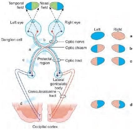
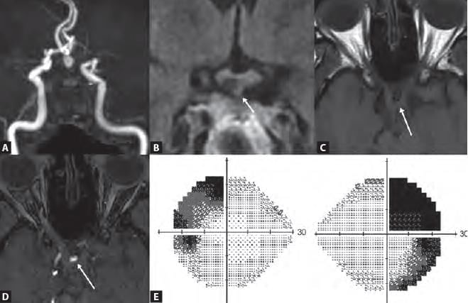
A patient presented with history of vision loss. On investigation an aneurysm was identified causing compression on the optic chiasma. Which is the following artery is most likely involved? (NEET-PG 2021)

Detailed Solution for Test: Anatomy - 2 - Question 19

An aneurysm located in the anterior communicating artery may exert pressure on the optic chiasma, resulting in bitemporal hemianopia (tunnel vision). The other options do not lead to compression of the optic chiasma.

Anterior communicating artery aneurysms (AcoA) typically manifest as subarachnoid haemorrhage (SAH) linked to focal deficits, particularly bitemporal hemianopsia (BTH).

It is important to note that an aneurysm in the posterior communicating artery can affect the oculomotor nerve.

Test: Anatomy - 2 - Question 20

The nerve supplying the marked muscle originates at the level of: (INI-CET July 2021)

Detailed Solution for Test: Anatomy - 2 - Question 20

The highlighted muscle in the illustration is the medial rectus. It is innervated by CN 3 (the Oculomotor nerve), which originates from the oculomotor nucleus, located in the rostral (upper) midbrain at the level of the superior colliculus/red nucleus.

The diagram depicts a dissected specimen with a transverse section in the head region, illustrating various structures:

  • Eyeballs and associated muscles
  • Nasal cavity
  • Parts of the brain in different cranial fossae

The four recti muscles originate from the common tendinous ring of Zinn and insert into the sclera of the eyeball, as shown in the illustration. This section of the midbrain is at the level of the superior colliculus. The Oculomotor nerve arises from the oculomotor nucleus, positioned in the rostral (upper) midbrain at the level of the superior colliculus/red nucleus.

Test: Anatomy - 2 - Question 21

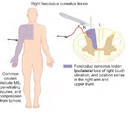
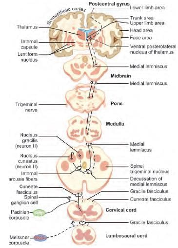
An injury to the lateral part of dorsal column at the cervical region results in loss of proprioception in: (NEET-PG 2023)

Detailed Solution for Test: Anatomy - 2 - Question 21

An injury to the lateral aspect of the dorsal column in the cervical area leads to a loss of proprioception in the ipsilateral upper limb, as it affects the fasciculus cuneatus, which runs ipsilaterally and transmits information from the upper body, including the brachial plexus (C5,6,7,8;T1). The dorsal column within the spinal cord is responsible for sensations such as:

  • Conscious proprioception
  • Pressure
  • Discriminative touch
  • Vibration
  • Stereognosis

It consists of two fasciculi:

  • Lateral: Fasciculus cuneatus – Transmits information from the upper body, for instance, sensations from the upper limb.
  • Medial: Fasciculus gracilis – Transmits information from the lower body, such as sensations from the lower limb.

Test: Anatomy - 2 - Question 22

Medial Lemniscus is continuation of: (AIIMS June 2020)

Detailed Solution for Test: Anatomy - 2 - Question 22

The dorsal column – medial lemniscal system transmits sensory data (such as pressure, touch, and vibration) towards the thalamus. The first order neurone located in the dorsal root ganglion travels as the dorsal column within the spinal cord. It is important to note that the dorsal column comprises two fasciculi:

  • Fasciculus cuneatus (upper body)
  • Fasciculus gracilis (lower body)

The dorsal column (including both the fasciculus cuneatus and gracilis) synapses onto the second order neurone (nucleus cuneatus and gracilis located in the lower medulla). The medial lemniscus is a tract that originates from the nuclei gracilis and cuneatus (internal arcuate fibres), crossing to the contralateral side in the lower part of the medulla oblongata to ascend through the brainstem, ultimately terminating in the thalamus (VPL – Ventral Posterior Lateral nucleus). The medial lemniscus transmits sensory impulses from the contralateral side of the body, and any damage to it results in sensory deficits on the opposite side of the body.

Test: Anatomy - 2 - Question 23

A lesion in the marked structure leads to: (AIIMS Nov 2019)

Detailed Solution for Test: Anatomy - 2 - Question 23

The structure referred to is the pyramid located on the front side of the medulla oblongata.

  • A lesion in this area can result in contralateral spastic hemiplegia, caused by damage to the pyramidal (corticospinal) tract that runs beneath it.
  • This condition may arise in cases of medial medullary syndrome, typically due to an obstruction in the anterior spinal artery.
  • Ataxia and vertigo are characteristics associated with lateral medullary syndrome.
  • This syndrome can occur from the occlusion of the PICA (Posterior Inferior Cerebellar Artery).
  • Additionally, ipsilateral facial nerve palsy may be observed in lateral pontine syndrome, resulting from a blockage in the AICA (Anterior Inferior Cerebellar Artery).
Test: Anatomy - 2 - Question 24

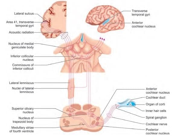
Arrange the following components of auditory pathway in peripheral to central order: (INI-CET May 2023)
1. Inferior colliculus
2. Cochlear nucleus
3. Lateral lemniscus
4. Medial geniculate body
5. Spiral ganglion

Detailed Solution for Test: Anatomy - 2 - Question 24

The auditory pathway follows this progression (SLIM-41, 42):

  • Organ of Corti (inner ear)
  • cochlear (spiral) ganglion and nerve
  • cochlear nuclei
  • trapezoid body
  • Superior olivary nucleus
  • Lateral lemniscus
  • Inferior colliculus
  • Medial geniculate body
  • sublentiform fibres of the internal capsule
  • auditory cortex (41, 42 Brodmann area) – Heschl’s gyrus

Test: Anatomy - 2 - Question 25

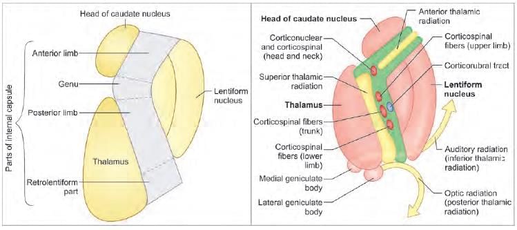
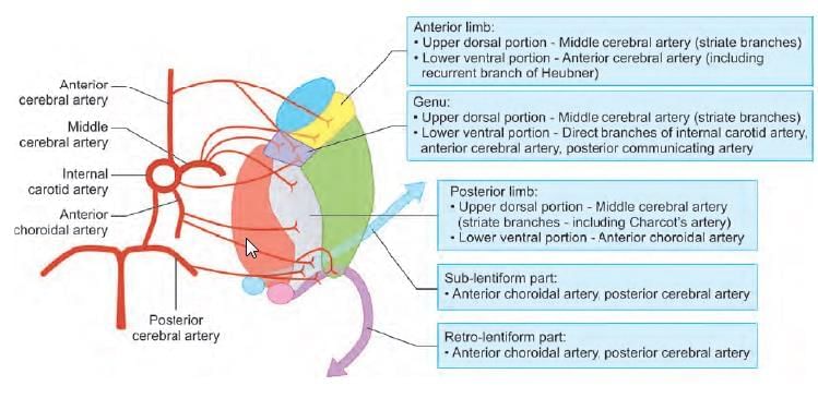
Which of the following statement is FALSE regarding internal capsule? (INI-CET Nov 2022)

Detailed Solution for Test: Anatomy - 2 - Question 25

Face fibres (axons) traverse the genu of the internal capsule, while upper limb fibres pass through the posterior limb (not the anterior) of the internal capsule. The internal capsule is a bundle of axons (white matter) that carries projection fibres, connecting higher brain centres with lower ones and vice versa. It contains both ascending (sensory) and descending (predominantly motor) fibres. It comprises several sections, each representing different body parts:

  • Anterior limb;
  • Genu (Head and neck fibres pass through here) -
    • Cortico-nuclear tract (motor)
    • Trigeminal lemniscus (sensory)
  • Posterior limb (Body is represented topographically from anterior to posterior) -
    • Upper limb fibres are more anterior
    • Lower limb fibres are more posterior
    • Cortico-spinal tract (motor)
    • Dorsal column – medial lemniscal system (sensory)
  • Sublentiform part (fibres run beneath the lentiform nucleus) -
    • Conveys the auditory pathway to the temporal auditory cortex
  • Retro-lentiform part (fibres move behind the lentiform nucleus) -
    • Transmits the visual/optic pathway to the occipital visual cortex

The arterial supply to the internal capsule primarily comes from branches of the middle cerebral artery, particularly the upper dorsal section. This artery supplies almost the entire anterior limb, posterior limb, and genu, but does not supply the sublentiform or retro-lentiform fibres. The anterior cerebral artery contributes a recurrent branch of Heubner, which supplies the anterior limb and genu. The anterior choroidal artery, a branch of the internal carotid artery, supplies the posterior limb and part of the sublentiform (and retro-lentiform) area of the internal capsule. The posterior cerebral artery supplies the sublentiform and retro-lentiform portions of the internal capsule.

Note: One of the lateral striate branches of the middle cerebral artery is larger and more prone to rupture (Charcot’s artery associated with cerebral haemorrhage). This branch supplies the posterior limb of the internal capsule, and its damage results in contralateral hemiplegia and hemiparesis.

Information about Test: Anatomy - 2 Page
In this test you can find the Exam questions for Test: Anatomy - 2 solved & explained in the simplest way possible. Besides giving Questions and answers for Test: Anatomy - 2, EduRev gives you an ample number of Online tests for practice
Download as PDF