NEET PG Exam  >  NEET PG Tests  >  Test: Anatomy - 3 - NEET PG MCQ

Test: Anatomy - 3 - NEET PG MCQ


Test Description

25 Questions MCQ Test - Test: Anatomy - 3

Test: Anatomy - 3 for NEET PG 2025 is part of NEET PG preparation. The Test: Anatomy - 3 questions and answers have been prepared according to the NEET PG exam syllabus.The Test: Anatomy - 3 MCQs are made for NEET PG 2025 Exam. Find important definitions, questions, notes, meanings, examples, exercises, MCQs and online tests for Test: Anatomy - 3 below.
Solutions of Test: Anatomy - 3 questions in English are available as part of our course for NEET PG & Test: Anatomy - 3 solutions in Hindi for NEET PG course. Download more important topics, notes, lectures and mock test series for NEET PG Exam by signing up for free. Attempt Test: Anatomy - 3 | 25 questions in 25 minutes | Mock test for NEET PG preparation | Free important questions MCQ to study for NEET PG Exam | Download free PDF with solutions
Test: Anatomy - 3 - Question 1

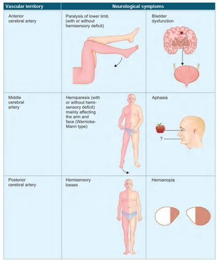
Ischemic stroke of which part of internal capsule leads to contralateral motor loss in upper and lower limb? (AIIMS June 2020)

Detailed Solution for Test: Anatomy - 3 - Question 1

Lesions in the posterior limb of the internal capsule result in harm to the corticospinal tract, causing contralateral hemiplegia, which is the loss of motor function in both the upper and lower limbs. The anterior limb of the internal capsule (ALIC) contains thalamic and brainstem fibres that originate from prefrontal cortical areas, linked to various components of emotion, motivation, cognitive processing, and decision-making.

  • The auditory pathway traverses the sub-lentiform fibres.
  • The visual pathway passes through the retro-lentiform fibres.

Test: Anatomy - 3 - Question 2

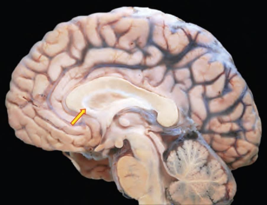
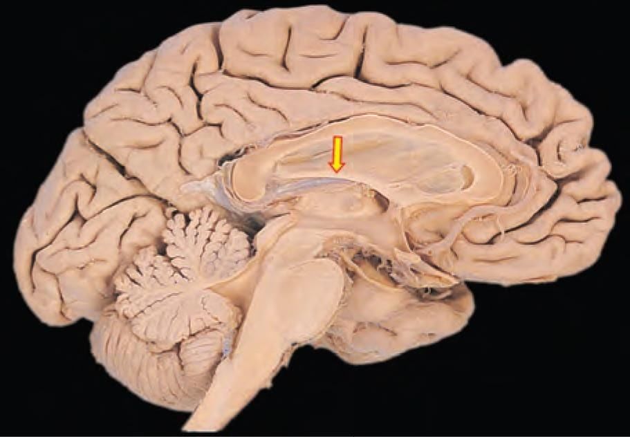
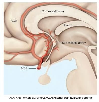
Mention the arterial supply of the marked structure: (INI-CET May 2022)

Detailed Solution for Test: Anatomy - 3 - Question 2

The arrow mark indicates the rostrum section of the corpus callosum, which is supplied by the anterior cerebral artery and the anterior communicating artery. The blood supply to the corpus callosum comes from:

  • A branch of the anterior communicating artery (the median callosal or subcallosal artery)
  • The pericallosal artery (the distal portion of the anterior cerebral artery)
  • The posterior pericallosal artery (typically a branch of the posterior cerebral artery)

Additional notes:

  • The splenium of the corpus callosum is supplied by the posterior pericallosal artery (a branch of the posterior cerebral artery).
  • The middle cerebral artery supplies the lateral aspect of the corpus callosum.

It is important to note that the rostrum of the corpus callosum (commissural fibres) connects the orbito-frontal cortex of both sides for inter-hemispheric transfer of information.

Test: Anatomy - 3 - Question 3

Arterial supply of marked structure: (AIIMS Nov 2019)

Detailed Solution for Test: Anatomy - 3 - Question 3

The fornix is primarily supplied by branches of the anterior cerebral artery. Additionally, it receives blood from branches of both the posterior cerebral artery and the internal carotid artery.

  • Anterior cerebral artery and anterior communicating artery – anterior columns of fornix.
  • Anterior choroidal artery (a branch of the internal carotid artery) – fimbria of fornix.
  • Posterior choroidal artery (a branch of the posterior cerebral artery) – posterior sections of fornix.

Test: Anatomy - 3 - Question 4

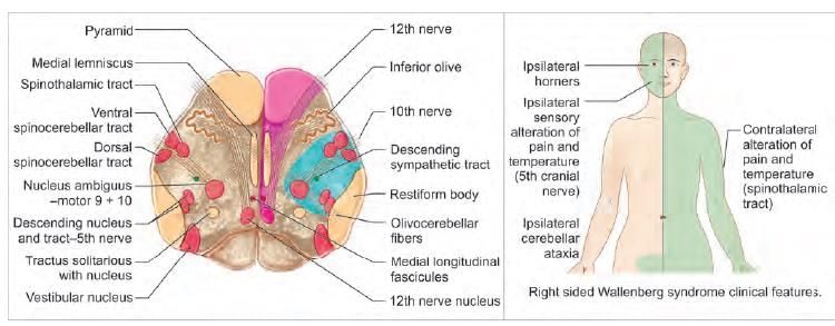
A patient presented with clinical features of ataxia and incoordination. It is most likely due to involvement of which artery among the following? (NEET-PG 2020)

Detailed Solution for Test: Anatomy - 3 - Question 4

Clinical characteristics of ataxia and in-coordination are indicative of cerebellar pathology, which may occur if the superior cerebellar artery is affected. Involvement of the cerebral arteries (anterior, middle, or posterior) typically does not manifest with signs and symptoms associated with the cerebellum.

Test: Anatomy - 3 - Question 5

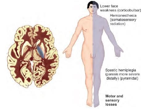
A patient presented with weakness of the right side of the face. There is associated ipsilateral loss of pain and temperature on face and contralateral body. The lesion is most likely located at: (NEET-PG 2020)

Detailed Solution for Test: Anatomy - 3 - Question 5

This case presents right lateral inferior pontine syndrome, which may have arisen from a condition affecting the right anterior inferior cerebellar artery.

  • Weakness on the right side of the face (lower motor neurone lesion of the facial nerve).
  • Loss of pain and temperature sensation on the same side of the face (involvement of the spinal sensory nucleus of the trigeminal).
  • Contralateral loss of pain and temperature sensation in the body (due to involvement of the crossed lateral spinothalamic tract).

Lesions in the medial pons affect the abducent nerve, which can cause squinting, and the dorsal column–medial lemniscal pathway, leading to contralateral loss of sensations such as pressure, touch, and vibration (not applicable in this case). Lesions in the medulla affect the hypoglossal nerve (causing tongue deviation) but do not lead to facial weakness. Lateral Inferior Pontine Syndrome (AICA occlusion):

  • Relevant cerebral artery: Associated neurologic deficit
  • Lateral spinothalamic tract: Loss of contralateral pain and body temperature sensations
  • CN VIII nuclei: Vertigo, hearing loss, tinnitus, nystagmus
  • CN VII: Bell's palsy without forehead sparing

Relevant cerebral artery:

  • Middle cerebellar peduncle: Ipsilateral ataxia
  • Spinal trigeminal nucleus/tract: Ipsilateral loss of pain/temperature sensation (face)
  • Descending sympathetic: Ipsilateral Horner syndrome (AICA: anterior inferior cerebellar artery)
Test: Anatomy - 3 - Question 6

A patient presents with sudden onset of dysarthria, sensory loss of right side of face, right upper limb ataxia, tendency to fall on the right side. There is sensory loss on left side of body (with no motor symptoms). Which artery is involved? (AIIMS June 2020)

Detailed Solution for Test: Anatomy - 3 - Question 6

This condition is an instance of Wallenberg syndrome, which may arise from a blockage in the ipsilateral posterior inferior cerebellar artery, or more commonly, the ipsilateral vertebral artery. Wallenberg (Lateral medullary) syndrome is caused by the occlusion of the vertebral artery exceeding that of the posterior inferior cerebellar artery.

  • Ipsilateral signs and symptoms (structures involved):
    • Loss of pain and temperature sensation on one side of the face (Descending tract and nucleus of the trigeminal nerve).
    • Cerebellar ataxia of the limbs, causing falls towards the side of the lesion (Cuneocerebellar and Spinocerebellar tract).
    • Nystagmus, vertigo, and nausea (Vestibular nucleus).
    • Horner’s syndrome—miosis, ptosis, and anhidrosis (Descending sympathetic tract).
    • Paralysis of the palate, pharynx, and larynx muscles, leading to difficulties in speech and swallowing (Nucleus ambiguus).
    • Loss of taste on one side of the tongue (Nucleus tractus solitarius).
  • Contralateral signs and symptoms (structure involved):
    • Loss of pain and temperature sensation on one side of the body (Lateral spinothalamic tract).

Ataxia refers to a deficiency in muscle control or coordination of voluntary movements, such as walking or grasping objects.

Test: Anatomy - 3 - Question 7

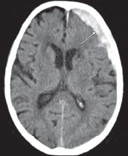
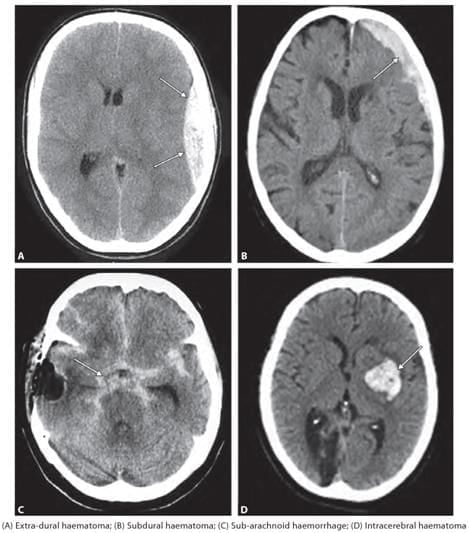
A 55-year-old patient presented with headache and confusion. There is a history of fall one week back. He also has personal history of chronic alcoholism. NCCT findings as shown below, point towards: (NEET-PG 2023)

Detailed Solution for Test: Anatomy - 3 - Question 7

The medical history of an older patient (experiencing brain atrophy) with chronic alcoholism, who does not present immediately in emergency, suggests a potential case of subdural haematoma (bleeding of bridging veins).

  • The NCCT findings should display a concavo-convex shape, with the bleeding not limited by the suture lines.

Extradural haematoma: Symptoms typically manifest immediately (within minutes to hours; not weeks), accompanied by a rapid decline in consciousness due to it being an arterial bleed that leads to a swift increase in intracranial pressure.

  • The NCCT reveals a hyperdense (whitish) biconvex bleeding pattern that does not cross the suture lines.

Subarachnoid haemorrhage: This may arise from the rupture of an aneurysm (for instance, a berry or saccular aneurysm), presenting acutely and rapidly, often described as ‘the worst headache of my life’, along with a related finding of blood in a lumbar puncture.

  • The CT scan indicates signs of acute (hyperdense/whitish) bleeding in the subarachnoid cisterns or ventricles.

Intracerebral haemorrhage: These bleeds might result from systemic hypertension, leading to the rupture of Charcot-Bouchard microaneurysms, typically found in the lenticulostriate branches that supply the basal ganglia (putamen, globus pallidus).

Test: Anatomy - 3 - Question 8
Which of the following is most likely seen due to rupture of saccular aneurysm in brain? (NEET-PG 2022)
Detailed Solution for Test: Anatomy - 3 - Question 8

Subarachnoid haemorrhage can arise from the rupture of an aneurysm, such as a berry or saccular aneurysm. These berry aneurysms are typically found in the anterior section of the circle of Willis, particularly at the junction of the anterior communicating artery. As the circle of Willis and its branches are located within the subarachnoid space, any bleeding collects in this area, resulting in subarachnoid haemorrhage. The symptoms present acutely, often in a younger patient who describes experiencing the “worst headache of my life.” A CT scan reveals evidence of acute (hyperdense or whitish) bleeding in the subarachnoid cisterns or ventricles, along with a corresponding finding of blood in a lumbar puncture.

  • Epidural/Extradural haematoma: This typically involves an arterial bleed from the anterior or frontal division of the middle meningeal artery, which ruptures beneath a vulnerable area of the skull known as the pterion. Due to the nature of the arterial bleed, most cases present immediately in an emergency setting.
  • Subdural haematoma: This may involve a classic history of an elderly individual (with brain atrophy) who has chronic alcoholism, not presenting immediately in an emergency. This suggests a case of subdural haematoma, which results from bleeding of bridging veins, such as the superior cerebral veins.
  • Intracerebral haemorrhage: These bleeds can occur due to systemic hypertension, often resulting from the rupture of Charcot-Bouchard microaneurysms, typically found in the lenticulostriate branches of the middle cerebral artery, which supply the basal ganglia region (including the putamen and globus pallidus).
Test: Anatomy - 3 - Question 9

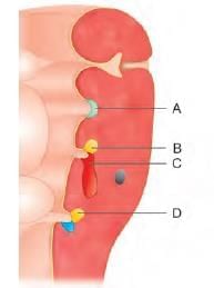
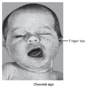
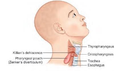
A newborn baby has presented with a pharyngeal pouch developmental anomaly. Facial twitching observed is due to involvement of which of the following marked region? (INI-CET July 2021)

Detailed Solution for Test: Anatomy - 3 - Question 9

The infant seems to have DiGeorge syndrome, which may arise from irregular development of the third and fourth pharyngeal pouches. The accompanying facial twitching, triggered by the Chvostek sign, could be a result of hypocalcaemia due to the abnormal formation of the inferior parathyroid gland (Marker B).

  • It is important to note that while DiGeorge syndrome involves both pharyngeal pouches 3 and 4, pouch 3 is referenced more frequently, as the thymus originates from it. Thymic aplasia is a characteristic feature of the syndrome.
  • Marker A: Pharyngeal pouch 2 (Derivative: Tonsillar epithelium).
  • Marker C: Pharyngeal pouch 3; Ventral portion (Derivative: cytoreticular cells of Thymus).
  • Marker D: Pharyngeal pouch 4; Derivative: Superior parathyroid.

DiGeorge syndrome results from abnormal migration of neural crest cells and is characterised by hypoparathyroidism, the absence of the thymus gland (leading to T-cell abnormalities), and cardiac anomalies involving the aorta and pulmonary septum.

Test: Anatomy - 3 - Question 10

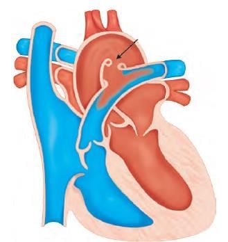
Congenital anomaly shown in the following diagram relates to which of the following pharyngeal arch artery? (NEET-PG 2023)

Detailed Solution for Test: Anatomy - 3 - Question 10

The congenital defect illustrated in the diagram is patent ductus arteriosus, which arises from the persistence of the ductus arteriosus, a remnant of the left 6th pharyngeal arch artery.

  • Chvostek sign.
  • Aortic (Pharyngeal) arch arteries give rise to the arteries that supply the head and neck, as well as the proximal arteries of the heart.
  • The ductus arteriosus experiences physiological closure (vasospasm) within 24–96 hours and anatomical closure (tunica intima proliferation) within 1–3 months.

It is important to note that normally, PGE (Prostaglandin E) maintains the patency of the ductus arteriosus during fetal circulation, and can also be beneficial post-birth in specific situations. Conversely, medications such as indomethacin, an inhibitor of prostaglandins, result in the closure of its lumen.

Test: Anatomy - 3 - Question 11

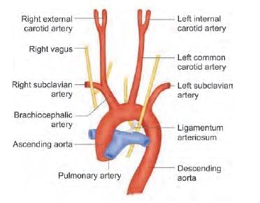
Left recurrent laryngeal nerve has a comparatively longer course due to the persistence of which pharyngeal arch artery? (NEET-PG 2020)

Detailed Solution for Test: Anatomy - 3 - Question 11

The left recurrent laryngeal nerve follows a relatively longer pathway because of the persistence of the ligamentum arteriosum, which is a vestigial remnant of the sixth pharyngeal arch artery on the left side. Initially, these nerves, which are branches of the vagus nerve, innervate the sixth pharyngeal arches. As the heart descends, they loop around the sixth aortic arches and then ascend again towards the larynx, which explains their recurrent trajectory (and name).

  • On the right side, the recurrent laryngeal nerve moves upwards and hooks around the right subclavian artery after the distal part of the sixth aortic arch and the fifth aortic arch regress.
  • Conversely, on the left side, the nerve does not ascend because the distal segment of the sixth aortic arch remains as the ductus arteriosus, which subsequently develops into the ligamentum arteriosum.

Test: Anatomy - 3 - Question 12

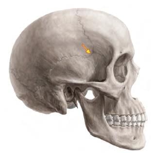
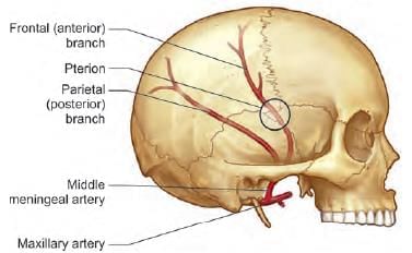
A fracture at the marked arrow (pterion) leads to bleeding of which artery? (AIIMS June 2020)

Detailed Solution for Test: Anatomy - 3 - Question 12

The highlighted area is the pterion, beneath which lies the anterior division of the middle meningeal artery, which may bleed if a fracture occurs at this location. The pterion marks the junction of four bones: the frontal, parietal, and temporal bones, along with the greater wing of the sphenoid bone. The central point of the pterion is referred to as the Sylvian point, where the cranium is notably thin.

  • Its immediate deep relationships include the frontal (anterior) branch of the middle meningeal artery, its accompanying vein, and the stem of the lateral (Sylvian) sulcus of the cerebrum.
  • The lateral sulcus extends posteroinferiorly from the Sylvian point, situated near the region of the pterion.

A fracture in this area can damage the middle meningeal artery, potentially leading to extradural haemorrhage. The pterion is crucial for the precise placement of burr holes for evacuating extradural haematomas. In neurosurgery, the pterional craniotomy is a frequently used technique to access the middle cranial fossa. Additionally, the pterion is the location of the anterolateral fontanelle, which closes at around six months after birth.

Test: Anatomy - 3 - Question 13

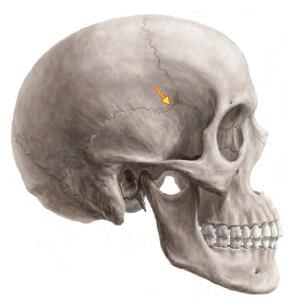
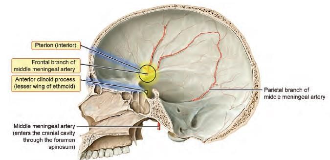
Which structure is deeply related to the arrow marked area? (INI-CET Nov 2020)

Detailed Solution for Test: Anatomy - 3 - Question 13

The highlighted area indicates the ‘H’ shaped suture – pterion, which serves as a significant anatomical reference.

  • Surgeons utilise this approach for planning anterior clinoidectomy.
  • As the anterior clinoid process forms part of the lesser wing of the sphenoid, it justifies the procedure.

Note: The anterior (frontal) branch of the middle meningeal artery is situated deep to the pterion.

  • This artery is susceptible to bleeding in the event of a skull fracture, which can result in a life-threatening extra-dural haematoma.

The anterior clinoid process (ACP) represents the apex of the lesser wing of the sphenoid bone.

  • Its anatomical positioning is crucial due to its proximity to surrounding structures such as the optic nerves, internal carotid artery (ICA), and other neurovascular components.

Anterior clinoidectomy (AC) is a vital technique that plays a key role in accessing lesions located at the central skull base, for instance, during the clipping of aneurysms in the para-clinoid region.

Test: Anatomy - 3 - Question 14

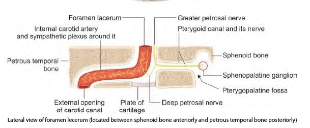
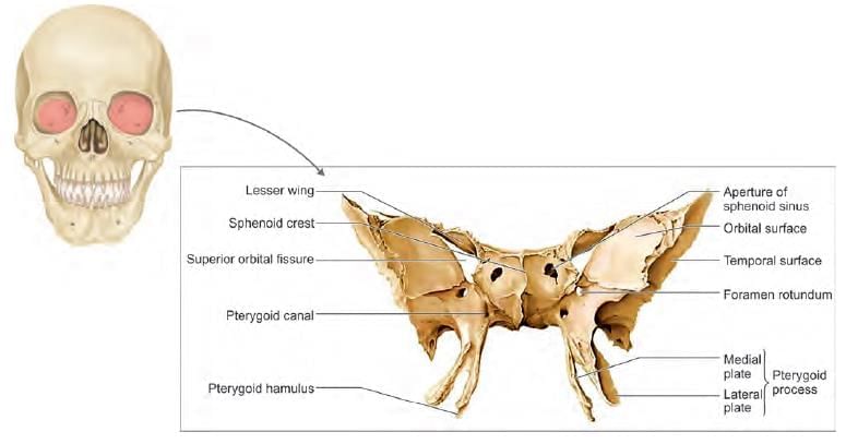
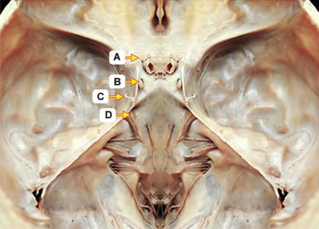
Which of the following foramen is located between three bones? (INI-CET May 2022)

Detailed Solution for Test: Anatomy - 3 - Question 14

Foramen lacerum is an opening found in the floor of the middle cranial fossa (at the base of the skull) where three bones meet:

  • Sphenoid bone (anteriorly)
  • Apex of the petrous temporal bone (postero-lateral)
  • Basilar part of the occipital bone (postero-medial)

Foramen ovale is located laterally in the greater wing of the sphenoid. The foramen lacerum is closed below by a fibrocartilaginous plate. The structures that traverse the floor of foramen lacerum include the internal carotid artery, which is surrounded by a sympathetic plexus. The greater petrosal nerve unites with the deep petrosal nerve to create the Vidian nerve of the pterygoid canal. The foramen lacerum links the extracranial pterygoid plexus with the intracranial cavernous sinus, providing a pathway for the spread of infections. It also serves as an entry point into the cranium for tumours, such as nasopharyngeal carcinoma and juvenile angiofibroma. Foramen rotundum is a circular opening in the greater wing of the sphenoid bone, situated in the floor of the middle cranial fossa. It connects the middle cranial fossa to the pterygopalatine fossa, allowing the maxillary nerve to pass through. The jugular foramen is located between two bones: the occipital bone and the petrous temporal bone. This foramen allows the passage of cranial nerves IX, X, and XI, along with the internal jugular vein and its first tributary, the inferior petrosal sinus. Foramen magnum is a large opening in the occipital bone through which the lower part of the brainstem (medulla oblongata) passes, along with the spinal part of the accessory nerve and the vertebral arteries.

Test: Anatomy - 3 - Question 15

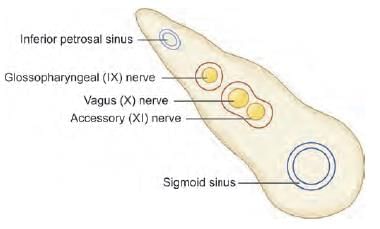
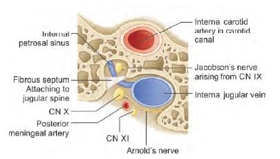
Which of the following structure passing through jugular foramen has separate canal? (INI-CET May 2022)

Detailed Solution for Test: Anatomy - 3 - Question 15

CN IX (the glossopharyngeal nerve) traverses the smaller anteromedial section of the jugular foramen, while the larger posterolateral section accommodates CN X and XI. CN IX is enveloped by a distinct sheath of the dura mater, whereas CN X and XI share a common dura mater sheath. This categorisation is based on an alternative imaging-based subclassification, defined by the jugular spine, a bony ridge that partially divides the jugular foramen into two sections, as previously noted. Details:

  • The jugular foramen is located at the base of the posterior cranial fossa, situated between the petrous temporal bone antero-laterally and the occipital bone postero-medially.
  • It is typically divided into three compartments, each containing different structures:
    • Anterior compartment: Inferior petrosal sinus.
    • Intermediate compartment: CN IX, X, and XI.
    • Posterior compartment: Sigmoid sinus (which becomes the internal jugular vein), meningeal branches from the occipital artery, and ascending pharyngeal arteries.

An alternative imaging-based subclassification also exists, which divides the jugular foramen into two sections, as follows:

  • The smaller, anteromedial, ‘pars nervosa’ compartment contains CN IX (the tympanic nerve, a branch of CN IX) and receives venous drainage from the inferior petrosal sinus.
  • The larger, posterolateral, ‘pars vascularis’ compartment includes CN X, CN XI, Arnold’s nerve (the auricular branch of CN X involved in the Arnold’s reflex, where stimulation of the external auditory meatus induces coughing), the jugular bulb, and the posterior meningeal branch of the ascending pharyngeal artery.

Clinical anatomy:

  • Jugular foramen syndrome may arise from certain enlargements (e.g., glomus tumour) or trauma to the skull base.
  • This could lead to haemorrhage or air embolism due to injury to the jugular vein.
  • Furthermore, it might affect the three cranial nerves IX, X, or XI, resulting in associated complications.
  • Damage to cranial nerve IX may lead to loss of taste and general sensation in the posterior one-third of the tongue and the surrounding area of the oro-pharyngeal isthmus on the ipsilateral side.
  • Injury to cranial nerve X can cause difficulties in speech and swallowing.
  • Impairment of cranial nerve XI, which affects the sternocleidomastoid and trapezius muscles, may result in shoulder droop and difficulty turning the head towards the opposite side.
Test: Anatomy - 3 - Question 16

The arrow marked foramen is: (INI-CET Nov 2020)

Detailed Solution for Test: Anatomy - 3 - Question 16

The bone shown in the illustration is a frontal (anterior) view of the sphenoid bone, with a marker positioned at the foramen rotundum. The maxillary nerve travels through the foramen rotundum anteriorly to access the pterygopalatine fossa. Note that there is another opening visible infero-medial to the marker, which is the Vidian (pterygoid) canal. The Vidian nerve from the pterygoid canal carries the combined axons of the greater petrosal nerve and the deep petrosal nerve. These axons, formed at the foramen lacerum, proceed anteriorly through the Vidian canal to reach the pterygopalatine fossa.

The sphenoid bone is characterised by its butterfly-like shape, comprising:

  • A central body, which contains pneumatic sphenoid air sinuses.
  • Two lesser wings, which are relatively smaller and extend supero-laterally from the body.
  • Two greater wings, which are comparatively larger and extend laterally from the body.
  • Two legs (pterygoid processes) with pterygoid plates that project inferiorly from the body.

The superior orbital fissure is located between the lesser wings (superiorly) and the greater wings (infero-laterally). A neurovascular bundle passes through this fissure, including cranial nerves III, IV, VI, and branches of the ophthalmic nerve (a division of the trigeminal nerve). The pterygoid muscles (medial and lateral) originate from the lateral pterygoid plate.

*Multiple options can be correct
Test: Anatomy - 3 - Question 17

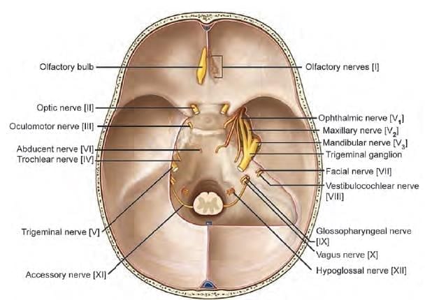
Which of the following marked nerves are associated with ocular movements? (INI-CET Nov 2020)

Detailed Solution for Test: Anatomy - 3 - Question 17

The provided diagram illustrates the base of the cranial fossae, highlighting the cranial nerves that emerge from different foramina. The movements of the eyes are primarily controlled by:

  • Cranial nerve 3 – the oculomotor nerve (Marker B)
  • Cranial nerve 4 – the trochlear nerve (Marker C)
  • Cranial nerve 6 – the abducent nerve (Marker D)

Marker A denotes the optic nerve, while Marker D represents the trigeminal nerve.

Test: Anatomy - 3 - Question 18

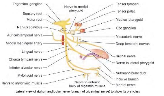
Which of the following is a CORRECT statement regarding mandibular nerve? (INI-CET Nov 2022)

Detailed Solution for Test: Anatomy - 3 - Question 18

The mandibular nerve, which is the primary trunk, supplies the medial pterygoid muscle. The branches from the posterior division are primarily motor and partially sensory. The buccal nerve originates from the anterior division. The auriculotemporal nerve is a branch of the posterior division. Details are as follows:

  • The mandibular nerve is associated with the first pharyngeal arch.
  • It has a main trunk that splits into two divisions: anterior and posterior.
  • The anterior division of the mandibular nerve is mainly motor, supplying the muscles of mastication that develop from the first pharyngeal arch, and is partly sensory.
  • A sensory branch, the buccal nerve, transmits general sensations from the oral mucosa.
  • The posterior division of the mandibular nerve is predominantly sensory and partly motor.

Branches (ALI):

  • A – Auriculotemporal nerve – transmits sensations from the auriculo-temporal region.
  • L – Lingual nerve – conveys sensations from the anterior two-thirds of the tongue (developing from the first pharyngeal arch).
  • I – Inferior alveolar nerve – carries sensations from the lower teeth.

It also supplies two muscles of mastication (developing from the first pharyngeal arch): the anterior belly of the digastric and the mylohyoid.

Test: Anatomy - 3 - Question 19

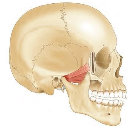
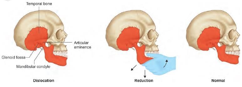
A child has presented with stuck open mouth, which resulted during yawning in the class, as per the given history. Which of the following muscle, attached to articular disc is under spasmodic contraction? (NEET-PG 2023)

Detailed Solution for Test: Anatomy - 3 - Question 19

Lateral pterygoid attaches to the pterygoid fovea on the neck of the mandible and can cause dislocation of the mandible during excessive yawning.

  • Dislocation of the mandible occurs when the mouth is open. In this state, the mandibular condyles advance and rest beneath the articular eminences.
  • This position represents the most unstable configuration of the temporomandibular joint.
  • If the mouth opens excessively, as happens during yawning, a sudden involuntary spasm of the lateral pterygoid muscles may occur.
  • This can lead to the head of the mandible on one or both sides slipping forwards and becoming locked in the infratemporal fossa, resulting in an inability to close the mouth – it remains stuck open.

To correct the dislocation, the condyle needs to be lowered and pushed back behind the peak of the articular eminence into the articular fossa. This is achieved by pressing down on the jaw with the thumb positioned on the last molar teeth while simultaneously elevating the chin.

The lateral pterygoid muscle inserts into the pterygoid fovea on the mandibular neck and can, during excessive yawning, pull the mandibular head out of its socket, leading to dislocation.

Test: Anatomy - 3 - Question 20

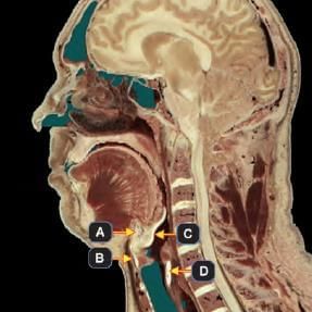
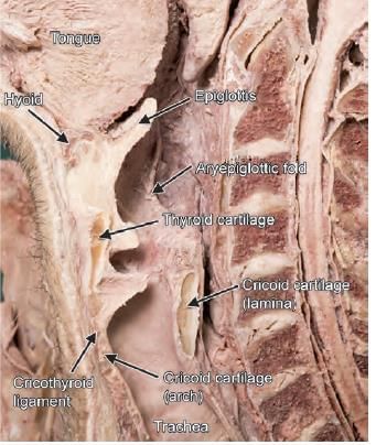
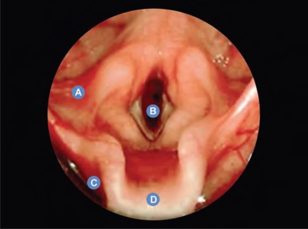
Which of the following marked structures does NOT constitute skeletal framework of larynx? (INI-CET Nov 2020)

Detailed Solution for Test: Anatomy - 3 - Question 20

Marker ‘A’ indicates the hyoid bone, which does not form part of the laryngeal skeleton. Marker B refers to the thyroid cartilage. Marker C denotes the epiglottis, a leaf-shaped cartilage that acts as a lid over the larynx. Marker D represents the cricoid cartilage (lamina). The laryngeal skeleton comprises nine cartilages:

  • Unpaired (midline): thyroid cartilage, cricoid cartilage, epiglottis.
  • Paired: arytenoid, corniculate, and cuneiform.

The hyoid bone is not included in the larynx, although the larynx is suspended from the hyoid.

Test: Anatomy - 3 - Question 21

A 70-year-old patient presented with history of fever, halitosis, night cough and regurgitation of food in the mouth. On examination there is a pulsatile swelling on left side of neck which produces gurgling sound on compression. What is the most likely diagnosis, by looking at the given barium swallow study of the patient? (NEET-PG 2020)

Detailed Solution for Test: Anatomy - 3 - Question 21

Zenker’s diverticulum is a false diverticulum located in the pharyngoesophageal region. This condition arises due to esophageal dysmotility, which leads to the herniation of mucosal tissue at the Killian triangle, situated between the thyropharyngeal and cricopharyngeal segments of the inferior pharyngeal constrictor. The symptoms typically presented include:

  • dysphagia
  • obstruction
  • gurgling sounds
  • aspiration
  • foul breath (halitosis)
  • neck mass

It is most frequently observed in elderly males.

Test: Anatomy - 3 - Question 22

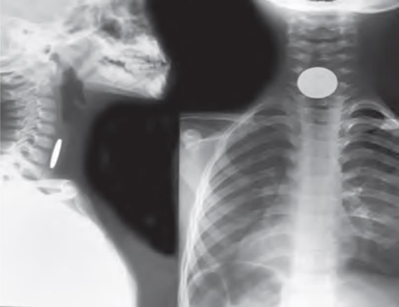
A 9-year-old male baby was brought to emergency by the mother with difficulty in swallowing for past few hours and a history of playing unsupervised. X-ray was taken. Identify the location of foreign body. (NEET-PG 2021)

Detailed Solution for Test: Anatomy - 3 - Question 22

The foreign object seems to be a coin, located just anterior to the vertebrae (where the oesophagus is situated). Additionally, there is a tracheal air shadow noted in front of the coin. This indicates that it is a coin lodged in the oesophagus. This is further supported by the patient presenting somewhat late with a complaint of difficulty in swallowing (indicating a foreign body in the gut tube) rather than in an acute emergency due to difficulty in breathing (suggesting a foreign body in the respiratory tube).

Test: Anatomy - 3 - Question 23
Identify pyriform fossa: (AIIMS June 2020)
Detailed Solution for Test: Anatomy - 3 - Question 23
The pyriform sinus (recess/fossa) is a pear-shaped area of the hypopharynx situated posterolaterally on either side of the laryngeal opening.
  • Applied anatomy: Occasionally, a fish bone may become lodged in the piriform fossa.
  • During removal, there is a risk of damaging the internal laryngeal nerve at the base, which can lead to a loss of the protective cough reflex and possible aspirations.
  • A malignant tumour in the laryngopharynx (hypopharynx) can develop within the space of the piriform fossa without causing symptoms until the patient shows signs of metastatic lymphadenopathy.
The piriform fossa is also referred to as the smuggler’s fossa, as it can be used to conceal valuable items such as diamonds.
Test: Anatomy - 3 - Question 24

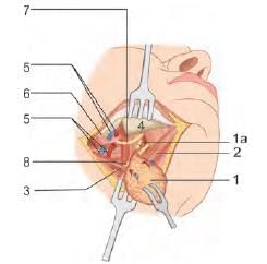
A patient has undergone submandibular gland excision and the Wharton’s duct was ligated in the process. Which of the following nerve is most likely to be damaged during the process of ligating the duct? (NEET-PG 2021)

Detailed Solution for Test: Anatomy - 3 - Question 24

The lingual nerve is located beneath the duct of the submandibular gland, and any surgical manipulation of this duct could potentially harm the nerve, as evidenced in the current case. The inferior alveolar nerve, which originates from the posterior division of the trigeminal nerve, is safeguarded within the mandible and provides sensation to the lower teeth.

  • The hypoglossal nerve may sustain damage during the excision of the submandibular gland, though this is less frequent.
  • The marginal mandibular branch of the facial nerve is the 'most common' nerve injured during the excision of the submandibular gland; however, this was not considered the answer due to the mention of a nerve injury associated with the surgical manipulation of the submandibular gland duct.

Surgical removal of the submandibular gland involves:

  1. Submandibular gland
    1a. Submandibular duct, ligated
  2. Anterior belly of the digastric muscle
  3. Stylohyoid muscle, concealed by the posterior belly of the digastric muscle
  4. Horizontal ramus of the mandible
  5. Facial artery and vein, ligated
  6. Marginal mandibular branch of the facial nerve (essential to preserve for lower lip muscle function)
  7. Lingual nerve
  8. Hypoglossal nerve
Test: Anatomy - 3 - Question 25

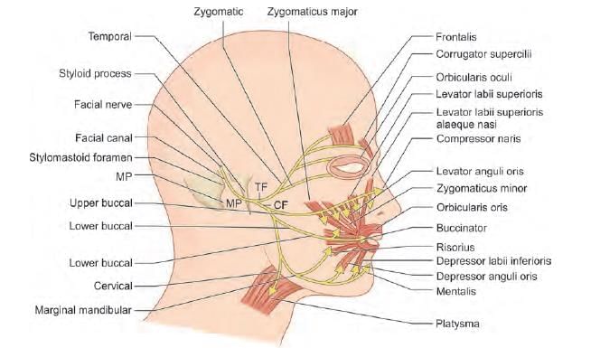
A patient on awakening after parotid surgery, noticed weakness of lower lip movement. Which of the following nerve is most likely injured? (AIIMS June 2020)

Detailed Solution for Test: Anatomy - 3 - Question 25

The cervical branch of the facial nerve supplies the platysma, which assists in the downward movement of the lip (such as in a horror expression). If this branch is damaged, the patient may experience weakness in the lower lip.

  • It is important to note that the muscles of the lower lip are primarily governed by the marginal mandibular nerve.
  • However, since this option is unavailable, we have selected the next most suitable answer.

Information about Test: Anatomy - 3 Page
In this test you can find the Exam questions for Test: Anatomy - 3 solved & explained in the simplest way possible. Besides giving Questions and answers for Test: Anatomy - 3, EduRev gives you an ample number of Online tests for practice
Download as PDF