NEET PG Exam  >  NEET PG Tests  >  Test: Anatomy and Physiology of Skin - NEET PG MCQ

Test: Anatomy and Physiology of Skin - NEET PG MCQ


Test Description

15 Questions MCQ Test - Test: Anatomy and Physiology of Skin

Test: Anatomy and Physiology of Skin for NEET PG 2025 is part of NEET PG preparation. The Test: Anatomy and Physiology of Skin questions and answers have been prepared according to the NEET PG exam syllabus.The Test: Anatomy and Physiology of Skin MCQs are made for NEET PG 2025 Exam. Find important definitions, questions, notes, meanings, examples, exercises, MCQs and online tests for Test: Anatomy and Physiology of Skin below.
Solutions of Test: Anatomy and Physiology of Skin questions in English are available as part of our course for NEET PG & Test: Anatomy and Physiology of Skin solutions in Hindi for NEET PG course. Download more important topics, notes, lectures and mock test series for NEET PG Exam by signing up for free. Attempt Test: Anatomy and Physiology of Skin | 15 questions in 20 minutes | Mock test for NEET PG preparation | Free important questions MCQ to study for NEET PG Exam | Download free PDF with solutions
Test: Anatomy and Physiology of Skin - Question 1

Desmosomes are helpful in connecting: 

Test: Anatomy and Physiology of Skin - Question 2

Spongiosis involves:

Test: Anatomy and Physiology of Skin - Question 3

In which of the following conditions does parakeratosis most frequently occur?

Test: Anatomy and Physiology of Skin - Question 4

Sweat glands of palm can be differentiated from others by the following:

Test: Anatomy and Physiology of Skin - Question 5

Acantholysis is seen in all except:

Detailed Solution for Test: Anatomy and Physiology of Skin - Question 5

Bullous pemphigoid is a chronic autoimmune skin condition characterised by:

  • Large, fluid-filled blisters on the skin, primarily affecting older adults.
  • Itching and discomfort, often preceding the appearance of blisters.
  • Blisters that tend to form on areas such as the abdomen, groin, and armpits.
  • The immune system mistakenly attacks the skin’s basement membrane, leading to separation of the epidermis from the dermis.

Management typically includes:

  • Topical or systemic corticosteroids to reduce inflammation.
  • Immunosuppressive agents in more severe cases.
  • Regular monitoring and supportive care to manage symptoms and prevent complications.
Test: Anatomy and Physiology of Skin - Question 6
Acantholysis is seen in:
Detailed Solution for Test: Anatomy and Physiology of Skin - Question 6

The epidermis is the outermost layer of skin, serving as a protective barrier for the body. It plays a crucial role in safeguarding against environmental factors such as pathogens and harmful substances. Additionally, the epidermis is responsible for regulating moisture and provides a surface for the synthesis of vitamin D when exposed to sunlight. This layer is primarily composed of keratinocytes, which produce keratin, a protein that helps in maintaining skin integrity and resilience.

Test: Anatomy and Physiology of Skin - Question 7

Acantholysis is due to destruction of: 

*Multiple options can be correct
Test: Anatomy and Physiology of Skin - Question 8

True about stratum lucidum: 

Detailed Solution for Test: Anatomy and Physiology of Skin - Question 8

Situated between the stratum corneum and the stratum granulosum, it comprises degenerated cells.

Test: Anatomy and Physiology of Skin - Question 9

All statements are true regarding skin except: 

Test: Anatomy and Physiology of Skin - Question 10

Which layer of epidermis is under­ developed in VLBW infants in the initial 7 days?

Detailed Solution for Test: Anatomy and Physiology of Skin - Question 10

The stratum corneum is the outermost layer of the skin, playing a crucial role in protecting the underlying tissues. It consists of dead skin cells that have undergone a process known as keratinisation. This layer serves several important functions:

  • It acts as a barrier, preventing the entry of pathogens and harmful substances.
  • It helps to retain moisture, thus maintaining skin hydration.
  • It contributes to the skin's overall appearance and texture.

Overall, the stratum corneum is essential for skin health and integrity.

Test: Anatomy and Physiology of Skin - Question 11

Normal turnover time of epidermis is:

Test: Anatomy and Physiology of Skin - Question 12

Melanocytes are present in:

Detailed Solution for Test: Anatomy and Physiology of Skin - Question 12

The stratum basale is the deepest layer of the epidermis. It plays a crucial role in the regeneration of skin cells by undergoing continuous division. This layer contains:

  • Keratinocytes, which are responsible for producing keratin, a protein that helps protect the skin.
  • Melanocytes that produce melanin, the pigment that gives skin its colour.
  • Merkel cells, which are involved in sensation.

As new cells are generated, they move upwards through the layers of the epidermis, eventually reaching the surface and shedding off. This process is vital for maintaining healthy skin.

Test: Anatomy and Physiology of Skin - Question 13

Layer of skin with maximum desmosome interconnection is depicted by which arrow?
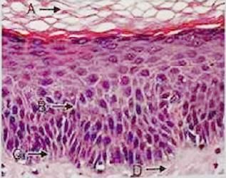
Detailed Solution for Test: Anatomy and Physiology of Skin - Question 13

The letter B indicates the spinous layer, which features the most notable desmosomes.

*Multiple options can be correct
Test: Anatomy and Physiology of Skin - Question 14

Desmosomes are associated with: 

Detailed Solution for Test: Anatomy and Physiology of Skin - Question 14

Pemphigus is traditionally linked to autoimmune desmosomal dysfunction. Desmosomes are also crucial for mechanical intercellular communication through cardiac coupling, which strengthens cardiomyocytes. These structures are vital for maintaining proper cardiac function.

Test: Anatomy and Physiology of Skin - Question 15

Meissner's corpuscles are found in: 

Detailed Solution for Test: Anatomy and Physiology of Skin - Question 15

The papillary dermis is a layer of skin located just beneath the epidermis. It is composed of loose connective tissue, which provides structural support and elasticity. This layer contains a rich supply of blood vessels, lymphatics, and nerve endings, which play a crucial role in thermoregulation and sensation.

  • The papillary dermis is primarily made up of thin collagen and elastin fibres.
  • It houses dermal papillae, small, nipple-like projections that interdigitate with the epidermis.
  • These projections increase the surface area for exchange of nutrients and waste products.
  • Additionally, they contribute to the unique patterns of fingerprints.
Information about Test: Anatomy and Physiology of Skin Page
In this test you can find the Exam questions for Test: Anatomy and Physiology of Skin solved & explained in the simplest way possible. Besides giving Questions and answers for Test: Anatomy and Physiology of Skin , EduRev gives you an ample number of Online tests for practice
Download as PDF