NEET PG Exam  >  NEET PG Tests  >  Test: Anatomy - 4 - NEET PG MCQ

Test: Anatomy - 4 - NEET PG MCQ


Test Description

25 Questions MCQ Test - Test: Anatomy - 4

Test: Anatomy - 4 for NEET PG 2025 is part of NEET PG preparation. The Test: Anatomy - 4 questions and answers have been prepared according to the NEET PG exam syllabus.The Test: Anatomy - 4 MCQs are made for NEET PG 2025 Exam. Find important definitions, questions, notes, meanings, examples, exercises, MCQs and online tests for Test: Anatomy - 4 below.
Solutions of Test: Anatomy - 4 questions in English are available as part of our course for NEET PG & Test: Anatomy - 4 solutions in Hindi for NEET PG course. Download more important topics, notes, lectures and mock test series for NEET PG Exam by signing up for free. Attempt Test: Anatomy - 4 | 25 questions in 25 minutes | Mock test for NEET PG preparation | Free important questions MCQ to study for NEET PG Exam | Download free PDF with solutions
Test: Anatomy - 4 - Question 1

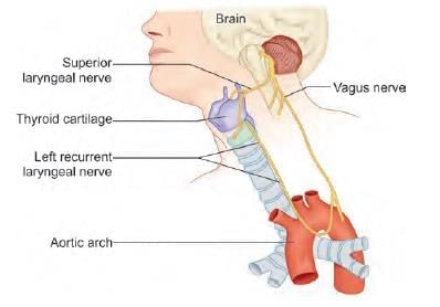
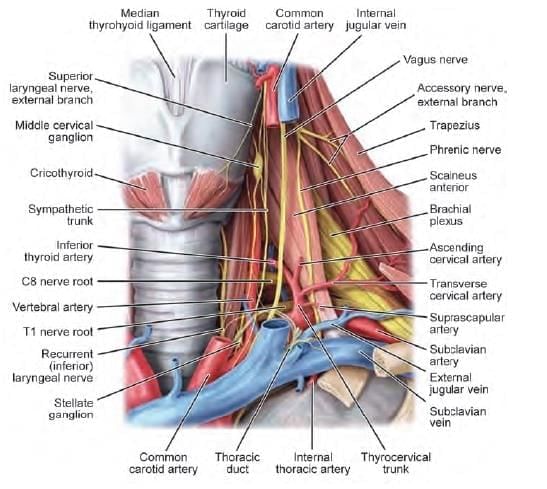
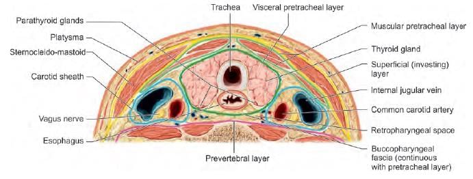
Which of the following nerve is least injured in thyroid surgery? (AIIMS June 2020)

Detailed Solution for Test: Anatomy - 4 - Question 1

The marginal mandibular nerve does not enter the operative field during thyroid surgery. The external laryngeal nerve, a branch of the superior laryngeal nerve from the vagus, is susceptible to injury during this procedure. Damage to this nerve results in the inability to lengthen a vocal fold, consequently leading to a failure to produce high-pitched sounds. This can be detrimental to singers or individuals whose professions depend on their vocal abilities.

The recurrent laryngeal nerve may also be at risk if proper precautions are not observed. During laryngoscopy, one may observe:

  • Posterior glottic rotation towards the affected side
  • Bowing of the vocal fold on the weaker side

In cases of unilateral vocal cord paralysis, the surgical options include:

  • Medialization (the most common procedure)
  • Reinnervation

For bilateral vocal cord paralysis, an emergency tracheotomy may be necessary. However, if feasible, it is advisable to first conduct endotracheal intubation. Procedures such as cordotomy and arytenoidectomy, commonly performed, help to enlarge the airway and may allow for the decannulation of a tracheostomy. Additionally, the ansa cervicalis may sustain damage while operating on the strap muscles, which are sometimes divided to gain access to the thyroid.

Test: Anatomy - 4 - Question 2

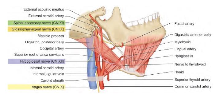
Patient undergoing surgery at the lateral part of skull. Postoperatively patient had aspirations without voice change. Nerve lesioned is: (AIIMS Nov 2019)

Detailed Solution for Test: Anatomy - 4 - Question 2

Injuries to the glossopharyngeal nerve can occur during surgical procedures in the lateral region of the neck, such as when dissecting the internal carotid artery above the hypoglossal nerve (carotid endarterectomy). The effects of glossopharyngeal nerve injury can vary significantly:

  • They may include mild dysphagia.
  • In more severe cases, they can lead to recurrent aspiration and respiratory failure.

It is important to note that damage to the vagus nerve or its branches typically manifests as changes in voice.

Test: Anatomy - 4 - Question 3

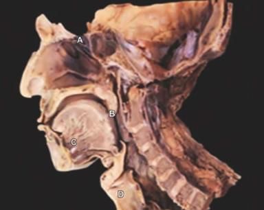
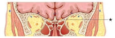
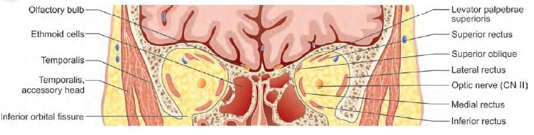
A patient was admitted with skull base trauma and difficulty in swallowing. The doctor was touching the marked structure. Which of the following cranial nerve is being tested? (NEET-PG 2021)

Detailed Solution for Test: Anatomy - 4 - Question 3

The dissected specimen is indicated with an arrow at the uvula (soft palate), which is stimulated to trigger the ‘gag reflex’. The glossopharyngeal nerve serves as the sensory component, which is the reason for this answer. Note: The gag reflex is typically assessed by stimulating the posterior pharyngeal wall, but it can also be provoked by contacting structures at the oropharynx entrance (soft palate, tonsillar area, base of the tongue).

  • Gag reflex: Stimulating the structures at or near the oropharynx leads to a reflex contraction of the muscles in the palate, pharynx, and larynx, effectively causing a swallowing movement.
  • Pathway: The afferent nerve (glossopharyngeal) transmits the sensation to the nucleus tractus solitarius (the sensory nucleus). This further activates the nucleus ambiguus (the motor nucleus), which sends impulses via the vagus nerve (motor/efferent), resulting in the contraction of the muscles in the palate, pharynx, and larynx.

Paradoxical gag reflex: In cases of glossopharyngeal nerve lesions, we would typically expect the ‘gag reflex’ to be absent, but it may be present in certain scenarios. This phenomenon is referred to as the paradoxical gag reflex, where, despite the lesion of the afferent (glossopharyngeal) nerve, the sensory input is transmitted by the maxillary (trigeminal) nerve. Note: The soft palate also receives additional sensory innervation from the maxillary (trigeminal) nerve.

The vagus nerve (CN X) is evaluated by observing the midline position of the uvula and noting any asymmetry when the patient is asked to say ‘Ah’ (which causes contraction of the soft palate muscles, leading to its elevation). In a unilateral vagus nerve lesion, the uvula shifts towards the unaffected side (because of the pull from the contraction of the palatal muscles), while there is sagging of the palatal half on the affected side (due to paralysis of the palatal muscles).

Test: Anatomy - 4 - Question 4

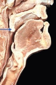
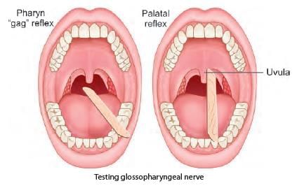
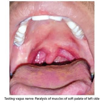
A patient with a fracture skull base at the temporo-occipital region, presented with difficulty in speaking post trauma. During investigation which structure should be tested which helps in diagnosis related with the points marked below? (INI-CET Nov 2021)

Detailed Solution for Test: Anatomy - 4 - Question 4

This illustration depicts a sagittal section of the head and neck region, with ‘marker B’ indicating the uvula (soft palate). This area is clinically assessed for lesions affecting cranial nerves IX and X. A patient suffering from a skull base fracture in the temporo-occipital region (where the jugular foramen is located) may present with Vernet syndrome, which results in damage to the IX, X, and XI cranial nerves that pass through the jugular foramen.

  • A range of lesions can affect the jugular foramen, including tumours, vascular issues, infections, and trauma (such as fractures).
  • Clinical manifestations will arise from the paralysis of the palate, pharynx, and larynx muscles (innervated by CN IX, X, and XI), leading to challenges in speech and swallowing, along with some sensory disturbances.
  • The most frequent initial symptom of jugular foramen syndrome (JFS) is either hoarseness or difficulty swallowing (dysphagia).
  • Pain is also a prevalent associated symptom, often presenting as persistent unilateral periauricular pain and headaches.

Clinical symptoms vary based on which structures are impacted by JFS:

  • Vagus nerve (CN X): Compression of the vagus nerve initially causes paralysis of the laryngeal muscles, resulting in hoarseness and a nasal tone. Further compression leads to unilateral paralysis of the soft palate and deviation of the uvula towards the unaffected side.
  • Glossopharyngeal nerve (CN IX): Involvement of this nerve results in:
    • Loss of sensation in the posterior ipsilateral area of the tongue,
    • Decreased secretions from the ipsilateral parotid gland, and
    • Loss of the gag reflex on the affected side.
  • Accessory nerve (CN XI): Dysfunction manifests as shoulder droop, difficulties in abducting the ipsilateral arm, and challenges in rotating the head to the opposite side due to weakness in the sternocleidomastoid and trapezius muscles.
  • Obstruction of venous sinuses and veins: This leads to headaches and papilledema caused by intracranial venous congestion, resulting in cerebral oedema and increased intracranial pressure.

When evaluating a patient with JFS, clinicians should take note of:

  • Palatal and gag reflexes (examine the uvula),
  • Shoulder droop,
  • Unilateral vocal cord paralysis (examine the larynx),
  • Strength of the sternocleidomastoid and trapezius muscles,
  • Fasciculation or winging of the scapula.
Test: Anatomy - 4 - Question 5

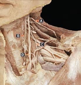
Which of the following arrow marked nerves, if injured present with paradoxical breathing? (INI-CET Nov 2020)

Detailed Solution for Test: Anatomy - 4 - Question 5

Marker ‘C’ indicates the phrenic nerve, which, when damaged, results in paradoxical breathing due to the paralysis of the corresponding side of the diaphragm.

  • Identification: The phrenic nerve originates from the C-3, 4, and 5 roots. It crosses the scalenus anterior (from lateral to medial, towards the front) and runs between the subclavian artery and vein before entering the thoracic region.
  • Paradoxical breathing: In cases of phrenic nerve paralysis and diaphragmatic eventration, one hemidiaphragm rises during inhalation and descends during exhalation, opposite to the movements of the other side.
  • Test: A fluoroscopic examination of the diaphragm, known as the sniff test, is beneficial for evaluating diaphragmatic function. This test entails a quick inspiratory effort and the observation of the hemidiaphragms. In healthy individuals, both hemidiaphragms move downwards during inspiration. However, in phrenic nerve paralysis, the affected side shows an abnormal upward movement.

Marker A – Spinal accessory nerve (Located in the posterior triangle of the neck, supplying the trapezius muscle) Marker B – Sympathetic chain (Identification: runs behind the carotid sheath, characterised by the presence of ganglia) Marker D – Vagus nerve (Identification: crosses the scalenus anterior and is positioned between the subclavian artery and vein)

*Multiple options can be correct
Test: Anatomy - 4 - Question 6

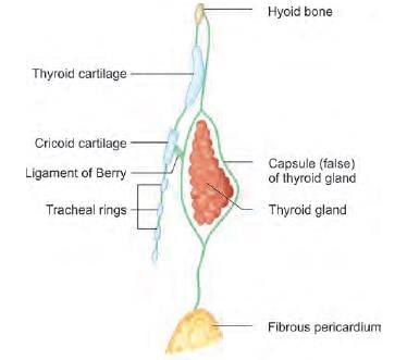
The upward extension of thyroid swelling is prevented by which of the following structure attached to thyroid cartilage? (NEET-PG 2020)

Detailed Solution for Test: Anatomy - 4 - Question 6

The upper extension of the thyroid gland is constrained by the superior connection of the pretracheal fascia and the strap muscles' attachment (such as the sternothyroid). A few authors note that:

  • The lateral aspect of the thyroid is enveloped by the sternothyroid muscle.
  • This muscle's connection to the oblique line of the thyroid cartilage inhibits the superior pole from extending upwards beneath the thyrohyoid muscle.

The pretracheal layer of deep cervical fascia covers the anterior and lateral sides of the trachea, dividing to encase the thyroid gland, thus forming its capsule. It is connected to the oblique line of the thyroid cartilage and to the cricoid cartilage's arch at the front.

The upper extension of the thyroid gland is restricted by the superior attachment of the pretracheal fascia. The lateral surface of the thyroid is covered by the sternothyroid muscle, and its attachment to the oblique line of the thyroid cartilage prevents the superior pole from extending upwards beneath the thyrohyoid muscle.

Test: Anatomy - 4 - Question 7

Saccule of inner ear develops from: (NEET-PG 2020)

Detailed Solution for Test: Anatomy - 4 - Question 7

The saccule of the inner ear originates from the pars inferior of the otic vesicle.

  • The terminal section of the tubotympanic recess, which develops from the first pharyngeal pouch, gives rise to buds that form sacci.
  • The saccus anticus and posticus play a role in the components of the middle ear. The ear begins to develop in week 4, arising from a thickening of cells in the surface ectoderm known as the otic placode.
  • The otic placode invaginates into the adjacent connective tissue (mesenchyme) near the rhombencephalon, forming the otic vesicle.
  • The otic vesicle subsequently separates into utricular and saccular sections.
Test: Anatomy - 4 - Question 8
Intractable cough reflex on scratching the floor of external auditory meatus is due to which nerve? (NEET-PG 2020)
Detailed Solution for Test: Anatomy - 4 - Question 8

In a small segment of the population, the auricular branch of the vagus nerve serves as the afferent limb of the Ear-Cough or Arnold Reflex. Physical stimulation, such as scratching, of the external acoustic meatus or its floor and posterior wall, which are innervated by the auricular nerve, triggers a cough. This response is similar to other cough reflexes linked to the vagus nerve.

  • The stimuli can reflexively lead to:
    • Persistent or intractable cough
    • Vomiting
    • Even fatal outcomes due to sudden cardiac inhibition
Test: Anatomy - 4 - Question 9

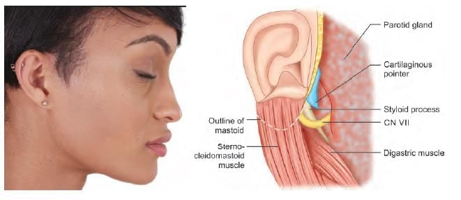
NOT an anatomical landmark of facial nerve identification in parotid surgery: (NEET-PG 2020p)

Detailed Solution for Test: Anatomy - 4 - Question 9

The inferior belly of the omohyoid is not associated with the path of the facial nerve, and therefore, it does not function as a reliable surgical landmark. The anatomical references for identifying the facial nerve during parotid surgery include:

  • Tragal cartilaginous pointer: This is a sharp triangular cartilage piece from the pinna that indicates the nerve's location.
  • Tympano-mastoid suture line: Situated between the tympanic and mastoid segments of the temporal bone, it is roughly 6–8 mm lateral to the stylomastoid foramen.
  • Styloid process: The nerve runs laterally to the styloid process.
  • Posterior belly of digastric: Following the upper border of this muscle back to its connection at the digastric groove, the nerve is located between it and the styloid process.

Test: Anatomy - 4 - Question 10

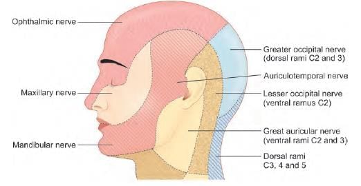
Post parotidectomy, patient feels numb while shaving. Which nerve was involved? (AIIMS May 2019)

Detailed Solution for Test: Anatomy - 4 - Question 10

The great auricular nerve (GAN) is frequently cut during parotidectomy, and one of the significant side effects is anaesthesia (numbness) in the area supplied by the nerve itself. Reports indicate that self-inflicted skin lesions can occur following the sacrifice of the GAN during parotidectomy.

  • In some patients, skin lesions from shaving are observed due to the numbness in the preauricular region.
  • In other cases, the lesions were found specifically at the earlobe.

Test: Anatomy - 4 - Question 11

Which layer maintains hydration of corneal stroma? (NEET-PG 2020p)

Detailed Solution for Test: Anatomy - 4 - Question 11

The endothelium supports Descemet's membrane and contains the most metabolically active cells of the cornea.

  • Na+/K+ ATPase pumps located in the basolateral membranes of these cells play a significant role in controlling the appropriate hydration level of the corneal stroma.
  • This regulation is essential for achieving maximum transparency and optimal light refraction.

Test: Anatomy - 4 - Question 12

The marked structure is supplied by: (AIIMS Nov 2019)

Detailed Solution for Test: Anatomy - 4 - Question 12

The identified structure is the superior oblique, which is positioned superiorly and medially within the orbit. It is innervated by the trochlear nerve.

Test: Anatomy - 4 - Question 13

Pretracheal layer covers all, except: (AIIMS June 2020)

Detailed Solution for Test: Anatomy - 4 - Question 13

Sternocleidomastoid is enveloped by the investing layer of deep cervical fascia (not the pretracheal layer). The pretracheal layer of deep cervical fascia comprises multiple sub-layers:

  • Visceral layer – surrounds the respiratory tube (larynx and trachea), gut tube (pharynx and oesophagus), and the thyroid gland located in front of the larynx and trachea.
  • Muscular layer – encases strap muscles such as sternohyoid and sternothyroid.

Test: Anatomy - 4 - Question 14
All are contents of carotid sheath, except: (AIIMS June 2020)
Detailed Solution for Test: Anatomy - 4 - Question 14

The sympathetic chain is located posterior to the carotid sheath. The carotid sheath encompasses four primary structures:

  • The common carotid artery, along with the internal carotid artery (medial)
  • The internal jugular vein (lateral)
  • The vagus nerve (CN X) (posterior)
  • The deep cervical lymph nodes

The carotid artery is positioned medial to the internal jugular vein, while the vagus nerve is found posteriorly between the two vessels. In the upper section, the carotid sheath also contains the glossopharyngeal nerve (IX), the accessory nerve (XI), and the hypoglossal nerve (XII), which briefly penetrate the superior part of the carotid sheath before exiting. The ansa cervicalis is situated within the anterior wall of the sheath, formed by “descendens hypoglossi” (C1) and “descendens cervicalis” (C2-C3). The cervical portion of the sympathetic chain (CSC) is embedded within the prevertebral fascia, directly posterior to the sheath.

Applied Anatomy: Unintentional injury to the cervical sympathetic chain during surgery could lead to Horner syndrome. The three main fascial layers in the neck that contribute to the carotid sheath include the investing fascia, the pretracheal fascia, and the prevertebral fascia.

Test: Anatomy - 4 - Question 15

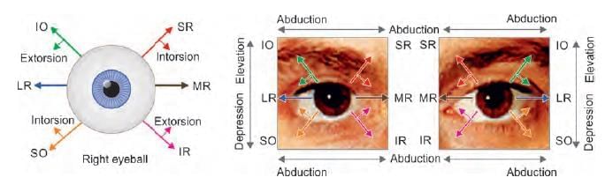
A patient presented with squint in right eye, as shown below. Identify the nerve lesioned in this case: (NEET-PG 2020p)

Detailed Solution for Test: Anatomy - 4 - Question 15

The patient exhibits an internal squint with the eyeball positioned elevated (up and in). This indicates hyperactivity of the muscles controlled by the oculomotor nerve. Such a condition may arise due to a lesion affecting the balancing nerve, specifically the trochlear nerve, which innervates the superior oblique muscle. Its actions include:

  • Abduction
  • Depression, particularly in an abducted eye

A lesion in the trochlear nerve leads to diplopia and a diminished ability to rotate the eye infero-laterally. Consequently, the eye deviates upward and slightly inward. The individual encounters challenges when walking downstairs.

Test: Anatomy - 4 - Question 16

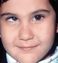
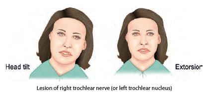
A child presented with left sided head tilt. On head straightening, there is right hypotropia, which increases on tilting head to right side. Which of the following muscle is paralysed? (AIIMS June 2020)

Detailed Solution for Test: Anatomy - 4 - Question 16

The patient exhibits right superior oblique palsy, characterised by:

  • Weakness of intorsion, leading to an extorsion deformity
  • Vertical diplopia, which is particularly noticeable when looking down in adduction

It is important to note that the superior oblique muscle is the primary muscle responsible for depressing the eyeball in the adducted position. If this muscle is paralysed, it causes a misalignment of the visual axes in the eyes, resulting in vertical diplopia.

The vertical diplopia resolves when the patient tilts their head to the contralateral side (opposite to the side of the lesion), which leads to a left-sided head tilt in this case. However, the diplopia re-emerges in a neutral head position and becomes more pronounced if the head is tilted towards the side of the lesion.

Test: Anatomy - 4 - Question 17

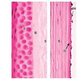
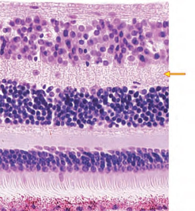
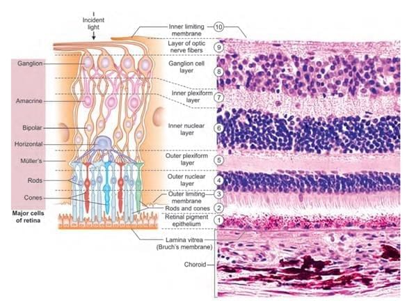
Identify the marked layer in the given histological section: (AIIMS Nov 2019)

Detailed Solution for Test: Anatomy - 4 - Question 17

This is a histological diagram of the retina, with the marker placed at the inner plexiform layer. The inner plexiform layer is a region of the retina composed of a dense network of fibrils created by the interwoven dendrites of retinal ganglion cells and the cells from the inner nuclear layer, which include bipolar and amacrine cells.

Test: Anatomy - 4 - Question 18

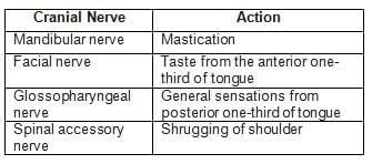
Match the following cranial nerves with their corresponding actions: (INI-CET May 2023)

Detailed Solution for Test: Anatomy - 4 - Question 18

The mandibular nerve innervates the muscles of the first pharyngeal arch responsible for mastication. The chorda tympani branch of the facial nerve transmits taste sensations from the anterior two-thirds of the tongue. The glossopharyngeal nerve conveys general and taste sensations from the posterior one-third of the tongue. The spinal accessory nerve innervates the trapezius muscle, which assists in shoulder shrugging.

Test: Anatomy - 4 - Question 19

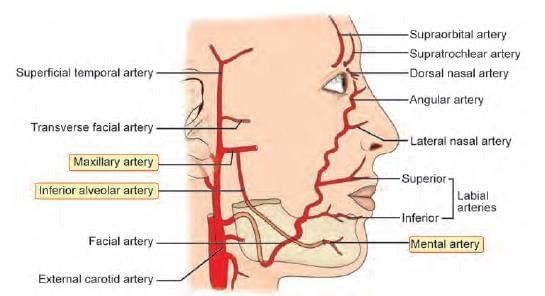
Which of the following is NOT a branch of facial artery? (INI-CET May 2022)

Detailed Solution for Test: Anatomy - 4 - Question 19

The mental artery is a branch of the inferior alveolar artery, which is itself a branch of the maxillary artery (not the facial artery). The facial artery is an anterior branch of the external carotid artery located in the carotid triangle (neck region). It originates just above the tip of the greater cornu of the hyoid bone; it travels through the cervical region and then takes a tortuous path on the face, moving antero-superiorly towards the medial angle of the eye.

The pulse of the facial artery can be palpated at the antero-inferior angle of the masseter, against the lower border of the mandible. The branches of the facial artery include:

  • Four cervical branches: ascending palatine, tonsillar, sub-mental, and glandular
  • Four facial branches: inferior labial artery, superior labial artery, lateral nasal artery, and angular artery (the terminal branch)

The transverse facial artery is a branch of the superficial temporal artery, which is the smaller terminal branch of the external carotid artery. The dorsal nasal artery is one of the two terminal branches of the ophthalmic artery (a branch of the internal carotid artery). It forms an anastomosis with the angular artery, providing a connection between branches of the internal carotid artery (dorsal nasal artery) and the external carotid artery (angular artery). Such anastomoses facilitate collateral circulation in the event of blockages.

Test: Anatomy - 4 - Question 20

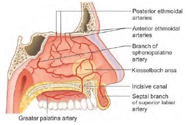
External carotid artery supplies nasal septum by all of the following branches, except: (AIIMS May 2019)

Detailed Solution for Test: Anatomy - 4 - Question 20

Anterior ethmoidal artery is a branch of the internal (not external) carotid artery.

*Multiple options can be correct
Test: Anatomy - 4 - Question 21

Inferior thyroid artery supplies: (INI-CET Nov 2021)
A. Oesophagus
B. Thyroid
C. Parathyroid
D. Thymus

Detailed Solution for Test: Anatomy - 4 - Question 21

The inferior thyroid artery (ITA) is a branch of the thyrocervical trunk (85%) or the subclavian artery (15%). It ascends to enter the thyroid gland on its posterior surface and supplies both the superior and inferior parathyroid glands.

  • The ITA also provides blood to the lower part of the oesophagus and the thymus.
  • It gives rise to the ascending cervical artery, the inferior laryngeal artery, and branches for the pharynx, trachea, and oesophagus.

The primary blood supply to the thymus comes from the internal thoracic arteries; however, it also receives blood from the inferior thyroid (and occasionally the superior) and pericardiophrenic arteries. Thymic veins drain into the left brachiocephalic, internal thoracic, and inferior thyroid veins.

*Multiple options can be correct
Test: Anatomy - 4 - Question 22

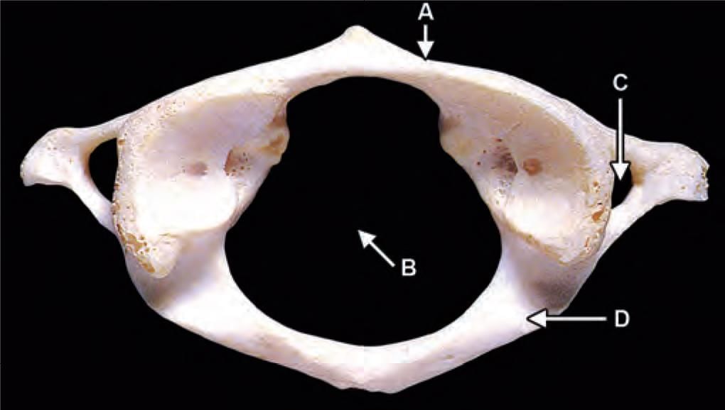
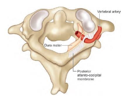
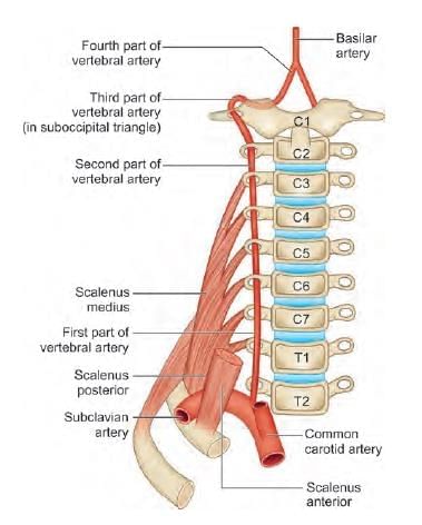
Third part of vertebral artery is related to which of the following marked region in the following diagram? (INI-CET Nov 2021)

Detailed Solution for Test: Anatomy - 4 - Question 22

This image depicts the C1 (atlas) vertebra, which is associated with V3 (the third segment of the vertebral artery). Most authors, including those of Gray’s Anatomy, indicate that it relates to the upper surface of the posterior arch of the C1 vertebra (marker D). However, some authors also suggest that it passes through the foramen transversarium (marker C).

  • Markers A: Anterior arch
  • Markers B: Vertebral foramen
  • Markers C: Foramen transversarium
  • Markers D: Posterior arch

The V3 (third segment of the vertebral artery) consists of two segments:

  • Vertical segment: This segment ascends from the transverse foramen of C2 (axis) and passes through the transverse foramen of C1 (atlas).
  • Horizontal segment: After exiting the C1 transverse foramen, V3 travels posteriorly and grooves the upper surface of the posterior arch of C1. It then enters the spinal canal by piercing the posterior atlanto-occipital membrane, dura mater, and arachnoid mater, continuing as V4 (the fourth part).

The horizontal segment of the third part of the vertebral artery rests over the superior surface of the posterior arch of the atlas (C1) vertebra.

Test: Anatomy - 4 - Question 23

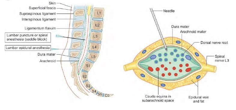
While doing lumbar puncture, which of the following structure is pierced before entering the lumbar cistern? (NEET-PG 2021)

Detailed Solution for Test: Anatomy - 4 - Question 23

The lumbar puncture needle penetrates the arachnoid mater to access the area beneath it: the sub-arachnoid space located in the lumbar cistern. The order in which the needle traverses to reach the lumbar cistern is as follows:

  • Skin
  • Superficial fascia
  • Supra-spinous ligament
  • Inter-spinous ligament
  • Ligamentum flavum (first pop sensation)
  • Dura mater (second pop sensation)
  • Arachnoid mater
  • Sub-arachnoid space (lumbar cistern)

Test: Anatomy - 4 - Question 24

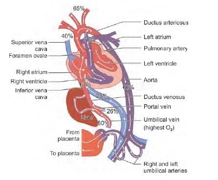
Which vessel carries deoxygenated blood back to placenta in fetal circulation? (NEET-PG 2021)

Detailed Solution for Test: Anatomy - 4 - Question 24

The umbilical arteries are responsible for transporting deoxygenated blood (oxygen concentration – 60%) back to the placenta from the fetus during placental circulation. The umbilical vein brings highly oxygenated blood (oxygen concentration – 80%) from the maternal side (placenta) towards the fetal side.

The descending aorta gathers blood from the pulmonary trunk (through the ductus arteriosus) in fetal circulation. This blood possesses a medium oxygen content (oxygen concentration – 60%).

In fetal circulation, the pulmonary arteries carry a very small volume of blood as the fetal lungs are non-functional. This blood has a medium oxygen content (oxygen concentration – 50%).

Test: Anatomy - 4 - Question 25

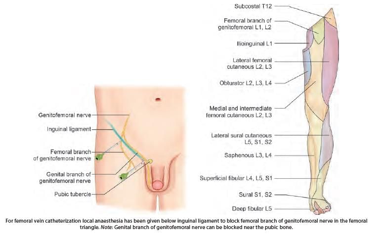
For femoral vein catheterization local anaesthesia has been given below inguinal ligament. Which of the following nerve is blocked?

Detailed Solution for Test: Anatomy - 4 - Question 25
  • Femoral Vein Catheterization: Local anesthesia is applied below the inguinal ligament to block the femoral branch of the genitofemoral nerve in the femoral triangle (L1 dermatome).
  • Genitofemoral Nerve:
    • A mixed (sensory and motor) nerve from the lumbar plexus, formed by anterior rami of L1-L2.
    • Splits into genital and femoral branches.
    • Femoral branch: Provides sensory innervation to the upper anterior thigh.
    • Genital branch: Innervates skin of the penile root and anterior scrotum (males) or mons pubis (females); supplies motor innervation to the cremaster muscle.
  • Femoral Triangle: Contains the femoral vein, with skin supplied by the femoral branch of the genitofemoral nerve (L1 dermatome).
  • Femoral Nerve (L2-L4):
    • Anterior division: Gives rise to intermediate and medial femoral cutaneous nerves, supplying anterior and medial thigh skin.
    • Posterior division: Forms the saphenous nerve, innervating anteromedial and posteromedial leg and medial foot dorsum.
  • Obturator Nerve (L2-L4): Supplies sensory innervation to the medial thigh skin.
  • Lateral Cutaneous Nerve of Thigh (L2-L3): Passes under the inguinal ligament, providing sensation to the lateral thigh via anterior and posterior branches.

Information about Test: Anatomy - 4 Page
In this test you can find the Exam questions for Test: Anatomy - 4 solved & explained in the simplest way possible. Besides giving Questions and answers for Test: Anatomy - 4, EduRev gives you an ample number of Online tests for practice
Download as PDF