NEET PG Exam  >  NEET PG Tests  >  Test: Aortic Dissection & Infective Endocarditis - NEET PG MCQ

Test: Aortic Dissection & Infective Endocarditis - NEET PG MCQ


Test Description

15 Questions MCQ Test - Test: Aortic Dissection & Infective Endocarditis

Test: Aortic Dissection & Infective Endocarditis for NEET PG 2025 is part of NEET PG preparation. The Test: Aortic Dissection & Infective Endocarditis questions and answers have been prepared according to the NEET PG exam syllabus.The Test: Aortic Dissection & Infective Endocarditis MCQs are made for NEET PG 2025 Exam. Find important definitions, questions, notes, meanings, examples, exercises, MCQs and online tests for Test: Aortic Dissection & Infective Endocarditis below.
Solutions of Test: Aortic Dissection & Infective Endocarditis questions in English are available as part of our course for NEET PG & Test: Aortic Dissection & Infective Endocarditis solutions in Hindi for NEET PG course. Download more important topics, notes, lectures and mock test series for NEET PG Exam by signing up for free. Attempt Test: Aortic Dissection & Infective Endocarditis | 15 questions in 15 minutes | Mock test for NEET PG preparation | Free important questions MCQ to study for NEET PG Exam | Download free PDF with solutions
Test: Aortic Dissection & Infective Endocarditis - Question 1

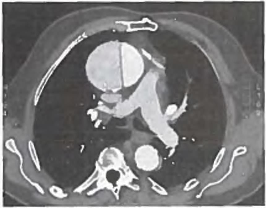
A 50-year male presented with high BP of 160/100 mm Hg and heart rate of 120/min. CECT is shown below. Which is best management of this condition? (AIIMS May 2018)

Detailed Solution for Test: Aortic Dissection & Infective Endocarditis - Question 1

The image displays a CT scan of the chest revealing an ascending aorta dissection, characterised by a tennis ball appearance.

  • Located behind it is the pulmonary artery bifurcation, which appears normal and shows no signs of embolism.
  • For acute ascending aortic dissections and intramural hematomas, urgent or emergent surgical intervention is the recommended treatment.
  • Type B aortic dissections affect the transverse and/or descending aorta.
  • In the case of uncomplicated or stable distal lesions and intramural type B hematomas, medical management is preferred.
  • For complicated type B dissections, surgical correction is necessary.

Test: Aortic Dissection & Infective Endocarditis - Question 2

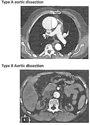
A 70-year-old man with Hypertension wakes up with severe chest pain and diaphoresis. On examination he has bounding pulses with wide pulse pressure. A diastolic murmur is heard along the right sternal border. Which of the following is the possible aetiology? (Recent Question 2019)

Detailed Solution for Test: Aortic Dissection & Infective Endocarditis - Question 2

The occurrence of hypertension and chest pain, accompanied by the emergence of a diastolic murmur, indicates the onset of aortic regurgitation.

  • Acute aortic regurgitation can arise from the retrograde propagation of aortic dissection.
  • Option B: Dysfunction of the papillary muscle would result in a systolic murmur.
  • Option C: Regurgitation results in a systolic murmur.
  • Option D: This is observed with renal artery stenosis and manifests as heart failure.
Test: Aortic Dissection & Infective Endocarditis - Question 3

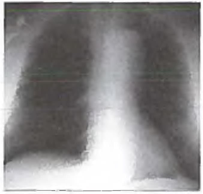
A 65-year old hypertension patient presents with Chest pain, difficulty in breathing for I hour. Chest X-ray done shows presence of:

Detailed Solution for Test: Aortic Dissection & Infective Endocarditis - Question 3

The X-ray of a patient with hypertension reveals the presence of mediastinal widening, which is the most frequent plain radiographic observation in cases of aortic dissection.

Test: Aortic Dissection & Infective Endocarditis - Question 4

Investigation of choice for aortic dissection with hypotension is ? (Recent Pattern 2014-15)

Detailed Solution for Test: Aortic Dissection & Infective Endocarditis - Question 4

The diagnosis of aortic dissection can be made using non-invasive methods such as echocardiography, CT, and MRI. Aortography is utilized less frequently due to the reliability of these non-invasive techniques.

  • Transthoracic echocardiography is easy to perform and can be done quickly, with an overall sensitivity of 60-85% for detecting aortic dissection.
  • Its sensitivity for identifying proximal ascending aortic dissections exceeds 80%, but it is not as effective for detecting dissections in the arch and descending thoracic aorta.
  • Transesophageal echocardiography requires more expertise and patient cooperation but is highly accurate, achieving 98% sensitivity and around 90% specificity for ascending and descending thoracic aorta dissections, though it is less effective for the arch.
  • This method also provides valuable insights into the presence and severity of aortic regurgitation and pericardial effusion.

Both CT and MRI are extremely accurate in detecting the intimal flap, the extent of the dissection, and involvement of major arteries, with each boasting a sensitivity and specificity of over 90%. They are beneficial for identifying intramural haemorrhage and penetrating ulcers.
MRI additionally allows for the assessment of blood flow, which can help differentiate between antegrade and retrograde dissection. The effectiveness of transesophageal echocardiography, CT, and MRI varies based on the resources and expertise available at specific institutions, as well as the patient’s haemodynamic stability, with CT and MRI being less suitable for patients who are unstable.

Test: Aortic Dissection & Infective Endocarditis - Question 5

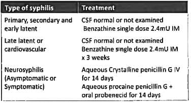
Treatment of choice for late cardiovascular syphilis is? (AIIMS May 2018)

Detailed Solution for Test: Aortic Dissection & Infective Endocarditis - Question 5

For the treatment of late latent syphilis, cardiovascular syphilis is managed with Penicillin G Benzathine administered at a dosage of 2.4 MU i.m. on a weekly basis for a duration of 3 weeks.

Test: Aortic Dissection & Infective Endocarditis - Question 6
Roth spots are seen in? (Recent Pattern 2018)
Detailed Solution for Test: Aortic Dissection & Infective Endocarditis - Question 6
Roth spots are characterised by white-centred haemorrhages. The causes of Roth spots include:
  • Bacterial endocarditis and sepsis
  • Lymphoproliferative disorders
  • Diabetes mellitus
  • Hypertension
  • Anaemia
  • Connective tissue disorders
Test: Aortic Dissection & Infective Endocarditis - Question 7
A 40-year-old patient came with complaints of spikes of fever and difficulty in breathing. T.E.E shows multiple vegetations on surface of heart valves. Blood culture showed growth of Burkholderia Cepacia. Which of the following is first line management for this patient? (AIIMS May 2017)
Detailed Solution for Test: Aortic Dissection & Infective Endocarditis - Question 7

Burkholderia cepacia exhibits resistance to the majority of antibiotics. Therefore, due to its intrinsic resistance, TMP-SMX, meropenem, and doxycycline are the most effective treatments. Fluoroquinolones and third-generation cephalosporins should only be administered once susceptibility to these agents has been established.

Burkholderia cepacia is responsible for cepacia syndrome, which is characterised by:

  • Rapidly progressive distress and sepsis
  • Recurrent pneumonia in cystic fibrosis
  • Ventilator-associated pneumonia
  • Catheter-associated infections
  • Wound infections
Test: Aortic Dissection & Infective Endocarditis - Question 8

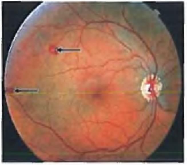
A 50 year man came with a 'grey spot' in his vision. He had recurrent fevers and weight loss for the past 3 months. He had a history of mitral valve infection. Vision is 6/6. This is the picture of his fundus. Choose the FALSE statement? (APPG 2016)

Detailed Solution for Test: Aortic Dissection & Infective Endocarditis - Question 8

The fundus examination of a patient with mitral valve infection reveals Roth spots. Choice B is correct.

  • The occurrence of recurrent fevers in a patient with pre-existing heart disease increases the risk of developing infective endocarditis.
  • Acute bacterial endocarditis is associated with Staphylococcus, while the subacute form is linked to Streptococcus viridans and Enterococci.
  • It can also arise from less virulent infections, making choice A accurate.

The causes of Roth spots include:

  • Infective endocarditis
  • SLE
  • Polyarteritis Nodosa
  • Severe anaemia
  • Leukaemia
  • Diabetes mellitus
  • Hypertension
Test: Aortic Dissection & Infective Endocarditis - Question 9

Which of the following is not a part of Duke Criteria for infective endocarditis? (AIIMS May 15)

Detailed Solution for Test: Aortic Dissection & Infective Endocarditis - Question 9

The Duke Criteria for diagnosing infective endocarditis include the following:

  • Major Criteria
    • Positive blood culture: A typical microorganism associated with infective endocarditis must be identified from two separate blood cultures. This includes:
      • Viridans streptococci
      • Streptococcus gallolyticus
      • HACEK group
      • Staphylococcus aureus
      • Community-acquired enterococci, in the absence of a primary focus
    • Persistently positive blood culture, defined as the recovery of a microorganism consistent with infective endocarditis from:
      • Blood cultures taken more than 12 hours apart
      • All three or a majority of at least four separate blood cultures, with the first and last taken at least one hour apart
    • A single positive blood culture for Coxiella burnetii or a phase I IgG antibody titer greater than 1:800
    • Evidence of endocardial involvement
  • Minor Criteria
    • Predisposition: A pre-existing heart condition or injection drug use
    • Fever exceeding 38.0°C (>100.4°F)
    • Vascular phenomena: Major arterial emboli, septic pulmonary infarcts, mycotic aneurysms, intracranial haemorrhages, conjunctival haemorrhages, and Janeway lesions
    • Immunologic phenomena: Glomerulonephritis, Osler's nodes, Roth's spots, and rheumatoid factor
    • Microbiological evidence: A positive blood culture that does not meet the major criteria as mentioned previously, or serological evidence of active infection with an organism consistent with infective endocarditis

Definite endocarditis is characterised by the documentation of:

  • Two major criteria, or
  • One major criterion and three minor criteria, or
  • Five minor criteria

Transesophageal echocardiography is advised for evaluating potential prosthetic valve endocarditis or complicated endocarditis. It is important to exclude single positive cultures for coagulase-negative staphylococci and diphtheroids, which are common contaminants, as well as organisms that infrequently cause endocarditis, such as gram-negative bacilli.

Test: Aortic Dissection & Infective Endocarditis - Question 10
Infective endocarditis is most commonly seen in? (Bihar PG IS)
Detailed Solution for Test: Aortic Dissection & Infective Endocarditis - Question 10
Infective endocarditis lesions are observed according to the pressure gradient. The highest occurrence in adults is associated with mitral regurgitation, whereas in children, it is linked to VSD. Infective endocarditis is least prevalent in cases of ASD. The greatest risk of adverse outcomes following infective endocarditis for which prophylaxis is advised includes:
  • Prosthetic cardiac valves
  • Unrepaired cyanotic congenital heart disease
  • Completely repaired defects involving prosthetic material
  • Rheumatic fever (including mitral stenosis and aortic regurgitation)
  • VSD and congenital aortic stenosis
Test: Aortic Dissection & Infective Endocarditis - Question 11

Osler's nodes are seen in? (Recent Pattern 2014-15)

Detailed Solution for Test: Aortic Dissection & Infective Endocarditis - Question 11

Peripheral manifestations of injective endocardites

  • Osler nodes
  • Subungual haemorrhages
  • Janeway lesions
  • Roth spots
  • Petechiae
Test: Aortic Dissection & Infective Endocarditis - Question 12

What is not true about infective endocarditis? (Recent Pattern 2014-15)

Detailed Solution for Test: Aortic Dissection & Infective Endocarditis - Question 12

With dental procedures, it is advisable to be cautious. Rose spots are bacterial emboli on the skin, observed in around 1/3 of typhoid fever cases. These spots are considered classic indicators of untreated disease; however, they may also be present in other conditions, such as Shigellosis and non-typhoidal salmonellosis.

  • They typically manifest as a rash between the seventh and twelfth day following the onset of symptoms.
  • These spots appear in clusters of five to ten lesions located on the lower chest and upper abdomen.
  • They are more prevalent after a paratyphoid infection.

Rose spots usually persist for three to four days. Hematuria may present as a sign of post-infectious glomerulonephritis in infective endocarditis (IE). Additionally, hemiplegia can arise following the formation of a brain abscess in IE.

Test: Aortic Dissection & Infective Endocarditis - Question 13

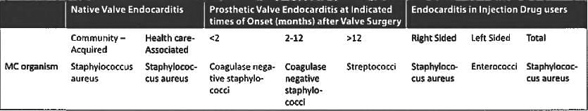
Most common cause of infective endocarditis is? (Recent Pattern 2014-15)

Detailed Solution for Test: Aortic Dissection & Infective Endocarditis - Question 13

Organisms Responsible for significant clinical Types of Endocarditis:

Test: Aortic Dissection & Infective Endocarditis - Question 14
Infective endocarditis where lifelong treatment is required: (Recent Pattern 2014-15)
Detailed Solution for Test: Aortic Dissection & Infective Endocarditis - Question 14

Fungal endocarditis is an exceptionally rare condition, representing only 2-4% of all endocarditis occurrences. Aspergillus was identified as the causative agent in 24% of fungal endocarditis cases. The crucial factor in the management of Aspergillus endocarditis is:

  • Early detection
  • Prompt surgical resection or debridement
  • Combination with antifungal therapy

This approach is vital due to the significant risk of embolic complications and cardiac decompensation. The Infectious Diseases Society of America suggests Voriconazole as the preferred treatment option. For candida endocarditis, the recommended regimen includes:

  • Liposomal amphotericin B or caspofungin for 6-8 weeks
  • With or without flucytosine
  • Followed by lifelong management with fluconazole

Due to the potential for recurrent infections, particularly after surgical intervention on infected prosthetic valves, it is advisable to consider lifelong prophylactic treatment with oral triazoles.

Test: Aortic Dissection & Infective Endocarditis - Question 15
Mitral valve vegetations do not usually embolise to: (AIIMS Nov 2001)
Detailed Solution for Test: Aortic Dissection & Infective Endocarditis - Question 15

Mitral valve vegetations would clearly not travel to the lungs, as this would entail a 'backward flow.'

Information about Test: Aortic Dissection & Infective Endocarditis Page
In this test you can find the Exam questions for Test: Aortic Dissection & Infective Endocarditis solved & explained in the simplest way possible. Besides giving Questions and answers for Test: Aortic Dissection & Infective Endocarditis, EduRev gives you an ample number of Online tests for practice
Download as PDF