NEET PG Exam  >  NEET PG Tests  >  Test: Arrhythmias and Emergency Medicine- 3 - NEET PG MCQ

Test: Arrhythmias and Emergency Medicine- 3 - NEET PG MCQ


Test Description

15 Questions MCQ Test - Test: Arrhythmias and Emergency Medicine- 3

Test: Arrhythmias and Emergency Medicine- 3 for NEET PG 2025 is part of NEET PG preparation. The Test: Arrhythmias and Emergency Medicine- 3 questions and answers have been prepared according to the NEET PG exam syllabus.The Test: Arrhythmias and Emergency Medicine- 3 MCQs are made for NEET PG 2025 Exam. Find important definitions, questions, notes, meanings, examples, exercises, MCQs and online tests for Test: Arrhythmias and Emergency Medicine- 3 below.
Solutions of Test: Arrhythmias and Emergency Medicine- 3 questions in English are available as part of our course for NEET PG & Test: Arrhythmias and Emergency Medicine- 3 solutions in Hindi for NEET PG course. Download more important topics, notes, lectures and mock test series for NEET PG Exam by signing up for free. Attempt Test: Arrhythmias and Emergency Medicine- 3 | 15 questions in 15 minutes | Mock test for NEET PG preparation | Free important questions MCQ to study for NEET PG Exam | Download free PDF with solutions
Test: Arrhythmias and Emergency Medicine- 3 - Question 1

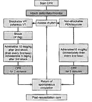
Unconscious child, CPR was started and ECG shows normal QRS but peripheral pulse was absent inspite of chest compressions. What should be done next? (JIPMER Nov 2015)

Detailed Solution for Test: Arrhythmias and Emergency Medicine- 3 - Question 1

Since the ECG displays a normal qRS but the pulse is absent, the child is experiencing Pulseless Electrical Activity (PEA). This condition is defined by a lack of responsiveness and the absence of a detectable pulse while exhibiting organised cardiac electrical activity.

  • In children, PEA is often associated with respiratory causes or shock.
  • In contrast, adults typically present with arrhythmias such as ventricular tachycardia (VT) or ventricular fibrillation.

Epinephrine should be given in 1 mg doses via IV/IO every 3-5 minutes during PEA arrest.

5Hs:

  • Hypoxia
  • Hypovolemia
  • Hydrogen ions (acidosis)
  • Hyperkalemia/hypokalaemia
  • Hypothermia

5Ts:

  • Tension pneumothorax
  • Cardiac tamponade
  • Coronary thrombosis
  • Pulmonary thrombosis
  • Drug overdose (tablets)
Test: Arrhythmias and Emergency Medicine- 3 - Question 2

New onset atrial flutter. Best treatment is? (JIPMER Nov 2015)

Detailed Solution for Test: Arrhythmias and Emergency Medicine- 3 - Question 2

In cases of atrial flutter, the use of class I agents may result in a reduction of the flutter rate to the point where 1:1 conduction occurs, which can cause the heart rate to rise to 200 bpm, leading to hemodynamic issues. Therefore, Ibutilide is advised, with a 1 mg infusion over 60 minutes being effective in 50-70% of patients, restoring them to normal sinus rhythm. However, this option is not available.

Electrical cardioversion with low intensity, ranging from 25 to 50 J, can convert 90% of patients to normal sinus rhythm and is considered the most effective treatment for recent onset atrial flutter. For those with recurrent atrial flutter, anti-arrhythmic medications such as sotalol and amiodarone may be utilised, although 70% of these patients will face recurrence. In such cases, catheter ablation at the cavo-tricuspid isthmus proves to be more effective.

Test: Arrhythmias and Emergency Medicine- 3 - Question 3

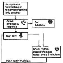
A 75-year-old patient clutches chest and falls down. A Physician arrives on the scene. What is the first thing to be done by the physician? (JIPMER Nov 2015)

Detailed Solution for Test: Arrhythmias and Emergency Medicine- 3 - Question 3


CPR Quality:

  • Push hard (approximately 5 cm) and at a rate of 100 compressions per minute.
  • Ensure complete chest recoil between compressions.
  • Limit interruptions to compressions.
  • Avoid excessive ventilation.
  • Change the compressor every two minutes.
  • If there is no advanced airway, use a 30:2 compression-ventilation ratio.

Utilise quantitative waveform capnography.

  • If ETCO2 is below 10 mm Hg, strive to enhance CPR quality.
  • Monitor intra-arterial pressure.
  • If the diastolic relaxation phase pressure is under 20 mm Hg, work to improve CPR quality.

Return of Spontaneous Circulation (ROSC):

  • Detect a pulse and blood pressure.
  • Observe a sudden consistent rise in ETCO2 (usually around 40 mm Hg).
  • Identify spontaneous arterial pressure waves with intra-arterial monitoring.
Test: Arrhythmias and Emergency Medicine- 3 - Question 4
In ECG lead II, III, and aVF are abnormal. Which of the following vessel is blocked? (JIPMER May 2015)
Detailed Solution for Test: Arrhythmias and Emergency Medicine- 3 - Question 4

Leads II, III, and aVF identify inferior wall ischemia, primarily supplied by the right coronary artery.

Test: Arrhythmias and Emergency Medicine- 3 - Question 5

A 55-year-old hypertensive patient has a standing BP 190/10 and sitting BP-180/100. He also has irregularly irregular rhythm, double apical impulse and bilateral basal crepitations. But no murmurs could be auscultated and heart rate could not be determined. What is the likely cause? (JIPMER Nov 2014)

Detailed Solution for Test: Arrhythmias and Emergency Medicine- 3 - Question 5

Left atrial myxoma is more frequently observed in young females, presenting with symptoms such as platypnea and reduced cardiac output, as the tumour restricts blood flow into the left ventricle.
Mitral regurgitation has been excluded since no murmur was detected during auscultation.
Cor pulmonale has also been ruled out as there are no indications of primary lung disease or signs of right ventricular failure.
Points supporting the diagnosis of left ventricular hypertrophy (LVH) include:

  • Hypertension may cause LVH, resulting in a double apical impulse.
  • High blood pressure can lead to diastolic dysfunction, affecting the atria and resulting in atrial fibrillation and an irregular pulse.
  • Basal crepitations can be accounted for by the presence of atrial fibrillation.
  • Blood pressure variations in both standing and supine positions are normal (within 10 mm Hg).

Hypertension is linked with left ventricular hypertrophy, impaired filling of the ventricle, enlargement of the left atrium, and a decrease in atrial conduction velocity. These alterations in cardiac structure and function promote the onset of atrial fibrillation and elevate the risk of thromboembolic events.

Test: Arrhythmias and Emergency Medicine- 3 - Question 6
A 70-year old hypertensive patient with complaint of palpitations and pre-syncope. On examination, his heart rate is 72 BPM and BP was 150/100. ECG done shows (Recent Question 2015-16)
Detailed Solution for Test: Arrhythmias and Emergency Medicine- 3 - Question 6

The ECG in lead II and other inferior leads reveals the existence of flutter waves. You can observe 3–4 flutter waves preceding each narrow QRS complex.

  • Given that the ventricular rate is approximately 75 bpm (RR interval), the atrial rate can be determined by multiplying the ventricular rate by 4, resulting in 300 bpm.
  • The presence of hypertension as a risk factor further supports the diagnosis of atrial flutter.
Test: Arrhythmias and Emergency Medicine- 3 - Question 7

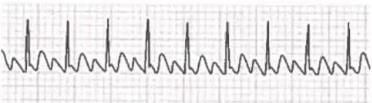
A 62-year-old male with underlying COPD develops a viral upper respiratory infection and begins taking an over-the­ counter decongestant. Shortly thereafter he experiences palpitations and presents to the emergency room, where the given rhythm strip is obtained, demonstrating: (Recent Question 2015-16)

Detailed Solution for Test: Arrhythmias and Emergency Medicine- 3 - Question 7

The rhythm strip presented indicates atrial flutter accompanied by a 2:1 atrioventricular (AV) block.

  • Atrial flutter is marked by an atrial rate ranging from 250 to 350 beats per minute.
  • The electrocardiogram usually displays a sawtooth baseline pattern caused by the flutter waves.
  • In this strip, every second atrial depolarisation passes through the AV node, leading to a ventricular rate of 75 beats per minute.
Test: Arrhythmias and Emergency Medicine- 3 - Question 8

A 40-year-old Lady intraoperatively develops HR 220 bpm and Blood pressure of 70/40, ECG shows qRS complex Ł 110 milliseconds. Best management is? (JIPMER 2014)

Detailed Solution for Test: Arrhythmias and Emergency Medicine- 3 - Question 8

Normal qRS complex duration is between 0.08 and 0.1 seconds. Since the qRS complex is extended, it indicates one of the following conditions:

  • Atrial fibrillation with ventricular pre-excitation (AVNRT)
  • Ventricular tachycardia

The diagnostic criteria for ventricular tachycardia include:

  • A qRS duration exceeding 140 ms in the absence of drug treatment
  • A superior and rightward qRS axis in the frontal plane
  • A bizarre qRS complex that does not resemble the typical qRS pattern seen in left or right bundle branch block
  • Slurring of the initial part of the qRS

Typically, the qRS complex is greater than 0.14 seconds; however, in this case, it measures 0.11 seconds. Therefore, the patient does not have ventricular tachycardia. For patients exhibiting pre-excitation and atrial fibrillation, treatment should focus on preventing a rapid ventricular response. In critical situations, DC cardioversion is recommended to terminate the atrial fibrillation. The acute management of AP-mediated macro-entrant orthodromic tachycardia resembles that for AV nodal reentry and aims to modify conduction within the AV node. Vagal stimulation through the Valsalva manoeuvre and carotid sinus pressure may provide enough AV nodal slowing to stop the AVRT. The initial pharmacological treatment involves intravenous adenosine, 6-12 mg; calcium channel blockers such as verapamil and diltiazem, or beta blockers, may also be beneficial.

Test: Arrhythmias and Emergency Medicine- 3 - Question 9

Most common arrhythmia in ICU patients: (Recent Pattern 2015-16)

Detailed Solution for Test: Arrhythmias and Emergency Medicine- 3 - Question 9

Atrial arrhythmias are the most prevalent type among patients with CHF, those who have undergone surgery and are admitted to the ICU, and individuals with hypertension.

Test: Arrhythmias and Emergency Medicine- 3 - Question 10

Most common mechanism of arrhythmia? (Recent Pattern 2014-15)

Detailed Solution for Test: Arrhythmias and Emergency Medicine- 3 - Question 10

The most prevalent mechanism for arrhythmia is re-entry. Essentially, re-entry is characterised by the circulation of an activation wave around a non-excitable barrier. This phenomenon arises from inconsistencies in myocardial conduction and/or recovery characteristics.

  • The existence of a unidirectional block combined with slow conduction permits the retrograde recovery of the obstructed myocardium.
  • This creates a circuit that can sustain a tachycardia if it continues.

Re-entry seems to underlie the majority of abnormal sustained Supra Ventricular Tachycardias (SVTs) and Ventricular Tachycardias (VTs). Two notable examples of primarily functional re-entry include:

  • Ventricular Fibrillation (VF) due to acute myocardial ischaemia
  • Polymorphic VT in individuals with a genetically inherited ion channel disorder, such as Brugada syndrome, Long QT Syndrome (LQTS), or catecholaminergic polymorphic VT.
Test: Arrhythmias and Emergency Medicine- 3 - Question 11
Patient of AV conduction episodes block complains of dizziness. Best treatment for this patient is? (Recent Pattern 2014-15)
Detailed Solution for Test: Arrhythmias and Emergency Medicine- 3 - Question 11

Temporary or permanent artificial pacing is the most dependable solution for individuals experiencing symptomatic AV conduction system disorders.

Test: Arrhythmias and Emergency Medicine- 3 - Question 12

All of the following electrocardiographic findings may represent manifestations of digitalis intoxication, except: (Recent Pattern 2014-I5)

Detailed Solution for Test: Arrhythmias and Emergency Medicine- 3 - Question 12

Ectopic rhythms observed in digoxin toxicity arise from increased automaticity, reentry, or a combination of both, and may encompass the following:

  • Ventricular bigeminy
  • Premature ventricular contractions
  • Bidirectional ventricular tachycardia: This occurs due to changes in intraventricular conduction, as well as junctional tachycardia with aberrant intraventricular conduction.
  • AV block
  • Non-paroxysmal atrial tachycardia with block.

Digoxin induces ST segment depression in leads V5-6 (Hockey stick sign). In cases of digoxin toxicity, any form of arrhythmia or block can manifest. The occurrence of ventricular ectopics bigeminy is the most prevalent. Non-paroxysmal atrial tachycardia with variable block is a distinctive feature.

Test: Arrhythmias and Emergency Medicine- 3 - Question 13

40-year-old female patient develops recurrent episodes of sudden palpitations with HR = 150/min, rhythm regular. After every episode she has diuresis. What could be the cause? (Recent Pattern 2014-15)

Detailed Solution for Test: Arrhythmias and Emergency Medicine- 3 - Question 13

Choices C and D are excluded as they would lead to an inconsistent rhythm.
Choice A is frequently observed in women aged between their 20s and 40s and occurs without any underlying structural heart disease. The simultaneous activation of both atria and ventricles against a closed tricuspid valve may create a fluttering sensation in the neck. Increased venous pressures cause the release of natriuresis, which can lead to post-tachycardia diuresis.
Choice B might be a normal reaction to stress, making it a less probable diagnosis.

Test: Arrhythmias and Emergency Medicine- 3 - Question 14

Atrial fibrillation may occur in all the following conditions, except- (Recent Pattern 2014-15)

Detailed Solution for Test: Arrhythmias and Emergency Medicine- 3 - Question 14

Atrial fibrillation arises in a heart that has structural damage, such as in cases of dilated cardiomyopathy or left atrial enlargement resulting from mitral valvular defects. It is also observed in thyrotoxicosis, where sympathetic overactivity induced by thyroid hormones can lead to the formation of micro-voltage gradients between the musculature of the pulmonary veins and the atrial muscle.

Test: Arrhythmias and Emergency Medicine- 3 - Question 15

Which of the following arrhythmia is most commonly associated with alcohol binge in alcoholics?  (Recent Pattern 2014-15)

Detailed Solution for Test: Arrhythmias and Emergency Medicine- 3 - Question 15

Patients with a history of alcohol use often have a heart that is structurally compromised due to atrial fibrillation. Engaging in binge drinking can trigger an episode of atrial fibrillation, leading to:

  • Palpitations
  • Subsequent syncope
  • A significant drop in blood pressure

This abrupt decline following heavy drinking is known as holiday heart syndrome. It is important to note that atrial fibrillation is the most prevalent sustained arrhythmia.

Information about Test: Arrhythmias and Emergency Medicine- 3 Page
In this test you can find the Exam questions for Test: Arrhythmias and Emergency Medicine- 3 solved & explained in the simplest way possible. Besides giving Questions and answers for Test: Arrhythmias and Emergency Medicine- 3, EduRev gives you an ample number of Online tests for practice
Download as PDF