NEET PG Exam  >  NEET PG Tests  >  Test: Malabsorption Syndrome, Pediatric Gastroenterology & MALT-oma - NEET PG MCQ

Test: Malabsorption Syndrome, Pediatric Gastroenterology & MALT-oma - NEET PG MCQ


Test Description

25 Questions MCQ Test - Test: Malabsorption Syndrome, Pediatric Gastroenterology & MALT-oma

Test: Malabsorption Syndrome, Pediatric Gastroenterology & MALT-oma for NEET PG 2025 is part of NEET PG preparation. The Test: Malabsorption Syndrome, Pediatric Gastroenterology & MALT-oma questions and answers have been prepared according to the NEET PG exam syllabus.The Test: Malabsorption Syndrome, Pediatric Gastroenterology & MALT-oma MCQs are made for NEET PG 2025 Exam. Find important definitions, questions, notes, meanings, examples, exercises, MCQs and online tests for Test: Malabsorption Syndrome, Pediatric Gastroenterology & MALT-oma below.
Solutions of Test: Malabsorption Syndrome, Pediatric Gastroenterology & MALT-oma questions in English are available as part of our course for NEET PG & Test: Malabsorption Syndrome, Pediatric Gastroenterology & MALT-oma solutions in Hindi for NEET PG course. Download more important topics, notes, lectures and mock test series for NEET PG Exam by signing up for free. Attempt Test: Malabsorption Syndrome, Pediatric Gastroenterology & MALT-oma | 25 questions in 25 minutes | Mock test for NEET PG preparation | Free important questions MCQ to study for NEET PG Exam | Download free PDF with solutions
Test: Malabsorption Syndrome, Pediatric Gastroenterology & MALT-oma - Question 1

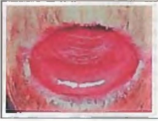
Which test is recommended for this patient? (Recent Question 2019)

Detailed Solution for Test: Malabsorption Syndrome, Pediatric Gastroenterology & MALT-oma - Question 1

The image reveals the occurrence of cheilosis, glossitis, and circumcorneal vascularisation. This indicates a deficiency in riboflavin. The suggested test is to measure RBC glutathione reductase levels.

Test: Malabsorption Syndrome, Pediatric Gastroenterology & MALT-oma - Question 2

Which is the most important complication of celiac sprue? (Recent Question 2016-17)

Detailed Solution for Test: Malabsorption Syndrome, Pediatric Gastroenterology & MALT-oma - Question 2

The primary complication associated with celiac sprue is the emergence of both intestinal and non-intestinal cancers, as well as lymphomas. The complications of celiac sprue include:
Complications of celiac sprue:

  • Intestinal neoplasms/lymphoma
  • Refractory sprue
  • Collagenous sprue

Tropical sprue is associated with:

  • Dermatitis herpetiformis
  • Type 1 diabetes mellitus
  • IgA deficiency
  • Down syndrome
  • Turner syndrome
Test: Malabsorption Syndrome, Pediatric Gastroenterology & MALT-oma - Question 3

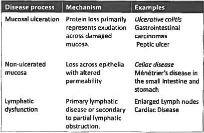
The following conditions can cause protein-losing enteropathy except? (UPSC 2015)

Detailed Solution for Test: Malabsorption Syndrome, Pediatric Gastroenterology & MALT-oma - Question 3

The causes of Protein losing enteropathy can be classified into three groups:

Test: Malabsorption Syndrome, Pediatric Gastroenterology & MALT-oma - Question 4
The following tests can be used for diagnosing celiac disease except: (UPSC 2015)
Detailed Solution for Test: Malabsorption Syndrome, Pediatric Gastroenterology & MALT-oma - Question 4
Antibody testing, particularly for immunoglobulin A anti-tissue transglutaminase antibody (IgA TTG), is regarded as the optimal initial test, although biopsies are required for definitive confirmation. In children under the age of 2, the IgA TTG test should be paired with tests for IgG-deamidated gliadin peptides. The most sensitive and specific antibodies for confirming coeliac disease include:
  • Tissue transglutaminase IgA
  • Endomysial IgA
  • Reticulin IgA
These antibodies correlate with the extent of mucosal damage. Anti-gliadin antibodies were among the earliest serological markers for coeliac disease. However, the typical sensitivity and specificity of anti-gliadin antibodies is approximately 85%.
Test: Malabsorption Syndrome, Pediatric Gastroenterology & MALT-oma - Question 5
Which cereal is not to be given in celiac sprue? (Recent Question 2015-16)
Detailed Solution for Test: Malabsorption Syndrome, Pediatric Gastroenterology & MALT-oma - Question 5
Coeliac disease results from a response to gliadin present in wheat, oats, rye, and barley. When gliadin is encountered, it triggers the formation of anti-tissue transglutaminase antibodies, which react with the tissue of the small intestine, leading to an inflammatory response. This inflammatory reaction results in villous atrophy and subsequently causes osmotic diarrhoea.
Test: Malabsorption Syndrome, Pediatric Gastroenterology & MALT-oma - Question 6

Malabsorption syndrome features include all, except: (Recent Pattern 2014-15)

Detailed Solution for Test: Malabsorption Syndrome, Pediatric Gastroenterology & MALT-oma - Question 6
  • Iron absorption occurs in duodenum which is affected in sprue.
  • Due to fat malabsorption steatorrhea and vitamin D deficiency results.
  • The symptoms & signs of malabsorption depend upon the length of small intestine involved & the age at which the patient presents.
  • Infants (< 2 years) are more likely to present with typical symptoms of malabsorption, including diarrhea, weight loss, abdominal distention, weakness, muscle wasting, or growth retardation.
  • However, they may also be watery and frequent in number (up to 10 - 12 daily).
  • Most patients report chronic diarrhea or flatulence due to colonic bacterial digestion of mal- absorbed nutrients, but the severity of weight loss is variable.
Test: Malabsorption Syndrome, Pediatric Gastroenterology & MALT-oma - Question 7

Anti - T.T.G antibodies are seen in: (Recent Pattern 2014-15)

Detailed Solution for Test: Malabsorption Syndrome, Pediatric Gastroenterology & MALT-oma - Question 7
  • Serological blood tests are the first-line investigation required to make a diagnosis of coeliac disease.
  • Anti-endomysial antibodies of the immuno-globulin A (IgA) type can detect coeliac disease with a sensitivity and specificity of 90% and 99%, respectively.
  • Serology for anli-T.T.G antibodies was initially reported to have a higher sensitivity (99%) and specificity (> 90%) for identifying coeliac disease.
  • Modern anti-tTG assays rely on a human recombinant protein as an antigen. tTG testing should be done first as it is an easier test to perform.
Test: Malabsorption Syndrome, Pediatric Gastroenterology & MALT-oma - Question 8
A 41-year-old patient presented with chronic diarrhea for 3 months. A D-xylose absorption test was order to look for: (Recent Pattern 2014-15)
Detailed Solution for Test: Malabsorption Syndrome, Pediatric Gastroenterology & MALT-oma - Question 8
D-Xylose is a monosaccharide, or simple sugar, that does not need enzymes for digestion before absorption. Its uptake only requires a healthy mucosa. In contrast:
  • Polysaccharides need enzymes, like amylase, to be broken down into monosaccharides for absorption.
In typical individuals, a 25 g oral dose of D-xylose is absorbed and excreted in the urine at roughly 4.5 g within 5 hours. A reduction in urinary excretion of D-xylose is observed in conditions affecting the gastrointestinal mucosa, such as:
  • Small intestinal bacterial overgrowth
  • Whipple's disease
In cases of bacterial overgrowth, D-xylose absorption values return to normal following antibiotic treatment. Conversely, if urinary excretion of D-xylose is normal, the issue is likely related to a non-mucosal cause of malabsorption, such as pancreatic insufficiency. This test was previously utilised but has since become obsolete due to the availability of antibody tests.
Test: Malabsorption Syndrome, Pediatric Gastroenterology & MALT-oma - Question 9

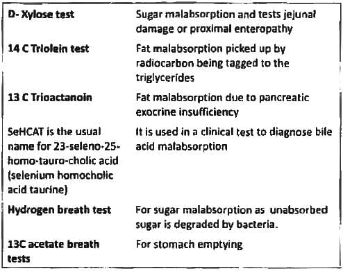
Xylose absorption test is used to assess: (Recent Pattern 2014-15)

Detailed Solution for Test: Malabsorption Syndrome, Pediatric Gastroenterology & MALT-oma - Question 9

Test: Malabsorption Syndrome, Pediatric Gastroenterology & MALT-oma - Question 10

Protein losing enteropathy is characterized by all except: (Recent Pattern 2014-15)

Detailed Solution for Test: Malabsorption Syndrome, Pediatric Gastroenterology & MALT-oma - Question 10

Obstruction of lymphatics for any reason can lead to increased pressure throughout the lymphatic system of the gastrointestinal (GI) tract. This causes lymph to stagnate and, if the pressure becomes sufficiently elevated, results in the escape of lymphatic fluid that is abundant in albumin and other proteins from the lacteals in the intestinal microvilli into the GI tract lumen. If the loss of albumin surpasses the synthesis rate, hypoalbuminaemia, and ultimately, oedema, occur. Moreover, along with albumin, other crucial components of lymph are also released into the bowel, such as:

  • lymphocytes
  • immunoglobulins
  • hydrophobic molecules, including cholesterol
  • lipids
  • fat-soluble vitamins
Test: Malabsorption Syndrome, Pediatric Gastroenterology & MALT-oma - Question 11

Alpha 1 anti-tryspin in stool is indicative of? (Recent Pattern 2014-15)

Detailed Solution for Test: Malabsorption Syndrome, Pediatric Gastroenterology & MALT-oma - Question 11

Alpha-1-antitrypsin (α-1-AT) is synthesised by the liver, intestinal macrophages, monocytes, and the mucous membrane cells in the gut. It is classified as an acute phase protein and is among the most significant proteinase inhibitors.

  • α-1-AT inhibits various proteinases, including trypsin and the elastase produced by neutrophils.
  • Only a minimal quantity of α-1-AT is either cleaved or absorbed in the gut.

Consequently, measuring α-1-AT levels in stool indicates the gut's permeability during inflammatory conditions.

Test: Malabsorption Syndrome, Pediatric Gastroenterology & MALT-oma - Question 12

Jejunal biopsy is diagnostic in: (Recent Pattern 2014-15)

Detailed Solution for Test: Malabsorption Syndrome, Pediatric Gastroenterology & MALT-oma - Question 12
  • Abetalipoproteinemia, or Bassen-Kornzweig syndrome,is a rare autosomal recessive disorder that interferes with the normal absorption of fat and fat-soluble vitamins from food.
  • It is caused by a mutation in microsomal triglyceride transfer protein resulting in deficiencies in the apolipoproteins B-48 and B-100, which are used in the synthesis and exportation of chylomicrons and VLDL respectively.
  • On intestinal biopsy, vacuoles containing lipids are seen in enterocytes. This disorder may also result in fat accumulation in the liver (hepatic steatosis). Because the epithelial cells of the bowel lack the ability to place fats into chylomicrons, lipids accumulate at the surface of the cell, crowding the functions that are necessary for proper absorption.
Test: Malabsorption Syndrome, Pediatric Gastroenterology & MALT-oma - Question 13

True about tropical sprue are A/E: (Recent Pattern 2014-15)

Detailed Solution for Test: Malabsorption Syndrome, Pediatric Gastroenterology & MALT-oma - Question 13

Small intestinal mucosal biopsy is not pathognomonic and mimics fundings of celiac spreua.

Test: Malabsorption Syndrome, Pediatric Gastroenterology & MALT-oma - Question 14

A 30-year-old lady presents with features of malabsorption and iron deficiency anaemia. Duodenal biopsy shows complete villous atrophy. Probable diagnosis is: (Recent Pattern 2014-15)

Detailed Solution for Test: Malabsorption Syndrome, Pediatric Gastroenterology & MALT-oma - Question 14

The presence of anti-endomyosial A/b is 90-05%.

Test: Malabsorption Syndrome, Pediatric Gastroenterology & MALT-oma - Question 15

In celiac sprue there is a deficiency of all except: (Recent Pattern 2014-15)

Detailed Solution for Test: Malabsorption Syndrome, Pediatric Gastroenterology & MALT-oma - Question 15

The proximal section of the gut plays a more significant role in celiac sprue than the distal section. Therefore, vitamin B12 levels are seldom low. Due to considerable damage to the duodenum and jejunum, deficiencies in iron and folic acid are prevalent. Vitamin A deficiency may arise in celiac disease through several mechanisms:

  • Upper digestive issues, such as insufficient stomach acid, can hinder the release of vitamin A from food sources.
  • Protein deficiency disrupts the absorption process across the intestinal lining.
  • Fat malabsorption affects the uptake of vitamin A into the lymphatic system.
Test: Malabsorption Syndrome, Pediatric Gastroenterology & MALT-oma - Question 16

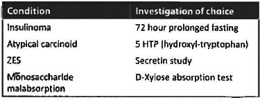
Not included in armamentarium of tests for malabsorption syndrome (AIIMS May 2013)

Detailed Solution for Test: Malabsorption Syndrome, Pediatric Gastroenterology & MALT-oma - Question 16

Test: Malabsorption Syndrome, Pediatric Gastroenterology & MALT-oma - Question 17

Malabsorption syndrome does not result from:  (Recent Pattern 2014-15)

Detailed Solution for Test: Malabsorption Syndrome, Pediatric Gastroenterology & MALT-oma - Question 17

Malabsorption syndrome occurs when the body cannot absorb nutrients properly. Here are the main causes that can lead to this condition:

  • Parasite infestation: Certain parasites can interfere with nutrient absorption.
  • Small bowel diverticulae: These are pouches that can form in the small intestine and may affect absorption.
  • Post gastrectomy: Surgery to remove part of the stomach can lead to difficulty in absorbing nutrients.

However, anterior resection of the colon typically does not cause malabsorption syndrome. This surgery mainly involves removing part of the colon but does not significantly impact the small intestine’s ability to absorb nutrients.

Test: Malabsorption Syndrome, Pediatric Gastroenterology & MALT-oma - Question 18

Non-tropical sprue is characterized by: (Recent Pattern 2014-15)

Detailed Solution for Test: Malabsorption Syndrome, Pediatric Gastroenterology & MALT-oma - Question 18

Nontropical sprue is alternative terminology to describe celiac sprue.
In non-tropical sprue (and to a lesser degree in tropical sprue), microscopic examination of the jejunum reveals

  1. Blunting or absence of the villi
  2. Substitution of cuboidal for columnar surface cells
  3. Infiltration of the lamina propria with lymphocytes and plasma cells
  4. Increased mitotic activity in the crypts of the unusually deep intestinal glands.

(The damage would lead to mal-absorption of carbohydrates and lipids).

Test: Malabsorption Syndrome, Pediatric Gastroenterology & MALT-oma - Question 19

Consider the following:
a. Visible gastric peristalsis
b. Bilious vomiting
c. Palpable mass
d. Melena
Which of the above is/are the feature/features of infantile hypertrophic pyloric stenosis? (UPSC 2015)

Detailed Solution for Test: Malabsorption Syndrome, Pediatric Gastroenterology & MALT-oma - Question 19

Non-bilious vomiting is the first sign of pyloric stenosis. Choice B is incorrect. The vomiting may initially be non-projectile but typically becomes progressive, occurring immediately after a feed. In healthy infants, feeding can assist in diagnosis.

  • After feeding, a visible gastric peristaltic wave may advance across the abdomen.

Choice A is accurate. Diagnosis has traditionally been made by feeling for the pyloric mass. Choice C is also correct. The mass is firm, movable, approximately 2 cm long, olive-shaped, hard, and best palpated from the left side, located above and to the right of the umbilicus in the mid epigastrium beneath the liver's edge. After the infant vomits, the abdominal muscles are more relaxed, making the 'olive' easier to feel. Vomiting typically begins after 3 weeks of age, although symptoms can arise as early as the 1st week of life or as late as the 5th month. As vomiting persists, there is a progressive loss of fluid, hydrogen ions, and chloride, resulting in hypochloraemic metabolic alkalosis.

Test: Malabsorption Syndrome, Pediatric Gastroenterology & MALT-oma - Question 20

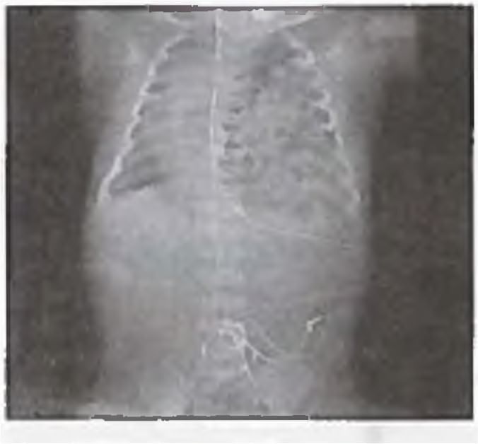
A Term neonate with respiratory distress since birth. The CXR shows?

Detailed Solution for Test: Malabsorption Syndrome, Pediatric Gastroenterology & MALT-oma - Question 20

Observe the air-filled loops of intestine in the left lung. Respiratory distress is a significant indicator in infants with congenital diaphragmatic hernia (CDH). This condition may manifest immediately, or there could be a 'honeymoon' phase lasting up to 48 hours during which the infant appears relatively stable. Early onset of respiratory distress within the first 6 hours of life is considered a negative prognostic sign.

  • The clinical manifestations of respiratory distress include tachypnoea, grunting, utilisation of accessory muscles, and cyanosis.
  • Children diagnosed with CDH will typically present with a scaphoid abdomen and an enlarged chest wall diameter.
  • Bowel sounds may also be audible in the chest alongside diminished breath sounds bilaterally.
Test: Malabsorption Syndrome, Pediatric Gastroenterology & MALT-oma - Question 21

A neonate has been diagnosed with necrotizing enterocolitis with X ray abdomen showing gas in the portal vein. The correct staging of the patient is ? (Recent Pattern 2014-15)

Detailed Solution for Test: Malabsorption Syndrome, Pediatric Gastroenterology & MALT-oma - Question 21

The Bell system is the most widely utilised staging system for describing necrotising enterocolitis (NEC). It categorises the disease as follows:

  • Stage IIA: presents with ileus and pneumatosis intestinalis.
  • Stage IIB: indicates the presence of portal vein gas.
  • Stage IIIA: exhibits characteristics of stage IIB in addition to ascites.
Test: Malabsorption Syndrome, Pediatric Gastroenterology & MALT-oma - Question 22

Exposure to which of the following drugs is incriminated in IHPS (infantile hypertrophic pyloric stenosis): (Recent Pattern 2014-15)

Detailed Solution for Test: Malabsorption Syndrome, Pediatric Gastroenterology & MALT-oma - Question 22

Infantile hypertrophic pyloric stenosis (IHPS) is a condition that affects infants, causing severe vomiting and dehydration. One of the drugs linked to this condition is erythromycin.
Here are some key points regarding this association:

  • Erythromycin is an antibiotic commonly used to treat infections.
  • It can lead to gastrointestinal issues in infants, including IHPS.
  • Recognising the symptoms early is vital for effective treatment.

Other medications like lithium, warfarin, and carbimazole are not associated with this condition.

Test: Malabsorption Syndrome, Pediatric Gastroenterology & MALT-oma - Question 23

All of the following are true regarding primary gastric lymphoma, except? (APPG 2016)

Detailed Solution for Test: Malabsorption Syndrome, Pediatric Gastroenterology & MALT-oma - Question 23

Helicobacter pylori is classified as a type A carcinogen linked to the development of mucosa-associated lymphoid tumour. The primary treatment modality for gastric MALTomas involves using a proton pump inhibitor (PPI) along with antibiotics to eliminate H. pylori.

  • Utilising a combination of amoxicillin, clarithromycin, and PPIs achieves eradication rates of 90%.
  • This combination leads to the regression of low-grade MALToma.

Traditional monotherapy regimens for MALTomas have included chlorambucil, cyclophosphamide, or fludarabine. The presence of t(11;18)(q21;q21) translocations has been identified as a predictor of a poor therapeutic response. MALTOMA is classified as a Non-Hodgkin lymphoma of the B cell type.
The median age at presentation is 65 years, and patients often exhibit non-specific features. Early gastric cancer typically presents with no associated symptoms; however, some individuals with incidental complaints are diagnosed with early gastric cancer. The majority of symptoms related to gastric cancer indicate advanced disease.

Test: Malabsorption Syndrome, Pediatric Gastroenterology & MALT-oma - Question 24

Which is the most common site of MALT? (AIMS Nov 2014)

Detailed Solution for Test: Malabsorption Syndrome, Pediatric Gastroenterology & MALT-oma - Question 24

Most prevalent location of MALT is the ileum. MALT lymphoma can develop in the following areas:

  • stomach
  • orbit
  • intestine
  • lung
  • thyroid
  • salivary gland
  • skin
  • soft tissues
  • bladder
  • kidney
  • CNS
Test: Malabsorption Syndrome, Pediatric Gastroenterology & MALT-oma - Question 25
What is wrong about GIST? (AIIMS Nov 2014)
Detailed Solution for Test: Malabsorption Syndrome, Pediatric Gastroenterology & MALT-oma - Question 25

Gastrointestinal stromal cell tumours (GISTs) were formerly categorised as gastrointestinal leiomyosarcomas and account for 1-3% of gastric neoplasms. Their cell of origin resembles the interstitial cell of Cajal, which regulates peristalsis. The majority of malignant GISTs possess activating mutations in the c-kit gene, resulting in ligand-independent phosphorylation and activation of the KIT receptor tyrosine kinase, which leads to tumour formation.

  • These tumours most commonly affect the anterior and posterior walls of the gastric fundus.
  • They frequently ulcerate and bleed.
  • GISTs seldom invade nearby organs and typically do not metastasise to lymph nodes.
  • However, they can spread to the liver and lungs.

The preferred treatment is surgical resection. GISTs do not respond to traditional chemotherapy; however, 50% of patients achieve an objective response and extended survival when treated with imatinib mesylate (Gleevec) (400-800 mg PO daily), a specific inhibitor of the c-kit tyrosine kinase. Many patients whose GISTs have become resistant to imatinib may benefit from sunitinib (Sutent), which is another inhibitor of the c-kit tyrosine kinase.

Information about Test: Malabsorption Syndrome, Pediatric Gastroenterology & MALT-oma Page
In this test you can find the Exam questions for Test: Malabsorption Syndrome, Pediatric Gastroenterology & MALT-oma solved & explained in the simplest way possible. Besides giving Questions and answers for Test: Malabsorption Syndrome, Pediatric Gastroenterology & MALT-oma, EduRev gives you an ample number of Online tests for practice
Download as PDF