NEET PG Exam > NEET PG Notes > Microbiology > Chapter Notes: Cestodes and Trematodes
Cestodes and Trematodes Chapter Notes | Microbiology - NEET PG PDF Download

Properties
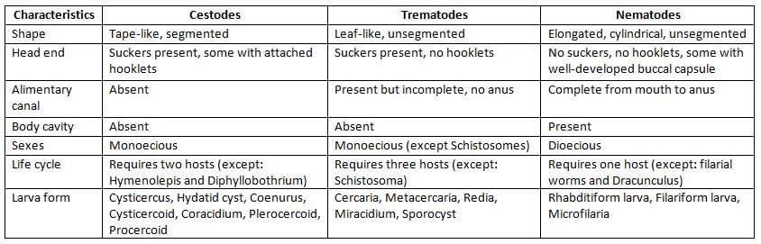
Cestodes
Cestodes or tapeworms represent segmented worms.Intestinal Cestodes — Humans are Definitive Host
- Taenia saginata and T. solium
- Diphyllobothrium
- Hymenolepis nana
- Dipylidium caninum
Tissue Cestodes —Humans are Intermediate Host
- Echinococcus: Causing hydatid disease (liver)
- Taenia solium: Causing cysticercosis (CNS)
- Taenia multiceps: Causing coenurosis (CNS)
- Spirometra: Sparganosis (muscle)
Cestodes Exist in three Morphological Forms
- Adult (tapeworm): Divided to head (scolex), neck and segments called as proglottids or strobila
- Some adults bear hooklets in scolex and called as armed tapeworm, e.g. T. solium, Echinococcus, H. nana
- Eggs: All cestodes eggs have an egg shell and three pair of hooklets except D. latum eggs (operculated)
- Larva: Eggs develop to larva which are called as:
- Cysticercus: Larval stage of Taenia [T. saginata-Cysticercus bovis, T. solium- C. cellulosae]
- Sparganum: Larval stage of Spirometra
- Hydatid cyst: Larval stage of Echinococcus
- Coenurus: Larval stage of Multiceps
- Cysticercoid: Larval stage of Hymenolepis, Dipyllidium
- Diphyllobothrium: has 3 larva stages, L1-Coracidium, L2-Procercoid, L3-Plerocercoid larva
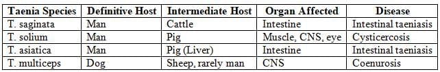
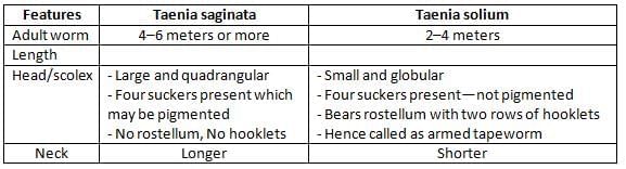
Taenia
Differences between Taenia saginata and Taenia solium:
Proglottids:
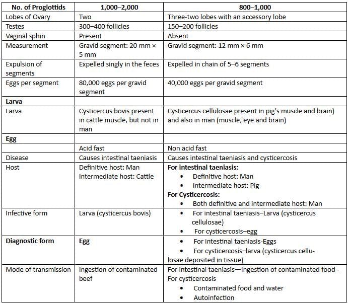
Cysticercosis
- Potentially dangerous systemic disease.
- Definitive host: Man, Intermediate host: Man
- Transmission: (i) Ingestion of food/water contaminated with eggs, (ii) autoinfection
- Eggs develop to larva (Cysticercus cellulosae) in human intestine
- Larvae penetrate the intestine and get deposited in-MC sites- CNS (60-90%) followed by Eye and muscle.
Neurocysticercosis (NCC)
- NCC: MC parasitic CNS infection of man and MC cause of adult onset epilepsy in world.
- MC site: Sub-arachnoid space followed by parenchyma.
- Seizure: MC manifestation (70% of cases). NCC accounts for 50% cases of late onset epilepsy.
- Hydrocephalus, intracranial hypertension and psychiatric disturbances.
- Four morphological stages: Vesicular, necrotic, nodular, calcified stages.
- Clinical feature depends on: (i) No. of cyst, (ii) Location-parenchymal or extraparenchymal, (iii) Size (small cyst-C. cellulosae, big cyst-C. racemosus), (iv) Morphological stage and (v) Host immune response.
- Lab diagnosis:
- Antibody detection by ELISA or Western blot (specific)
- Detection of the cyst by CT/MRI
- CT scan is superior to detect calcified cysts (appears as hyperdense dots).
- MRI is superior to CT scan to detect the Extra parenchymal cysts in ventricle and cisterns, inflammatory changes, vesicular, necrotic lesions and noncystic lesions.
- Del Brutto’s criteria used for neurocysticercosis.
- Treatment: Albendazole, Praziquantel, Surgery (for ocular and spinal and ventricular lesions).
Taenia Multiceps
- Definitive host: Dog, fox and wolf
- Intermediate host: herbivorous animals like sheep (or man)
- Transmission: Ingestion of food/water contaminated with eggs
- Eggs develop to larva (Coenurus) which is a unilocular cyst with multiple scolices.
- Larva penetrate the intestine and get deposited in CNS (Coenurosis)
- Man space occupying lesions in CNS (headache, vomiting, paralysis, seizure, etc.)
- In animals, it causes gid (CNS lesion)
- Epidemiology: African countries (like Uganda, Kenya).
Echinococcus Granulosus
Life Cycle
- Definitive host: Dog and wild carnivores.
- Intermediate hosts: Man and other herbivorous animals.
- Man is an accidental host (dead end).
- Eggs: Infective stage of the parasite.
- Eggs transform to larva (hydatid cyst) that penetrate GIT and migrates to various organs like liver.
Clinical Features
- Hydatid disease: Hepatomegaly (60—70% of cases), then lungs
- E. multilocularis:
- Causes Alveolar Hydatid disease because cyst has multiple locules but has no fluid/free brood capsule
- 90% liver involvement, rapidly metastasizes (mimic malignant tumor)
- E. oligarthrus and E. vogeli: Causes Polycystic Hydatid disease.
Diagnosis
- Hydatid fluid microscopy:
- Wet mount examination to demonstrates protoscolices and brood capsule
- Acid fast staining of centrifuged deposit
- Histological examination
- Casoni’s skin test: Example of immediate hypersensitivity reaction
- Antibody: Indicates past infection, used for seroepidemiology:
- Screening: IHA, CIEP, ELISA
- Confirm: Western Blot (against antigen B fragment )
- Detection of antigen: Indicates Recent infection
- Imaging methods like USG, MRI and X-ray: Demonstrates size, exact location and extension of the cysts
- Water lily sign in USG: Due to collapsed cyst (floating membrane) floating in the abdomen
- Tests to monitor the response to treatment: Imaging methods and Antigen detection methods.
Treatment
- Treatment of choice: Surgery
- DOC: Albendazole and mebendazole
- Commonly preferred method: Percutaneous Aspiration Injection Reaspiration (PAIR) of the cyst.
Diphyllobothrium Latum
- Largest tapeworm in human GIT: Adult is >10 meters with long > 3000 proglottids
- Scolex bears two longitudinal groove called bothria
- Also k/a-fish tapeworm or human broad tapeworm
- Definitive host: Man
- Intermediate host:
- 1st intermediate host: Cyclops/diaptomus
- 2nd intermediate host: Fresh water fish
- There are three larval stages: L1 (coracidium), L2 (procercoid) and L3 (plerocercoid)
- Infective form- Plerocercoid: (L3 stage larva)
- Mode: Ingestion of raw fish
- Life cycle: Ingestion of Plerocercoid (L3) in fish → develop to Adult → Eggs released in feces → Eggs transform to coracidium (L1) in feces → Cyclops (forms Procercoid) → ingested by fish (forms pleurocercoid)
- Causes Megaloblastic anemia (adult worm absorbs B12)
- Diagnostic form: Operculated eggs in stool.
Sparganosis
- Caused by Spirometra and other nonhuman Diphyllobothrium tapeworms
- Definitive hosts: Dogs and cats (rarely man), 1st intermediate host: Cyclops and 2nd intermediate host: Frog, snakes and birds.
- Sparganosis: Sparganum or plerocercoid (L3) larva get deposited in SC tissues, muscles, eyes, lymphatics and visceral organs like brain.
Hymenolepis Nana
- Also called as Dwarf tapeworm
- Egg is infectious to man
- Only one host involved
- Autoinfection seen
- Armed scolex
- Larva form called Cystecercoid larva
- Egg smaller, bile non stained and has polar filament: Diagnostic form.
H. diminuta
- Rat tapeworm
- Mode: Ratflea infected with cystecercoid larva
- Diagnosed by the detection of eggs in the stool: Egg larger and lack polar filament.
Dipylidium Caninum (Double Pored Tapeworm)
- Host: Definitive host—dogs and cats (rarely man), intermediate host—insects (flies)
- Man acquires infection by ingestion of flea containing cysticercoid larva
- GIT symptoms
- Diagnostic form:
- Eggs in packets
- Proglottid has two common genital pore
- Barrel shaped Proglottid.
Treatment of Cestodes
Praziquantel is the DOC of all cestodes followed by Niclosamide except. Hydatid disease and neurocysticercosis: Albendazole.
Trematodes
General Features
- Agents:
- Schistosoma (blood fluke)
- Fasciola hepatica (liver fluke), Fasciolopsis buski (intestinal fluke)
- Paragonimus westermani (lung fluke)
- Clonorchis, Opisthorchis
- Infective form: Metacercaria larva for all except: (Cercaria larva for Schistosoma)
- Definite host: Man
- Intermediate host:
- 1st – Snail
- 2nd–Aquatic plants (F. hepatica and F. buski) Cray fish/crab Fish (Paragonimus, Clonorchis, Opisthorchis)
- Mode of transmission:
- For all: Ingestion of 2nd intermediate host containing Metacercaria larva
- Schistosoma: Skin penetration by cercaria larva present in contaminated water
- All trematodes are Oviparous (lays eggs)
- Diagnostic form:
- For all: Demonstration of operculated Eggs
- Schistosoma: Demonstration of nonoperculated Eggs
- All trematodes are hermaphrodite (except Schistosoma: sexes are separate)
- DOC: Praziquantel is DOC of all trematodes except F. hepatica (Triclabendazole).
Schistosoma Hematobium – (Blood Fluke)
- Resides in: Vesical and pelvic venous plexus
- Associated with:
- Hematuria
- Hydroureter and hydronephrosis
- Bladder Carcinoma: Sqamous cell Ca (in high worm burden) > transitional cell Ca (low worm burden)
- Egg has terminal spine
- Antibody detection:
- HAMA-FAST: ELISA (Falcon assay screening test ELISA) using S. haematobium adult worm microsomal antigen (HAMA).
- HAMA Western blot: Specific
- Other methods: Cercarial Huller reaction, IFA, IHA
- Antigen detection:
- Circulating cathodic antigen (CCA) in urine and circulating anodic antigen (CAA) in serum.
- It indicates recent infection and can be used for monitoring the treatment.
S. Mansoni
- Common in Africa including Caribbean Islands (West Indies), South America
- Resides in mesenteric veins draining sigmoido-rectal region.
- Clinical manifestation:
- Swimmer’s itch (cercarial dermatitis): Type 1 hypersensitivity reaction
- Dysentery and Eosinophilic diarrhea
- Acute schistosomiasis (Katayama fever) Serum sickness like illness: Type III hypersensitivity
- Chronic schistosomiasis: due to fibrosis and granuloma formation as a result of egg deposition in various sites like intestinal wall, liver, spleen and lungs.
- Secondary bacterial infection especially with Salmonella spp.
- Diagnostic form: Egg has lateral spine (feces), eggs of S. mansoni are acid fast.
- Quantitations of eggs in stool by Kato Katz thick smear technique.
S. Japonicum
- Resides in mesenteric veins draining the ileocecal region.
- Clinical feature: Similar to S. mansoni but it is more severe due to higher egg production and smaller size of the eggs (easy dissemination).
- Diagnostic form: Eggs in stool (has rudimentary lateral spine).
Fasciola Hepatica - Sheep Liver Fluke
- Definitive host: Sheep or man, Intermediate host- 1st -Snail and 2nd -Water cress
- Mode of transmission: Ingestion of aquatic plant contaminated with encysted metacercaria.
- Clinical manifestation:
- Hepatomegaly
- Halzoin (laryngeal edema): d/t eating sheep liver
- Bile duct obstruction
- Diagnostic form: Operculated eggs in feces.
Fasciolopsis Buski
- Intestinal fluke → Largest fluke
- Definite host: Man/pig
- Mode of transmission: Ingestion of aquatic plant contaminated with encysted metacercaria
- GIT symptoms
- Diagnostic form: Operculated eggs in feces.
Paragonimus Westermani: (Lung Fluke)
- Definitive: host man; Intermediate host- 1st –snail, 2nd - Cray/Crab fish
- Mode of transmission: Ingestion of crab/crey fish contaminated with encysted metacercaria
- Cyst in Right lung (granuloma formation due to egg deposition)
- Cerebral and cutaneous paragonimiasis
- Golden brown sputum
- Causes endemic hemoptysis
- Diagnostic form: Operculated eggs in early morning, deeply coughed sputum
- Endemic in Manipur.
Clonorchis Sinensis
- Oriental/Chinese liver fluke
- Definitive: host man; Intermediate host: 1st –snail, 2nd - Cray/crab fish
- Mode of transmission: Ingestion of crab/crey fish contaminated with encysted metacercaria
- Causes:
- Cholangitis, dilatation of the bile duct and ductal epithelial hyperplasia and fibrosis
- Cholangiocarcinoma
- Diagnostic form: Flask shaped operculated egg in stool.a
The document Cestodes and Trematodes Chapter Notes | Microbiology - NEET PG is a part of the NEET PG Course Microbiology.
All you need of NEET PG at this link: NEET PG
|
75 docs|5 tests
|
FAQs on Cestodes and Trematodes Chapter Notes - Microbiology - NEET PG
| 1. What are the main morphological forms of cestodes? |  |
Ans. Cestodes exist in three primary morphological forms: Taenia, Cysticercosis, and Neurocysticercosis (NCC). Each form represents a different stage or type of the organism, which can lead to various clinical manifestations and complications in infected hosts.
| 2. What is cysticercosis and how does it relate to Taenia? | |
Ans. Cysticercosis is an infection caused by the larval stage of the Taenia solium (pork tapeworm). When humans ingest the eggs of Taenia solium, the larvae can develop in tissues, leading to cysticercosis. This condition can affect multiple organs, including muscles and the central nervous system, potentially causing serious health issues.
| 3. What are the clinical features of neurocysticercosis (NCC)? | |
Ans. Neurocysticercosis (NCC) can present with a variety of clinical features, including seizures, headaches, and neurological deficits. Symptoms vary depending on the number and location of cysts in the brain. In some cases, it can lead to more severe complications, such as hydrocephalus or increased intracranial pressure.
| 4. How is Echinococcus granulosus related to cestodes, and what condition does it cause? | |
Ans. Echinococcus granulosus is a type of cestode that causes hydatid disease (or hydatidosis) in humans. Infection occurs when humans accidentally ingest Echinococcus eggs, leading to the formation of cysts in various organs, particularly the liver and lungs. These cysts can cause significant health problems if they grow large or rupture.
| 5. What are the common diagnostic methods for detecting cestode infections? | |
Ans. Common diagnostic methods for detecting cestode infections include serological tests, imaging techniques (such as CT or MRI scans for NCC), and stool examinations to identify eggs or segments of the tapeworm. Accurate diagnosis is crucial for effective treatment and management of the infections.
About this Document

Oct 10, 2025
Last updated
Document Description: Chapter Notes: Cestodes and Trematodes for NEET PG 2025 is part of Microbiology preparation.
The notes and questions for Chapter Notes: Cestodes and Trematodes have been prepared according to the NEET PG exam syllabus. Information about Chapter Notes: Cestodes and Trematodes covers topics
like Properties, Cestodes, Trematodes and Chapter Notes: Cestodes and Trematodes Example, for NEET PG 2025 Exam. Find important definitions, questions, notes, meanings, examples, exercises and tests below for Chapter Notes: Cestodes and Trematodes.
Introduction of Chapter Notes: Cestodes and Trematodes in English is available as part of our Microbiology
for NEET PG & Chapter Notes: Cestodes and Trematodes in Hindi for Microbiology course.
Download more important topics related with notes, lectures and mock test series for NEET PG
Exam by signing up for free. NEET PG: Cestodes and Trematodes Chapter Notes | Microbiology - NEET PG
Description
Full syllabus notes, lecture & questions for Cestodes and Trematodes Chapter Notes | Microbiology - NEET PG - NEET PG | Plus excerises question with solution to help you revise complete syllabus for Microbiology | Best notes, free PDF download
Information about Chapter Notes: Cestodes and Trematodes
In this doc you can find the meaning of Chapter Notes: Cestodes and Trematodes defined & explained in the simplest way possible. Besides explaining types of
Chapter Notes: Cestodes and Trematodes theory, EduRev gives you an ample number of questions to practice Chapter Notes: Cestodes and Trematodes tests, examples and also practice NEET PG
tests

Related Searches
Summary
,Semester Notes
,video lectures
,Objective type Questions
,past year papers
,ppt
,Previous Year Questions with Solutions
,Extra Questions
,study material
,Important questions
,Cestodes and Trematodes Chapter Notes | Microbiology - NEET PG
,Exam
,mock tests for examination
,MCQs
,Cestodes and Trematodes Chapter Notes | Microbiology - NEET PG
,Cestodes and Trematodes Chapter Notes | Microbiology - NEET PG
,practice quizzes
,Free
,shortcuts and tricks
,Sample Paper
,Viva Questions
;


Additional Information about Chapter Notes: Cestodes and Trematodes for NEET PG Preparation
Chapter Notes: Cestodes and Trematodes Free PDF Download
The Chapter Notes: Cestodes and Trematodes is an invaluable resource that delves deep into the core of the NEET PG exam.
These study notes are curated by experts and cover all the essential topics and concepts, making your preparation more efficient and effective.
With the help of these notes, you can grasp complex subjects quickly, revise important points easily,
and reinforce your understanding of key concepts. The study notes are presented in a concise and easy-to-understand manner,
allowing you to optimize your learning process. Whether you're looking for best-recommended books, sample papers, study material,
or toppers' notes, this PDF has got you covered. Download the Chapter Notes: Cestodes and Trematodes now and kickstart your journey towards success in the NEET PG exam.
Importance of Chapter Notes: Cestodes and Trematodes
The importance of Chapter Notes: Cestodes and Trematodes cannot be overstated, especially for NEET PG aspirants.
This document holds the key to success in the NEET PG exam.
It offers a detailed understanding of the concept, providing invaluable insights into the topic.
By knowing the concepts well in advance, students can plan their preparation effectively.
Utilize this indispensable guide for a well-rounded preparation and achieve your desired results.
Chapter Notes: Cestodes and Trematodes
Chapter Notes: Cestodes and Trematodes Notes offer in-depth insights into the specific topic to help you master it with ease.
This comprehensive document covers all aspects related to Chapter Notes: Cestodes and Trematodes.
It includes detailed information about the exam syllabus, recommended books, and study materials for a well-rounded preparation.
Practice papers and question papers enable you to assess your progress effectively.
Additionally, the paper analysis provides valuable tips for tackling the exam strategically.
Access to Toppers' notes gives you an edge in understanding complex concepts.
Whether you're a beginner or aiming for advanced proficiency, Chapter Notes: Cestodes and Trematodes Notes on EduRev are your ultimate resource for success.
Chapter Notes: Cestodes and Trematodes NEET PG Questions
The "Chapter Notes: Cestodes and Trematodes NEET PG Questions" guide is a valuable resource for all aspiring students preparing for the
NEET PG exam. It focuses on providing a wide range of practice questions to help students gauge
their understanding of the exam topics. These questions cover the entire syllabus, ensuring comprehensive preparation.
The guide includes previous years' question papers for students to familiarize themselves with the exam's format and difficulty level.
Additionally, it offers subject-specific question banks, allowing students to focus on weak areas and improve their performance.
Study Chapter Notes: Cestodes and Trematodes on the App
Students of NEET PG can study Chapter Notes: Cestodes and Trematodes alongwith tests & analysis from the EduRev app,
which will help them while preparing for their exam. Apart from the Chapter Notes: Cestodes and Trematodes,
students can also utilize the EduRev App for other study materials such as previous year question papers, syllabus, important questions, etc.
The EduRev App will make your learning easier as you can access it from anywhere you want.
The content of Chapter Notes: Cestodes and Trematodes is prepared as per the latest NEET PG syllabus.
|
© EduRev
|
Education Revolution
|


|


Signup to see your scores
go up within 7 days!
Access 1000+ FREE Docs, Videos and Tests
Takes less than 10 seconds to signup