Miscellaneous viruses Chapter Notes | Microbiology - NEET PG PDF Download
| Table of contents |
|
| Rodent-borne Viral Infections |
|
| Filoviruses |
|
| Coronaviruses |
|
| Slow Viruses and Prions |
|
| Rotavirus |
|

Rodent-borne Viral Infections
Rodent-borne viruses, known as roboviruses, spread from rodents to humans via direct contact with bodily fluids or waste, without involving insect carriers, distinguishing them from arboviruses. They primarily fall into two categories.
Hantaviruses
These viruses are round, covered with an envelope, and possess three parts of negative-sense single-stranded RNA, classified under Bunyaviridae family.- Rodents serve as the natural hosts.
- Humans get infected by breathing in particles from rodent droppings.
- They lead to two serious illnesses in people.
- Hemorrhagic fever accompanied by kidney syndrome (involving inflammation in kidney tissues) results from various hantavirus types including Hantaan virus, Dobrava virus, Puumalavirus (causing nephropathia epidemica), and Seoul virus.
- Hantavirus lung syndrome is triggered by Sin Nombre virus.
- For example, in regions with high rodent populations, such as rural areas, inhaling contaminated dust during cleaning can lead to hantavirus infection, illustrating how everyday activities can pose risks.
Arenaviruses
These viruses vary in shape, measure 50–300 nm, have an envelope with prominent club-like spikes, and include two segments of single-stranded RNA.- New world types: Junin, Machupo, Guanarito, and Sabia viruses, responsible for hemorrhagic fevers in South America.
- Old world types: Lassa fever virus (found in Africa, treated primarily with ribavirin) and Lymphocytic choriomeningitis (LCM) virus.
- For instance, Lassa fever often spreads in West African households through contact with infected rodent urine, demonstrating the role of poor sanitation in transmission.
Filoviruses
- Comprise two groups: Ebolavirus and Marburgvirus, both inducing African hemorrhagic fever.
- They vary in form, often appearing as extended threads, sized from 80–1000 nm, averaging 665 nm for Marburg and 805 nm for Ebola.
- Extremely deadly, among all viral bleeding fevers, with fatality rates ranging from 25–90%.
Ebola virus
- Global Threat: The Ebola virus became a serious global concern during its outbreak in 2014, which the World Health Organization (WHO) declared a public health emergency affecting the whole world.
- History: The virus first appeared in humans in 1976 near the Ebola River in Africa, which is how it got its name.
- Species:There are five known types of the Ebola virus:
- Zaire
- Sudan
- Taï Forest
- Reston
- Bundibugyo
- Geographical Distribution: Since its discovery, the Ebola virus has led to multiple outbreaks in African nations, with over 17,000 reported cases and nearly 7,000 deaths. The largest outbreak took place in 2014, affecting Guinea, Liberia, and Sierra Leone.
- As of January 2016, there were about 28,637 suspected cases and 11,315 deaths recorded.
- Case Statistics:
- Sierra Leone reported the highest number of cases.
- However, Liberia had the most deaths.
- Currently, Guinea is the only country experiencing widespread transmission, while control measures are in place in Sierra Leone and Liberia.
- Reservoir: The exact source of the virus is unknown, but it is believed to come from infected animals, likely fruit bats or primates (such as apes and monkeys).
- Transmission:
- The virus spreads through direct contact with the blood, bodily fluids, or organs of infected animals or humans, and also through contaminated surfaces and materials (like bedding or clothing).
- Health care workers and family members of infected individuals are at higher risk of infection.
- Ebola can remain in semen for up to 3 months, but sexual transmission has not been reported.
- Clinical Manifestations:
- The incubation period for Ebola is approximately 2 to 12 days, with an average of 8 to 10 days.
- Common symptoms include:
- Fever
- Headache
- Muscle pain
- Sore throat
- Abdominal pain
- Vomiting
- Diarrhea
- Rash
- Hemorrhaging (bleeding or bruising), which can lead to shock and death.
- Laboratory Diagnosis:
- Antibody Detection: ELISA tests can identify both IgM and IgG antibodies using recombinant nucleoprotein and glycoprotein antigens.
- Serum Antigen Detection: Capture ELISA tests target proteins such as NP, VP40, and GP.
- Molecular Methods: Reverse transcriptase PCR (RT-PCR) and real-time RT-PCR are used to find viral RNA.
- Electron Microscopy: This can show typical filamentous viruses in specimens.
- Virus Isolation: Specimens must be processed in biosafety level-4 cabinets to prevent laboratory spread.
- Treatment: Supportive care, such as rehydration, is necessary. There is currently no specific treatment or vaccine available.
- Measures Taken in India:
- As of now, there have been no confirmed Ebola cases in India.
- Due to the risk of infection from travelers, strict monitoring is happening at Indian airports.
- Individuals arriving with fever and a recent history of travel to Guinea, Liberia, Sierra Leone, or Mali are quarantined at the airport until they test negative for the Ebola virus.
Marburg Virus
Marburg illness first noted in Germany and Yugoslavia in 1967 among lab personnel handling African green monkey tissues from Africa.
- Subsequently, more than 450 instances in African countries like Kenya, South Africa, Democratic Republic of Congo, Uganda, and Angola.
- Latest epidemic in Angola (2005), involving 252 individuals and 227 deaths (90% fatality).
Coronaviruses
These are covered viruses with petal or club-shaped spikes resembling a solar crown. Mainly affect animals and birds; human cases are very uncommon.
- Most human strains are common globally, causing light upper airway infections and sometimes loose stools.
- Exceptions: SARS-CoV and MERS-CoV, limited to specific areas, person-to-person spread, causing severe lung disease outbreaks with elevated death rates.
- Transmission:
- Human coronaviruses can spread through coughing and sneezing.
- Close personal contact, like touching your mouth, nose, or eyes, can also lead to the spread.
- Shaking hands is another way these viruses can be passed from one person to another.
- The virus known as SARS-CoV can spread through droplets released when an infected person coughs or sneezes.
- In rare cases, the virus might spread through the air, which is referred to as airborne spread.
SARS-CoV (Severe Acute Respiratory Syndrome Coronavirus)
- History: SARS was first identified in China in 2003 by Dr. Carlo Urbani, a doctor from the WHO. He diagnosed the illness in a businessman who had traveled from China to Hong Kong and then to Hanoi, Vietnam. Both the businessman and the doctor who diagnosed SARS did not survive the illness.
- Epidemiology: During the outbreak in 2003, the SARS virus spread from Asia to many parts of the world, leading to nearly 8,098 cases in 29 countries and resulting in over 774 deaths that year. However, India reported no cases. Since 2004, there have been no reported cases worldwide.
- Source: It is thought that humans contract the SARS-CoV virus from animals, including monkeys, Himalayan palm civets, raccoon dogs, cats, dogs, and rodents.
- Clinical Manifestation: SARS typically causes a serious infection in the lower respiratory tract, which includes symptoms such as:
- Muscle pain
- Headache
- Sore throat
- Fever
MERS-CoV (Middle East Respiratory Syndrome Coronavirus)
MERS-CoV has been known to cause a serious type of lower respiratory illness, with a mortality rate of 30%.
- Epidemiology: This virus was first reported in Saudi Arabia in 2012. Since then, hundreds of cases have been documented in various countries around the Arabian Peninsula, including:
- Saudi Arabia
- UAE
- Qatar
- Oman
- Jordan
- Kuwait
- Yemen
- Lebanon
- Iran
- The exact source of the virus is unknown, but it is believed to be transmitted from camels and bats.
- People who are at greater risk for MERS-CoV infection include:
- Those who have traveled to the Arabian Peninsula in the last 14 days
- Close contacts of confirmed MERS cases
- Healthcare workers not following safety precautions
- Individuals who have been around infected camels
- Clinical Manifestation: The incubation period for MERS-CoV is typically between 2 to 14 days. Symptoms can include:
- High fever
- Cough
- Shortness of breath
Slow Viruses and Prions
Slow virus diseases, including prion diseases, are a set of neurodegenerative disordersthat impact both humans and animals. These diseases are marked by:
- A long incubation period, which can last from months to years.
- A tendency to affect the central nervous system (CNS): Slow viruses primarily target the CNS.
- The vacuuming of neurons (known as spongiform changes), leading to the buildup of amyloid-like plaques and gliosis.
- Symptoms that include:
- Loss of muscle control
- Shivering
- Tremors
- Dementia
- These diseases are always fatal.
- A strong genetic predisposition is often observed.
- Slow viruses and prions are not antigenic; as a result, there is:
- A lack of immune response and interferon production against the viral proteins.
- A lack of inflammation associated with the disease.
- They do not cause cytopathologic effects in laboratory conditions (in vitro).
- Slow virus diseases can be caused by:
- Conventional viruses
- Unconventional viruses, known as prions.
Slow viral diseases
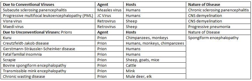
Prion Diseases
- Prions are protein-based infectious agents without nucleic acids, filterable like viruses but resistant to many sterilization methods.
- Numerous prion conditions affect humans and animals.
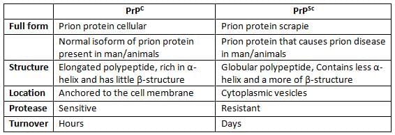
Prion Proteins have Two Isoforms
- PrPSc is the disease-causing prion protein, named after scrapie where it was first found.
- PrPC is the regular cell form of prion protein on mammal membranes, coded on chromosome 20, precursor to PrPSc, differing in various aspects.
Mechanism of Prion Diseases
The theory put forth by Stanley B. Prusiner, who won the Nobel Prize in 1997, explains the process in detail.
- When an individual becomes infected, the prion proteins (referred to as PrPSc) move to the neurons. They attach to the normal PrPC proteins on the surface of the cells.
- This attachment leads to the release of PrPC from the cell surface, which is then transformed into the harmful form known as PrPSc. This transformation is a post-translational modification that changes the long polypeptide PrPC into a globular form called PrPSc.
- The cell produces new PrPC, and this cycle continues, resulting in the accumulation of a large amount of PrPSc.
- The PrPSc proteins group together to form amyloid-like plaques in the brain. Since these plaques are made up of the host's proteins, there is minimal immune response or inflammation.
- PrPSc proteins are taken up by neurons and build up inside the cytoplasmic vacuoles, giving the cells a spongiform look.
Laboratory Diagnosis
- Detection of PrPSc can be performed using a conformation-dependent immunoassay, which is a reliable diagnostic method.
- Brain MRI results show that over 90% of patients have increased brightness in the basal ganglia and observe cortical ribboning.
- Neuropathological diagnosis from brain biopsies reveals spongiform degeneration and astrocytic gliosis without any signs of inflammation under a light microscope.
- Sequencing the PRNP gene helps find mutations, which is crucial for identifying familial cases of prion diseases.
- The stress protein 14-3-3 is found to be elevated in the CSF (cerebrospinal fluid).
- An abnormal EEG may be observed in the late stages of the disease, characterized by high-voltage, triphasic sharp waves.
Treatment
- No proven therapy to stop or cure prion conditions.
- Trials with quinacrine, anti-PrP antibodies cleared PrPSc in cells but not in living models.
Decontamination
Prions are very tough and do not easily break down with regular cleaning methods. The following are suggested ways to sterilize items that have been in contact with prion proteins:
- Use an autoclave at a temperature of 134 °C for 1 to 1.5 hours.
- Treat with 1 N NaOH for 1 hour.
- Soak in a solution of 0.5% sodium hypochlorite for 2 hours.
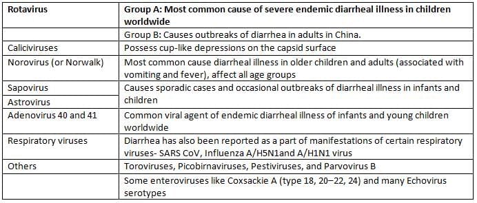
Rotavirus
Rotaviruses are part of the Reoviridae family, which is the only family of RNA viruses that has double-stranded RNA (dsRNA).
- They are enclosed by a triple-layered capsid.
- Rotaviruses contain 11 segments of segmented dsRNA.
- There are a total of 12 proteins:
- 6 structural viral proteins (VPs)
- 6 nonstructural proteins (NSPs)
Classification of Rotaviruses
- Traditional Classification: Based on the group-specific VP6 antigen, rotaviruses are classified into seven groups (A-G). Group A is the primary cause of diarrhea in humans, with groups B and C being less common.
- Binary Typing System: Rotaviruses are typed using VP7 (a glycoprotein, or G-type antigen) and VP4 (a protease-sensitive protein, or P-type antigen).
- Methods for typing include:
- Serotyping (via neutralization tests)
- Genotyping (via sequencing)
- Currently, there are 19G and 28[P] types identified, with the most prevalent type worldwide, including India, being G1P [8], accounting for about 70% of isolates.
Clinical Manifestation
- Rotaviruses are the leading cause of diarrheal illness in young children.
- The incubation period is approximately 1 to 3 days.
- Symptoms typically begin suddenly with:
- Vomiting
- Watery diarrhea
- Fever
- Abdominal pain
Laboratory Diagnosis
- Direct detection of the virus can be done through:
- Immunoelectron microscopy (IEM): Shows rotaviruses with sharp-edged triple-shelled capsids resembling spokes around a wheel hub.
- Isolation of rotavirus: This is challenging, but tissue cultures may be used to help with replication.
- Detection of viral antigen in stool can be done using:
- ELISA
- Latex agglutination methods
- RT-PCR is the most sensitive test for detecting rotavirus in stool samples.
- Serologic tests (ELISA) can help identify increases in antibody levels, useful for studying seroprevalence.
Treatment and Control
- Treatment focuses on supportive care, mainly to replace lost fluids and electrolytes, which can be administered either orally or through injections.
- Vaccine: The human live attenuated rotavirus vaccine, known as Rotarix, is available.
- This vaccine contains live attenuated strains of rotavirus from serotypes G1, G2, G3, G4, and G9.
- Administration schedule: The vaccine is given orally in two doses at 2 and 4 months of age.
- The most serious complication associated with the rotavirus vaccine is intussusception.
Viral agents of Gastroenteritis and their features
|
75 docs|5 tests
|
FAQs on Miscellaneous viruses Chapter Notes - Microbiology - NEET PG
| 1. What are hantaviruses and how do they affect human health? |  |
| 2. How do arenaviruses differ from other virus families? | |
| 3. What are the key differences between Ebola virus and Marburg virus? | |
| 4. What are the main transmission routes for SARS-CoV and MERS-CoV? | |
| 5. What are prion diseases and how do they differ from viral infections? | |


Exam
,mock tests for examination
,Important questions
,Objective type Questions
,ppt
,Miscellaneous viruses Chapter Notes | Microbiology - NEET PG
,shortcuts and tricks
,Miscellaneous viruses Chapter Notes | Microbiology - NEET PG
,Extra Questions
,MCQs
,Viva Questions
,Free
,Semester Notes
,Sample Paper
,Previous Year Questions with Solutions
,Miscellaneous viruses Chapter Notes | Microbiology - NEET PG
,past year papers
,video lectures
,study material
,practice quizzes
,Summary
;


Chapter Notes: Miscellaneous viruses Free PDF Download
Importance of Chapter Notes: Miscellaneous viruses
Chapter Notes: Miscellaneous viruses
Chapter Notes: Miscellaneous viruses NEET PG Questions
Study Chapter Notes: Miscellaneous viruses on the App
|
© EduRev
|
Education Revolution
|


|


