NEET PG Exam > NEET PG Notes > Microbiology > Chapter Notes: Vibrio
Vibrio Chapter Notes | Microbiology - NEET PG PDF Download
| Table of contents |
|
| Classification of Vibrio cholerae |
|
| Pathogenesis of Cholera |
|
| Clinical Manifestations of Cholera |
|
| Epidemiology |
|
| Laboratory Diagnosis |
|
| Cholera Treatment |
|
| Cholera Vaccines |
|
| Halophilic Vibrios |
|

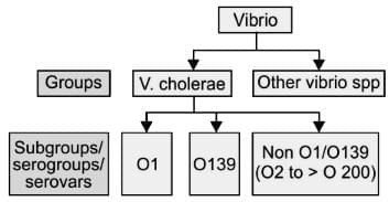
Classification of Vibrio cholerae
1. Based on Salt Requirement:
- Non-halophilic vibrios: These vibrios cannot survive in high salt levels. Examples include V. cholerae and V. mimicus.
- Halophilic vibrios: These vibrios require salt for their growth and thrive in salt concentrations of 7–10%. Examples include V. parahaemolyticus, V. alginolyticus, and V. vulnificus.
2. V. cholerae is Further Classified
- Serogrouping: V. cholerae is divided into over 200 serogroups or serovars based on the O antigen side chains of lipopolysaccharides.
- The O1 serogroup has been responsible for all cholera pandemics and most epidemics.
- Non-agglutinable vibrios (NAG vibrios) are those that do not belong to the O1 serogroup.
- The O139 serogroup was identified in 1992 and is known for causing cholera outbreaks in India and Bangladesh.
- Non-O1/O139 serogroups have occasionally caused isolated cases of diarrhea and other issues but have not led to epidemics.
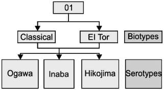
- Serotyping: The O1 serogroup can be further divided into three serotypes: Inaba, Ogawa, and Hikojima, based on small differences in the O antigen.
- Ogawa is the most common serotype found in clinical samples, followed by Inaba.
- During outbreaks, a shift from Ogawa to Inaba can occur.
- Hikojima is a transitional state showing both Inaba and Ogawa antigens.
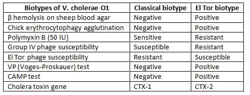
- Biotyping: The O1 serogroup has two biotypes: classical and El Tor, distinguished by various biochemical reactions.
- The classical biotype caused the first six cholera pandemics.
- Currently, most cholera cases are linked to El Tor, although some classical isolates still exist.
- Some isolates do not fit into either biotype and are termed El Tor variants.
- El Tor variants: Recent variants of the El Tor biotype, found in Bangladesh and other regions, show characteristics similar to the classical biotype, such as:
- Matlab variants (El Tor hybrid): These strains couldn't be classified into types because they have features from both classical and El Tor strains. They were first identified in Bangladesh in 2002.
- Mozambique variant (2004–2005): This variant shows typical characteristics and the genetic makeup of El Tor, but the cholera toxin and its gene (CTX) belong to the classical type.
Pathogenesis of Cholera
Cholera is primarily caused by the cholera toxin produced by V. cholerae O1 and O139.
- Transmission: Cholera spreads through contaminated water or food.
- Infective Dose: high dose of about 10 8 bacilli is needed to survive the acidic environment of the stomach.
- Factors Promoting Transmission: Conditions like low stomach acidity (hypochlorhydria) or the use of antacids increase the risk of infection.
- In the small intestine, V. cholerae overcomes the protective mucus layer due to its strong motility and the production of mucinase and hemagglutinin protease (cholera lectin).
- Adhesion: The bacteria adhere to the intestinal lining with the help of fimbriae known as the toxin co-regulated pilus (TCP).
- Cholera Toxin (CT)
- CT consists of two peptide fragments: A and B.
- Fragment B: This fragment binds to GM1 ganglioside receptors on the intestinal lining.
- Fragment A: The active fragment (27 kDa) modifies a G protein, increasing the activity of adenylate cyclase and raising cyclic AMP levels within the cell.
- The increase in cyclic AMP levels leads to:
- Accumulation of Sodium Chloride: This occurs in the intestinal lumen.
- Water influx into the lumen, resulting in isotonic fluid accumulation and watery diarrhea.
- Fluid and Electrolyte Loss: This can cause shock due to severe dehydration and acidosis from bicarbonate loss.
- The gene for cholera toxin (CTX) is encoded by a phage. The ToxR gene regulates the expression of CT, TCP, and other virulence factors. V. cholerae has two circular chromosomes, unlike most bacteria with a single chromosome.
- Other Virulence Factors
- Zonula Occludens Toxin: Disrupts tight junctions between mucosal cells.
- Siderophore: Essential for iron acquisition.
- Bacterial Endotoxin (LPS): The LPS of V. cholerae does not contribute to pathogenesis but triggers an immune response and is used in killed vaccines.
Clinical Manifestations of Cholera
- Infection with Vibrio cholerae O1 or O139 leads to a range of symptoms, including:
- Asymptomatic: 75% of cases
- Mild Diarrhoea: 20% of cases
- Explosive Diarrhoea: 5% of cases (cholera gravis)
- Incubation Period: Typically ranges from 24 to 48 hours.
- Watery Diarrhoea: Usually begins suddenly and is painless.
- Rice Water Stool:
- Non-bilious, slightly cloudy, grey, and watery.
- Contains mucus flakes but no blood or pus cells.
- Has a mild, fishy odour.
- Other symptoms may include vomiting and muscle cramps, while fever is generally absent.
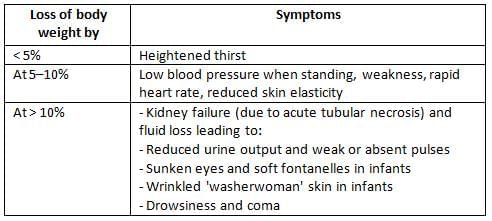
- Complications: These are associated with fluid loss and result in a decrease in body weight.
Loss of Body Weight:
Epidemiology
History of Pandemics
Cholera can manifest in various forms, including sporadic cases, limited outbreaks, endemic situations, epidemics, or pandemics.
- The disease originates from the delta region of the Ganges and Brahmaputra rivers, located in West Bengal (India) and Bangladesh.
- The first six pandemics, spanning from 1817 to 1923, were caused by the classical biotype of V. cholerae. This biotype spread from Bengal, leading to thousands of deaths worldwide.
- The seventh pandemic,which began in 1961, was distinct from the earlier ones.
- It was the only pandemic that originated outside India, starting from Indonesia (specifically Sulawesi, formerly Celebes Island) in 1961. India was affected in 1964, and by 1991, the pandemic had spread globally.
- This pandemic was caused by the El Tor biotype, which resulted in milder cases of cholera with a higher rate of carriers compared to the classical form. El Tor is more resilient but less harmful than the classical vibrios.
- The O139 strain, also known as the Bengal strain, was first identified in Chennai in 1992. It was classified as a new serogroup, O139, as it was not agglutinated by any available antisera at that time (O1 to O138) .
- O139 appears to be a variation of O1 El Tor, differing due to a unique lipopolysaccharide (LPS) and being capsulated. This allows it to invade the body and cause conditions beyond just cholera.
- There is no cross-protection between O1 and O139. Currently, O139 accounts for a minority of cholera cases in India and Bangladesh.
Seventh Pandemic
- The seventh pandemic is notable for being the only one that originated outside of India.
- It is also unique for being the only pandemic caused by the El Tor biotype.
- This pandemic resulted in milder cases of cholera, characterized by a higher rate of carriers compared to previous pandemics.
Classical vs El Tor
- El Tor biotype is associated with a higher carrier rate.
- The classical biotype, on the other hand, is more virulent.
- The case-carrier ratio further illustrates this difference:
- For the classical biotype, the ratio is 1:50.
- For the El Tor biotype, the ratio is 1:90.
Epidemiological Determinants of Cholera
- Reservoir: Humans are the only reservoir for V. cholerae, as there is no animal reservoir.
- Source of Infection: Infections may originate from asymptomatic cases or carriers.
- Carriers: Healthy individuals who shed the bacilli in their feces. Carriers can be:
- Incubatory Carriers: These are less common due to the short incubation period of cholera, which is typically 1 to 2 days.
- Persistence of V. cholerae:
- During Epidemics: V. cholerae is maintained by carriers.
- During Inter-Epidemic Periods: The bacteria can be found in sea water, crustaceans, and plankton.
- Resistance of V. cholerae:
- V. cholerae is acid-labile but stable in alkaline conditions.
- It is heat-labile but remains stable when refrigerated, lasting in ice for 4 to 6 weeks.
- Selective Media for V. cholerae:
- Alkaline bile salt agar (BSA)
- Monsoor's GTTTA medium
- TCBS agar
- Types of Carriers:
- Convalescent Carriers: Recovered patients who continue to shed the bacilli for 2 to 3 weeks.
- Contact Carriers: Healthy individuals with subclinical infections who shed bacilli for less than 10 days.
- Chronic Carriers: small number of convalescent carriers become chronic. The El Tor biotype generally has a higher carrier rate than the classical biotype. The case-carrier ratio can vary depending on the outbreak context.
- Cholera Season: Maximum transmission occurs during heavy rainfall and flooding.
- Factors Determining Severity:
- Lack of pre-existing immunity.
- Blood group susceptibility: Individuals with blood group O are at higher risk of severe disease if infected, while those with blood group AB are at lower risk.
- Age: All age groups are equally affected during inter-epidemic periods, but children are more commonly affected during epidemics.
- Habitat: V. cholerae is naturally found in coastal salt water and brackish estuaries.
- Persistence of V. cholerae:
- During outbreaks, the bacteria are kept alive by carriers and people who show no symptoms.
- In between outbreaks, it survives in sea water, crustaceans, and plankton.
- Resistance:
- V. cholerae is sensitive to acid but can withstand alkaline conditions.
- It does not survive well in heat but can be kept in refrigeration and can last in ice for 4 to 6 weeks.
Laboratory Diagnosis
Specimens
- Freshly collected watery stool should be obtained before starting antibiotics.
- A rectal swab is the best specimen for patients recovering or for carriers.
Transport/Holding Media
- Venkatraman-Ramakrishnan (VR) medium
- Alkaline salt transport medium
- Cary-Blair medium
- Autoclaved sea water
Direct Microscopy
- Gram staining of the stool smear shows short, curved, comma-shaped, gram-negative rods in parallel rows, like fish in a stream.
- Darting motility, also known as shooting star motility, describes organisms that move actively and often change direction; this type of movement is also seen in Campylobacter and Aeromonas.
Culture
- V. cholerae is not picky about its growing conditions and thrives in oxygen-rich environments.
- This bacteria can break down blood when grown on blood agar.
- It grows better in alkaline environments, with the best growth occurring at a pH of 8.2.
- Low levels of NaCl (between 0.5% and 1%) help promote its growth, but high levels of NaCl (greater than 6%) can hinder it.
Culture Medium
- Enrichment Broth:
- Alkaline Peptone Water (APW): This medium is commonly used for the isolation of V. cholerae due to its favorable alkaline conditions.
- Monsur’s Taurocholate Tellurite Peptone Water: This selective medium, with a pH of 9.0, facilitates the isolation of V. cholerae by inhibiting competing microorganisms.
- Selective Agar:
- Monsur’s Gelatin Taurocholate Trypticase Tellurite Agar (GTTTA): Another medium used for the selective isolation of V. cholerae.
- TCBS Agar (Thiosulfate, Citrate, Bile Salts, Sucrose): This medium is set at a pH of 8.6 and is used to differentiate V. cholerae from other vibrios. V. cholerae ferments sucrose, producing yellow colonies, whereas nonfermenters like V. mimicus and V. parahemolyticus produce green colonies.
Biochemical Reactions
- Catalase Test: V. cholerae is positive for the catalase test, indicating the presence of the enzyme catalase, which breaks down hydrogen peroxide into water and oxygen.
- Oxidase Test: The bacterium is also positive for the oxidase test, suggesting the presence of cytochrome c oxidase, an enzyme involved in the electron transport chain.
- Indole Test: V. cholerae tests positive for the indole test, which is part of the Cholera red reaction, indicating the ability to produce indole from tryptophan.
- Nitrate Test: The nitrate test is positive for V. cholerae, further confirming its biochemical profile.
- String Test: This test is positive for V. cholerae, indicating the presence of the polysaccharide capsule.
- Susceptibility to O/129: V. cholerae is susceptible to O/129, a vibriostatic agent used to differentiate between vibrios.
Cholera Treatment
- For Adults: Macrolides like Azithromycin and Erythromycin, followed by Doxycycline.
- For Children: Cotrimoxazole is the preferred treatment.
- During Pregnancy: Furazolidone is considered safe and effective.
- For Chemoprophylaxis: Tetracycline is the drug of choice.
Cholera Vaccines
- Injectable Killed Vaccines: These are no longer used because they offer limited protection and can cause adverse effects.
- Oral Cholera Vaccines (OCV): There are two main types currently available:
- Killed Whole-Cell Vaccine: This includes the Whole Cell (WC) vaccine and the Whole Cell Recombinant B Subunit Cholera Vaccine (WC/rBS), such as Dukoral.
- Vaccination Schedule: Two oral doses are given with a 7-day interval. This vaccine is not recommended for children under 2 years of age.
- Duration of Protection: The protection is short-lived, with 58% effectiveness for the WC vaccine and 85% for the WC/rBS vaccine at 6 months. Children tend to have better protection than adults.
- WHO Recommendations: The World Health Organization advises using this vaccine during community epidemics rather than in the inter-epidemic period.
- Oral Live Attenuated Vaccines: These include CVD 103-HgR, Peru-15, and V. cholerae 638 for classical and/or El Tor biotypes of V. cholerae O1. Ongoing trials for CVD-112 and Bengal-15 vaccines for V. cholerae O139.
Halophilic Vibrios
Halophilic vibrios are capable of thriving in environments with high salt concentrations, typically exceeding 6%
V. parahaemolyticus
Manifestations
- The most prevalent condition caused by halophilic vibrios is food-borne gastroenteritis, which often occurs after the consumption of raw or undercooked seafood, such as oysters. This condition is characterized by watery diarrhea, and in some cases, dysentery and abdominal cramps.
- Extraintestinal infections, including wound infections, ear infections, and sepsis, are relatively rare.
Laboratory Diagnosis
- Morphology: Halophilic vibrios are encapsulated bacteria that exhibit bipolar staining in fresh samples, and their shape can change in older cultures. They are motile due to the presence of peritrichous flagella, although they do not exhibit darting movement.
- On TCBS agar, these bacteria form green colonies because they do not ferment sucrose.
- The Kanagawa phenomenon is a characteristic feature of halophilic vibrios, causing β hemolysis on Wagatsuma agar.
- Biochemical reactions: Various biochemical tests can be conducted to identify specific traits of halophilic vibrios, such as the urease test, which is positive in some strains, and the salt tolerance test, which demonstrates the organism's ability to withstand up to 8% NaCl.
- Kanagawa Phenomenon: This phenomenon is associated with β hemolysis on Wagatsuma agar and is a characteristic feature of certain Vibrio species.
- Swarming on Blood Agar: Some Vibrio species may exhibit swarming behavior on blood agar plates.
- Urease Test: The urease test may yield positive results in specific strains of Vibrio.
- Salt Tolerance Test: This test assesses the organism's ability to tolerate high salt concentrations, up to 8% NaCl.
V. vulnificus
- L+ Vibrio: This species is known for its ability to ferment lactose and is associated with severe infections among vibrios.
- Primary Sepsis: This condition is more likely to occur in individuals with liver disease, iron overload, renal issues, or immunosuppression.
- Primary Wound Infections: This species can cause primary wound infections, although gastroenteritis is less common.
V. alginolyticus
- V. alginolyticus: This species may cause eye, ear, and wound infections, with rare cases of bacteremia in immunocompromised individuals.
- V. parahaemolyticus: This species is known for its high salt tolerance, thriving in salt concentrations exceeding 10%.
The document Vibrio Chapter Notes | Microbiology - NEET PG is a part of the NEET PG Course Microbiology.
All you need of NEET PG at this link: NEET PG
|
75 docs|5 tests
|
FAQs on Vibrio Chapter Notes - Microbiology - NEET PG
| 1. What are the key characteristics that classify V. cholerae and its salt requirement? |  |
Ans.Vibrio cholerae is classified based on its salt requirement, specifically its ability to grow in environments with varying salt concentrations. This organism is halophilic, meaning it thrives in saline conditions, which is significant for its survival in marine environments and estuaries. The classification also includes its pathogenicity and biochemical reactions, which differentiate it from other Vibrio species.
| 2. What is the historical significance of cholera pandemics? | |
Ans.The historical significance of cholera pandemics lies in their impact on public health and societal changes. The first recorded pandemic occurred in the early 19th century, with subsequent waves influencing sanitation and water treatment practices worldwide. The pandemics prompted advances in epidemiology and the establishment of health measures to control outbreaks, which are still relevant in managing infectious diseases today.
| 3. What are the primary epidemiological determinants of cholera? | |
Ans.Primary epidemiological determinants of cholera include access to clean drinking water, sanitation facilities, and the presence of V. cholerae in the environment. Socioeconomic factors, population density, and climate conditions also play critical roles in the transmission of cholera. Understanding these determinants is essential for effective prevention and control strategies.
| 4. What types of specimens are used for diagnosing cholera, and what transport media are recommended? | |
Ans.Specimens for diagnosing cholera typically include stool samples from affected individuals. It is crucial to transport these specimens in appropriate holding media, such as Cary-Blair medium or alkaline peptone water, to preserve the viability of V. cholerae during transport to the laboratory for culture and analysis.
| 5. How is V. cholerae identified through direct microscopy and culture methods? | |
Ans.V. cholerae can be identified using direct microscopy by examining stained smears under a microscope, where the characteristic curved rods can be observed. Culture methods involve inoculating selective media, such as thiosulfate-citrate-bile salts-sucrose (TCBS) agar, allowing for the growth and identification of V. cholerae based on its biochemical reactions, such as sucrose fermentation and acid production.
About this Document

Oct 10, 2025
Last updated
Document Description: Chapter Notes: Vibrio for NEET PG 2025 is part of Microbiology preparation.
The notes and questions for Chapter Notes: Vibrio have been prepared according to the NEET PG exam syllabus. Information about Chapter Notes: Vibrio covers topics
like Classification of Vibrio cholerae, Pathogenesis of Cholera, Clinical Manifestations of Cholera, Epidemiology, Laboratory Diagnosis, Cholera Treatment, Cholera Vaccines, Halophilic Vibrios and Chapter Notes: Vibrio Example, for NEET PG 2025 Exam. Find important definitions, questions, notes, meanings, examples, exercises and tests below for Chapter Notes: Vibrio.
Introduction of Chapter Notes: Vibrio in English is available as part of our Microbiology
for NEET PG & Chapter Notes: Vibrio in Hindi for Microbiology course.
Download more important topics related with notes, lectures and mock test series for NEET PG
Exam by signing up for free. NEET PG: Vibrio Chapter Notes | Microbiology - NEET PG
Description
Full syllabus notes, lecture & questions for Vibrio Chapter Notes | Microbiology - NEET PG - NEET PG | Plus excerises question with solution to help you revise complete syllabus for Microbiology | Best notes, free PDF download
Information about Chapter Notes: Vibrio
In this doc you can find the meaning of Chapter Notes: Vibrio defined & explained in the simplest way possible. Besides explaining types of
Chapter Notes: Vibrio theory, EduRev gives you an ample number of questions to practice Chapter Notes: Vibrio tests, examples and also practice NEET PG
tests

Related Searches
MCQs
,past year papers
,Important questions
,Semester Notes
,Sample Paper
,Free
,Vibrio Chapter Notes | Microbiology - NEET PG
,mock tests for examination
,study material
,shortcuts and tricks
,Viva Questions
,Summary
,Previous Year Questions with Solutions
,video lectures
,Objective type Questions
,Extra Questions
,Vibrio Chapter Notes | Microbiology - NEET PG
,Vibrio Chapter Notes | Microbiology - NEET PG
,practice quizzes
,ppt
,Exam
;


Additional Information about Chapter Notes: Vibrio for NEET PG Preparation
Chapter Notes: Vibrio Free PDF Download
The Chapter Notes: Vibrio is an invaluable resource that delves deep into the core of the NEET PG exam.
These study notes are curated by experts and cover all the essential topics and concepts, making your preparation more efficient and effective.
With the help of these notes, you can grasp complex subjects quickly, revise important points easily,
and reinforce your understanding of key concepts. The study notes are presented in a concise and easy-to-understand manner,
allowing you to optimize your learning process. Whether you're looking for best-recommended books, sample papers, study material,
or toppers' notes, this PDF has got you covered. Download the Chapter Notes: Vibrio now and kickstart your journey towards success in the NEET PG exam.
Importance of Chapter Notes: Vibrio
The importance of Chapter Notes: Vibrio cannot be overstated, especially for NEET PG aspirants.
This document holds the key to success in the NEET PG exam.
It offers a detailed understanding of the concept, providing invaluable insights into the topic.
By knowing the concepts well in advance, students can plan their preparation effectively.
Utilize this indispensable guide for a well-rounded preparation and achieve your desired results.
Chapter Notes: Vibrio
Chapter Notes: Vibrio Notes offer in-depth insights into the specific topic to help you master it with ease.
This comprehensive document covers all aspects related to Chapter Notes: Vibrio.
It includes detailed information about the exam syllabus, recommended books, and study materials for a well-rounded preparation.
Practice papers and question papers enable you to assess your progress effectively.
Additionally, the paper analysis provides valuable tips for tackling the exam strategically.
Access to Toppers' notes gives you an edge in understanding complex concepts.
Whether you're a beginner or aiming for advanced proficiency, Chapter Notes: Vibrio Notes on EduRev are your ultimate resource for success.
Chapter Notes: Vibrio NEET PG Questions
The "Chapter Notes: Vibrio NEET PG Questions" guide is a valuable resource for all aspiring students preparing for the
NEET PG exam. It focuses on providing a wide range of practice questions to help students gauge
their understanding of the exam topics. These questions cover the entire syllabus, ensuring comprehensive preparation.
The guide includes previous years' question papers for students to familiarize themselves with the exam's format and difficulty level.
Additionally, it offers subject-specific question banks, allowing students to focus on weak areas and improve their performance.
Study Chapter Notes: Vibrio on the App
Students of NEET PG can study Chapter Notes: Vibrio alongwith tests & analysis from the EduRev app,
which will help them while preparing for their exam. Apart from the Chapter Notes: Vibrio,
students can also utilize the EduRev App for other study materials such as previous year question papers, syllabus, important questions, etc.
The EduRev App will make your learning easier as you can access it from anywhere you want.
The content of Chapter Notes: Vibrio is prepared as per the latest NEET PG syllabus.
|
© EduRev
|
Education Revolution
|


|


Signup to see your scores
go up
within 7 days!
within 7 days!
Takes less than 10 seconds to signup