Implantation & Placenta | Biology for Grade 12 PDF Download
| Table of contents |

|
| Implantation |

|
| Extra Embryonic Membranes and Placenta |

|
| Placenta |

|
| Hormones of Human Placenta |

|
Implantation
The attachment of developing embryo to the appropriate body layer or surface to obtain nutrition is called implantation. This phenomenon is a common event in most mammals (except prototheria) in which embryo (blastocyst stage) after reaching in uterus attaches itself with the wall of the uterus. In other animals like fishes, reptiles, birds, prototherian mammals etc., this nutritive connection is established with the yolk present in egg. In higher mammals including men, the blastocyst on its contact with endometrium of uterus gets completely buried in the wall of the uterus.
 Process of Implantation
Mechanism of Implantation
Initially the oocyte after its release from ovary, comes into fallopian tube where the process of fertilization is completed. Just after fertilization, embryonic development starts and a blastocyst is formed after cleavage and morulation. In human being, the blastocyst gets attached with the uterine endometrium in about four days after entering in uterus. At the same time , the cells of endometrium of implantation area separate out and adhere with embryonic cells with the help of certain enzymes secreted by the cells of trophoblast. In human, the site of implantation is generally mid-dorsal/fundus part of uterus. Implantation of blastocyst takes about 7-8 days after fertilization in human and by 12th day it is completely buried in the wall of the uterus. The place of entry through which the embryo enters into the wall, is completely closed by a fibrous and cellular plug, known as closing coagulum.
Extra Embryonic Membranes and Placenta
Extra Embryonic Membranes in Human
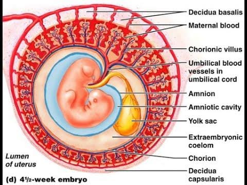
 Fig: Extraembryonic membrane in human
The process of gastrulation in embryo results into the formation of endoderm or hypoblast, ectoderm or epiblast, amniotic cavity, yolk sac, extra embryonic parietal and visceral mesoderm, connecting stalk etc. Extra embryonic membranes are also formed during this process. Each extra embryonic membrane is derived from two layers.
1. Amnion- It is formed by the layer of amniogenic cells present around the amniotic cavity and the extra embryonic mesoderm. Extra embryonic mesoderm layer surrounds the amnion. The connecting stalk is also attached with it. With a gradual increase in size the amnion covers the embryo from all sides. After about eight weeks of fertilization, amnion is completely incorporated into connecting stalk, which finally forms the umbilical cord. Embryo, in this stage, is called as foetus remains hanging in amniotic fluid. Fig: Formation of extraembryonic membrane in human2. Chorion- It is formed by the extra embryonic parietal layer of mesoderm and the cell of trophoblast.
After implantation of blastocyst, the trophoblast gives out several finger like processes, the chorionic villi which get embedded into uterine endometrium Mesoderm also contributes in the formation of these villi. After a period of four month these villi disappear from all parts except the connecting stalk where they grow rapidly and participate in the formation of placenta.
3. Yolk sac- Yolk sac is formed by the cells of extra embryonic visceral mesoderm and endoderm. Initially the size of yolk sac is larger as compared to that of the embryo. About eight weeks after fertilization, the yolk is reduced in size and changes into a tubular structure. Ultimately a placenta is developed with the incorporation of yolk sac and mesodermal connecting stalk with the amnion and chorion.
4. Allantois- It is a solid and cylindrical mass formed by embryonic mesoderm. A small cavity lined by endodermal cells develops in it. The mesoderm of allantois forms many small blood vessels in this region. These vessels connect the embryo with placenta and ensure nutritional and respiratory supply to embryo. In human, allantois does not function to store the excretory wastes as it does in reptiles and birds.
Placenta
The eggs of viviparous animals are unable to develop into their embryos outside the uterus independently. This is because of the very little or negligible amount of yolk present in these eggs, which can not fulfill the nutritional and other physiolgical demands of a developing embryo. Here the embryo depends upon maternal tissues for shelter, nutrition, respiration etc. These animals therefore, have developed adaptation, respiratory and other physiological requirements from mother's body.
Placenta is found in all viviparous (except sub-class-prototheria; oviparous) animals.
Structure of Placenta
Placenta is not a simple membrane.It is made up of the tissues from two different sources
Maternal tissue - These include uterine epithelium, connective tissues and blood capillaries.
Embryonic tissue - These include extra embryonic membranes (mainly chorion). Yolk sac and allantois may also take part in placenta formation. Embryonic connective tissues and blood capillaries are also constituents of it. Fig: The placental villi embryology
Hormones of Human Placenta
The placenta of human mainly secretes two steroid hormones like estradiol and progesterone, and two protein hormones like human chorional gonadotropin HCG and human placental somatomammotropin HCS. Large amount of HCG hormone is secreted, during early pregnancy, from the placenta. Because of this reason its quantity increases in the urine of pregnant lady. On the basis of this fact, pregnancy test is performed. The above hormones are also held responsible for keeping the corpus luteum active, protection of embryo, prevention of abortion and growth of mammary glands.
Functions of Placenta
1. Exchange of important materials between foetal and maternal blood.
2. The essential materials are exchanged by diffusion, pinocytosis or active transport.
3. The small molecules like O2, CO2, H2O etc. and other inorganic substances like chlorides, phosphates, sodium, potassium, magnesium etc. are also diffused through placenta.
4. Large molecules like lipids, polysaccharides, carbohydrates, proteins etc. are obtained by pinocytosis process.
5. The nutritional substances are supplied to embryo from the mother through placenta.
6. Placenta also serves as a respiratory medium for exchange of O2 and CO2 between embryo and mother.
7. The nitrogenous and metabolic wastes from foetus are released into the blood of mother by diffusion through placenta.
8. The antibodies for measles, chickenpox, polio etc. present in the blood of mother reach the embryo through placenta.
9. Pathogenic viruses may also enter in embryo through placenta.
10. If a female takes some harmful chemicals, liquor, drugs etc.during pregnancy, these may cross the placenta and on reaching into foetus may cause deformity during organo genesis.(eg. Thallidomide)
11. Placenta itself secretes some hormones like progesterone, estrogen, lactogen, HCG, HCS etc.
12. Progesterone, maintains and supports the foetus during the whole pregnancy period . At the time of parturition, relaxin is secreted by placenta which lubricates, and widens the birth canal to facilitate child birth.
Old NCERT Syllabus
➢Types of Implantation
On the basis of the position of attachment in the uterus, implantation is of three types-
1. Central or Superficial Implantation- In this type the blastocyst attaches superficially with the wall of uterus, and remains suspended in the lumen of the uterus.
Example: cow, pig, dog etc.
 Fig: Types of implanatation
2. Interstitial Implantation- The blastocyst is buries deeply inside the wall of uterus and covered by the endometrial tissues lying under epithelium. This type of implantation occurs in human being.
3. Eccentric Implantation- It occur in rat, squirrel etc. In this type of implantation, the blastocyst settles in in the folds of epithelium of uterus. After some time it is completely surrounded by these folds.
➢ Summary of Developmental Stages in Human
Day 1 - Fertilization; the diameter of fertilized egg is about 0.15 mm.
Day 2 - Two cell stage
Day 3 - 16 cell stage, morula
Day 4 - Entry of blastocyst in the lumen of uterus, disappearance of zona pellucida, diameter of blastocyst is about 0.3 mm.
7-8 Days - Partial entry of blastocyst inside the endometrium of uterus; implantation
Day 12 - Complete entry of blastocyst into endoderm, extra-embryonic mesoderm, amnion and yolk sac.
Day 14 - primitive streak formation.
Day 18 - Formation of 3-5 pairs of somites

Day 24 – Indication of the formation of head and tail region; 21-23 pairs of somites formed,formation of heart continues at ventral side.
Day 28 – Heart starts beating, neural tube formed, 3 pairs of visceral arch and 30-31 pairs of somites formed. Blood islands appear
Day 32 – 30-39 pairs of somites formed.
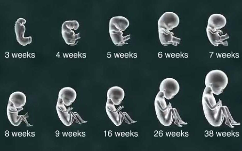
7 Weeks – Jaws, fingers and external ears begin to appear. The CR length(crown-rump length; the length from head to the bottom of hips) is 19-20 mm.
8 Weeks – The embryo is completely surrounded by amnion, fingers and toes clearly visible, almost all organs formed with continuing development, at the end of 8thweek the embryo appears like a little human, now called as foetus; C-R length is 28 to 30 mm.
5 months – Blood formation starts in bone marrow Decidua capsularis and parietalis connect together, hairs appear.
9 months – placenta attains maximum size, nails on fingers appear. In the next 10 days the foetus is ready to born as a little baby.
The above mentioned timing are approximate time periods. Some times, due to some reasons, certain babies are born before stipulated time. The babies born in 7th month may also survive as normal babies.
➢Extra Embryonic Membranes in Chordates
In chordates like reptiles, birds and prototherian mammals, blastula is a disc shaped structure called as blastodisc.The cellular layer formed of blastomeres remains as blastoderm. The central part of blastoderm gives rise to embryo proper, while the peripheral portion does not take part in the formation of embryo. This peripheral part is known as extra embryonic region. This region takes part in the formation of certain membranes called extra embryonic membranes. These extra embryonic membranes provide facilities for nutrition, respiration and excretion to the embryo. Extra embryonic membranes are of four types-
1. Amnion
2. Chorion
3. Yolk sac
4. Allantois
On the basis of presence or absence of amnion, two groups of vertebrates are categorized
1. Amniota - This group is characterized with the presence of amnion in the embryos of its members.
For example members of class Reptilia, Aves and Mammalia.
2. Anamniota - Animals of this group are devoid of amnion in their embryos. For example class cyclostomata, pisces and amphibian.
➢Types of Placenta
On the basis of extra embryonic membranes, the placenta is of three types.
1. Yolk sac placenta- It is formed by yolk sac and uterine epithelium.For example, Elasmobranchs (Sharks), Mustelus etc.
2. Chorio-vitelline placenta- It is formed by chorion and yolk sac combinedly. Hence it is called as choriovitelline placenta. For example, Didelphis, Macropus and other metatherian mammals.
3. Chorio-allantoic placenta- This type of placenta is formed by embryonic chorion and allantoic membranes.
It is also referred to as a true placenta. It is found in eutherian mammals.
Chorio-allantoic placenta in mammals:
1. In this type of placenta, allantoic mesodern and the mesoderm of umbilical cord jointly form the blood vessels of umbilical cord. The endodermal part of the allantois remains as a very small cavity.
2. To obtain nutrition from maternal blood several finger like processes or villi are formed by chorion which penetrate deeply into the crypts of uterus.Initially the villi are scattered over the whole surface of chorion but later they become restricted in the decidua besalis region . The chorionic villi on the remaining surface disappear shortly. The part of chorion, which helps in placenta formation is known as chorionic frondosum.
Classification of Placenta
On the basis of different characters, the placenta are classified in following manner:-
1. On the basis of intimacy After implantation, the wall of uterus is called as decidua, instead of endometrium. The part of decidua, where placenta is formed is called decidua basalis whereas, the part separating the embryo from lumen of uterus is called decidua capsularis. The remaining part of lumen of uterus is called decidua parietalis. Decidua also comes out from uterus at the time of parturition. On the basis of intimacy between embryo and uterine wall the placenta is classified into three classes
(i) Non-deciduate or Semi placenta- In this type of placenta, there is no close and rigid association between embryo and the wall of uterus. Hence, at the time of parturition, there is no bleeding as the chorionic villi are easily pulled out from the crypts of uterus. For example, cow, buffalo, horse, pig.
(ii) Contra-deciduate placenta- There is a close association between embryonic and maternal tissues. However at parturition, the damaged maternal and embryonic tissues along with the part of placenta remain inside the uterus which are absorbed in situ by leukocytes.
For example- Parameles, Talpa etc.
(iii) Deciduate placenta- This type of placenta is found in human, dog, hare etc.It is characterized with a very close association between chorionic villi and uterine wall. At the time of birth, the mucosal covering of the uterus is also damaged and discarded outside. This results in an extensive bleeding at child birth. This placenta is known as a true placenta.
2. On the basis of implantation
Three types of placenta are found on the basis of implantaion.
(i) Superficial- When the placenta is situated in the lumen of uterus. For example, Parameles, pig, cow, cat etc.
(ii) Eccentric- The placenta is situated in the fold or pocket of the cavity of uterus. For example- rat, Squirrel etc.
(iii) Interstitial- This type of placenta is found in man, guinea pig, apes etc. The chorionic sac (placenta) penetrates deep inside the wall of uterus. Hence, the association between embryo and maternal part becomes very close.
3. On the basis of distribution of villi
On this basis, the placenta are of four types.
(i) Diffused placenta- The villi are scattered on the whole surface of placenta. For example pig, horse, lemur etc.
(ii) Cotyledonary placenta- The villi are distributed in small isolated groups on the chorionic surface. These groups of villi are called as cotyledons. For example, cow, buffalo, sheep, deer etc.
(iii) Zonary placenta- This type of placentae have the villi distributed in a belt shaped zone which is large sized and circular.
Zonary placenta is of two types-
(a) Complete Zonary placenta - The belt of villi is complete and ring shaped init. For example- dog, cat, lion etc.
(b) Incomplete Zonary placenta- The belt of villi is incomplete in it. For example- racoon.
(iv) Discoidal placenta- In this type of placenta, whole of the chorionic surface is covered by villi in initial stage, but the villi disappear later from most area except the region of implantation, that is only a disc like region is left with villi.
Discoidal placenta is also of two types–
(a) Mono discoidal placenta– The villi are present only on dorsal surface in a single circular disc like area.
For example- human, hare etc.
(b) Bidiscoidal placenta- If the villi are distributed in two disc like areas, the placenta is called as bidiscoidal,
Example: monkeys.
 Fig: Different types of placenta
Types of placenta on the basis of distribution of villi
4. On the basis of histology
The blood of maternal and embryonic do not mix together through placenta. The blood circulations of the two sides are kept separated by one or more layers described below–


The transportation of various materials takes place by diffusion through these six layers. The intimacy between maternal and embryonic tissues in different mammals is determined by the presence or absence of these layers in placenta. Therefore, on the basis of presence of above layers, the placenta is of five types.
1. Epitheliochorial- It is the most primitive type of placenta in which all the six layers, mentioned earlier, remain intact.
For example - pig, horse etc.
2. Syndesmochorial- In this type of placenta the uterine epithelium is eroded by chorionic villi, so only two maternal layers remain functional. Therefore, along with three foetal layers, total five layers are present in this placenta.
For example - sheep, goat, cow etc.
3. Endotheliochorial- Here uterine connective tissue layer is also damaged along with uterine epithelial layer. Therefore only four layers (3 foetal and one maternal) are found in this placenta
For example - dog, cat, etc.
4. Haemochorial- All the three maternal layers are penetrated in this placenta. The chorionic epithelium comes in direct contact with uterine blood sinusoids.
For example- man, monkey, bat etc. Fig: Classification of placenta
5. Haemo-endotheliochorial/Haemo-endothelial : it is the most typical placenta in which the tropho-blastic epithelium of embryo is also eroded along with all three maternal layers. The foetal capillaries are in direct contact with maternal blood.
For example: rat, guinea pig, rabbit etc.
HISTORY
- Aristotle is known as "Father of Embryology" he first studied the development in chick and other embryos. He gave its description in his book "Historia Animalia".
- Leeuwenhoek (1671)
(a) He observed and described human sperm for the first time.
(b) According to Hartsoeker and Leeuwenhoek there is a small model of developing animal present in the head of the sperm of that animal. This small model is called homunculus. Both these scientists are called spermists, and this theory is called "Theory of spermist". - Swammer Dame, Haller, Bonette & Malpighi:- According to these scientists, small model of animal is always present in the egg. These scientists are called Ovists, and their theory is known as 'Ovists theory'
- Schleiden & Schwann:- Both the scientists established the cellular structure of egg and sperm.
- Pander:- He described the presence of three germinal layers in chick embryo.
- Fredrich Wolff:- He first presented the "theory of epigenesis".
- Muller :- He gave the recapitulation theory.
- Haeckel :- He gave the details of Recapitulation theory and named it as the bio-genetic law. - Bio-genetic Law :- According to this each organism during its embryonal development, passes through all stages, through which its species has evolved or embryo repeats its ancestry. i.e. Ontogeny recapitulates its Phylogeny.
- "Carl Ernest Von Baer":- He is known as the "father of modern embryology." He gave the Baer's Law which in turn proves the recapitulation theory.
According to this law, during embryonal development, the development of general structures takes place earlier and specific structures develop at last or later on. - A. Weismann:- He gave the theory of germplasm or the theory of continuity of germplasm. According to him, there are 2 types of protoplasm in the body of animals :-
(a) Somatoplasm
(b) Germplasm.
Somatoplasm dies but the germplasm is never destroyed, rather it is transferred to the progenies. - Wilhelm:- He studied embryonal development in frog and gave the mosaic theory.
He said that there are some presumptive areas in the eggs of frog. These areas form specific structures during embryonal development. This is termed as "Promorphology". and these type of eggs are termed as the mosaic eggs. - Hans Driesch:- He studied embryonal development in sea-urchin and gave the regulative theory. - In the eggs of Sea-Urchin presumptive areas are not found i.e. promorphology is not found. So, each part of the egg is capable of forming the complete embryo. These type of eggs are termed as regulative eggs.
- Boveri & Child:-
They gave the gradient theory to explain the mosaic development in eggs.
According to them, a metabolic gradient is present inside the eggs.
Different parts of the egg have different metabolic rates.
The rate of metabolism is faster at the animal- pole of the egg and is slower at the vegital pole of the egg.
Due to different metabolic rates, different structures are formed from different parts of the egg. - Spemann:- He gave the "Theory of organizers". -
(a) According to it embryo has some special type of tissues, which induce development of some specific structures.
(b) These are termed as the organizers.
(c) These organizers secrete some special chemicals called evocators which induce the formation of specific structures.
(d) Spemann got the Nobel prize for his theory of organizers. - R.V. Graff:-
He studied a follicle in human ovary and termed it as "Graafian follicle".
|
124 videos|210 docs|207 tests
|
FAQs on Implantation & Placenta - Biology for Grade 12
| 1. What is implantation and why is it important in the development of an embryo? |  |
| 2. What are extra-embryonic membranes and what role do they play in the development of the placenta? |  |
| 3. What is the placenta and what functions does it perform during pregnancy? |  |
| 4. How does the placenta form and what are the key components involved in its development? |  |
| 5. Can problems with implantation or the placenta affect the outcome of a pregnancy? |  |

|
Explore Courses for Grade 12 exam
|

|


