Important Diagrams: Tissues | Science Class 9 PDF Download
Q1: Answer the following questions based on the diagram given below:
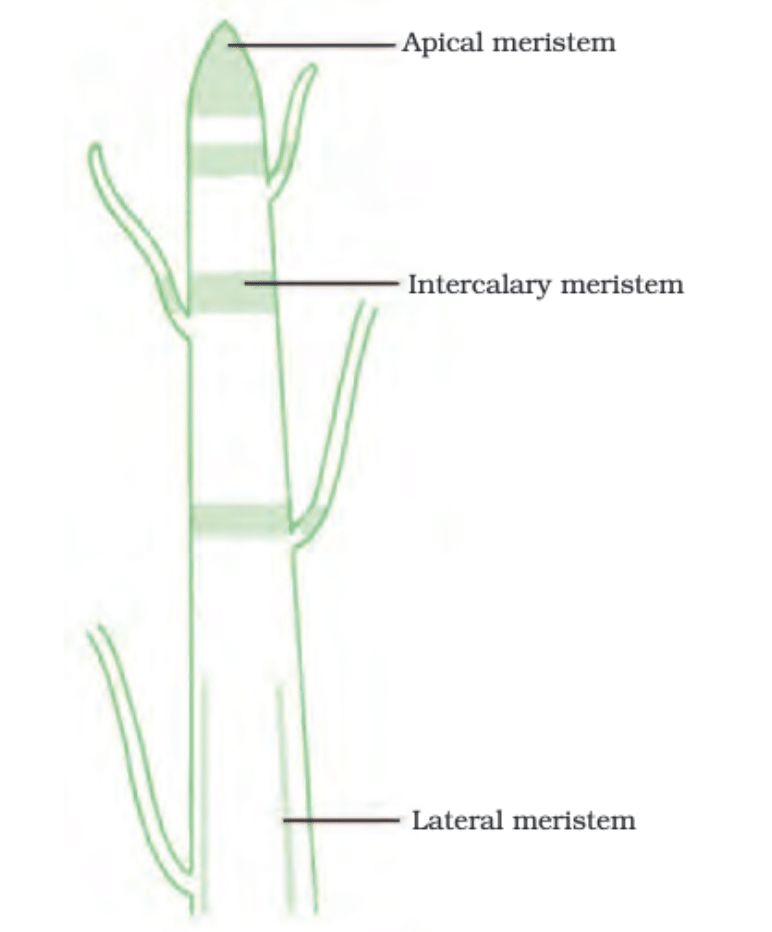
(i) Where is apical meristem found in a plant?
Ans: Apical meristem is located at the growing tips of stems and roots.
(ii) Which meristem increases the length of the stem and root?
Ans: Apical meristem increases the length of the stem and root.
(iii) What is the function of lateral meristem (cambium) in a plant?
Ans: Lateral meristem (cambium) increases the girth or thickness of the stem or root.
(iv) Where is intercalary meristem typically located in a plant?
Ans: Intercalary meristem is found near the node in some plants.
(v) Describe the characteristics of cells in meristematic tissue.
Ans: Cells in meristematic tissue are highly active, possess dense cytoplasm, thin cellulose walls, prominent nuclei, and lack vacuoles.
Q2: Answer the following questions based on the diagram given below:
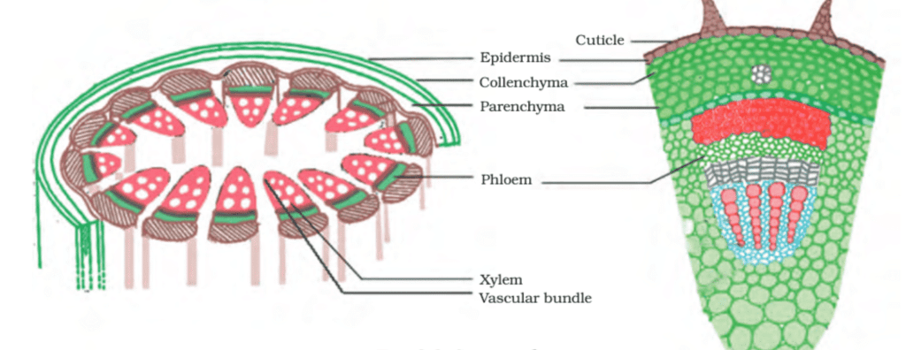
(i) What is the process by which meristematic cells become permanent tissues called?
Ans: The process by which meristematic cells become permanent tissues is called differentiation.
(ii) What is the most common type of simple permanent tissue, and what is its function?
Ans: The most common type of simple permanent tissue is parenchyma. Its function includes storing food, sometimes containing chlorophyll for photosynthesis (chlorenchyma), and in aquatic plants, it has large air cavities (aerenchyma) to aid in floating.
(iii) Which permanent tissue in plants allows for flexibility and bending without breaking?
Ans: Collenchyma is the permanent tissue in plants that allows for flexibility and bending without breaking.
(iv) What is the primary function of sclerenchyma tissue in plants?
Ans: Sclerenchyma tissue makes plants hard and stiff, providing strength to plant parts.
(v) What is the function of epidermal tissue in plants, and why do its cells form a continuous layer without intercellular spaces?
Ans: Epidermal tissue in plants serves to protect against the loss of water, mechanical injury, and invasion by parasitic fungi. Its cells form a continuous layer without intercellular spaces because it has a protective role to play.
Q3: Answer the following questions based on the diagram given below:
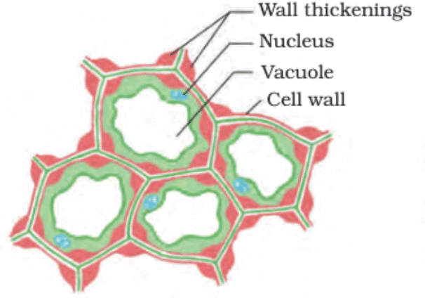
(i) What is collenchyma and where is it found in a plant?
Ans: Collenchyma is a type of simple permanent plant tissue. It is primarily found in the cortex of stems and leaves.
(ii) Describe the structure of collenchyma cells.
Ans: Collenchyma cells have thickened cell walls, often irregular in shape, and they provide flexibility and mechanical support to the plant.
(iii) How does collenchyma differ from parenchyma and sclerenchyma?
Ans: Collenchyma has unevenly thickened cell walls, while parenchyma has thin and uniform walls, and sclerenchyma has very thick, rigid walls.
(iv) What is the function of collenchyma cells in a plant?
Ans: Collenchyma cells provide support to the plant by maintaining flexibility and helping in the elongation of plant parts.
Q4: Answer the following questions based on the diagram given below:
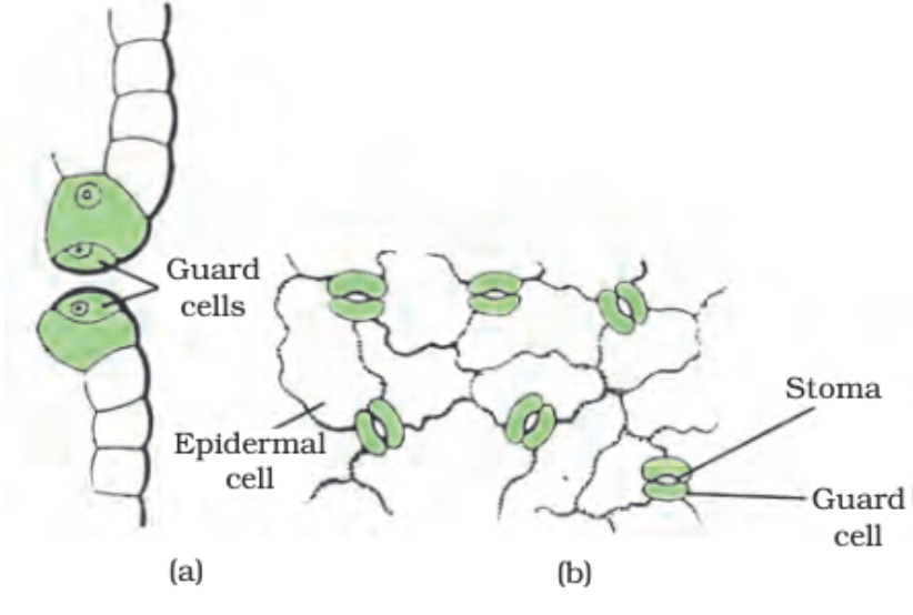
(i) What is the role of stomata in a leaf?
Ans: Stomata regulate the exchange of gases (like oxygen and carbon dioxide) and water vapour between the plant and its environment.
(ii) How do guard cells control the opening and closing of stomata?
Ans: Guard cells control the opening and closing of stomata through their shape and turgor pressure. When they are turgid, the stomata open, and when they lose water and become flaccid, the stomata close.
(iii) What factors influence the opening and closing of stomata?
Ans: Factors like light, water availability, carbon dioxide concentration, and temperature influence the opening and closing of stomata.
(iv) What is the importance of regulating water loss through stomata for a plant?
Ans: Regulating water loss through stomata helps the plant to balance its water uptake and prevent excessive loss, maintaining proper hydration and supporting various physiological processes.
(v) How does the structure of guard cells facilitate their function in controlling stomatal opening and closing?
Ans: Guard cells have a kidney or bean shape, which enables them to change shape and control the size of the stomatal pore. When they swell, the stomata open, and when they shrink, the stomata close.
Q5: Answer the following questions based on the diagram given below:

(i) What are the main components of xylem and their functions?
Ans: Xylem consists of tracheids, vessels, xylem parenchyma, and xylem fibres. Tracheids and vessels transport water and minerals, while xylem fibres provide support.
(ii) How do tracheids and vessels contribute to water transport in plants?
Ans: Tracheids and vessels have thick walls and a tubular structure, allowing them to transport water and minerals vertically in plants.
(iii) What is the function of xylem parenchyma?
Ans: Xylem parenchyma stores food in plants.
(iv) How does the xylem differ from the phloem in terms of structure and function?
Ans: The xylem and phloem are both complex tissues, but the xylem primarily transports water and minerals while the phloem transports organic nutrients like sugars.
(v) What is the difference between simple permanent tissue and complex tissue? Provide an example of each.
Ans: Simple permanent tissues are made of a single type of similar cells, whereas complex tissues are made of different types of cells working together. The xylem and phloem are examples of complex tissues.
Q6: Answer the following questions based on the diagram given below:
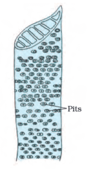
(i) What are the main components of the phloem?
Ans: The main components of the phloem are sieve cells, sieve tubes, companion cells, phloem fibres, and phloem parenchyma.
(ii) What is the function of sieve tubes in the phloem?
Ans: Sieve tubes transport food from leaves to other parts of the plant through their perforated walls.
(iii) Name a type of phloem cell that is not a living cell.
Ans: Phloem fibres are the type of phloem cells that are not living cells.
(iv) Why is the transport of food important in a plant?
Ans: The transport of food is important in a plant as it helps distribute nutrients and energy to different parts of the plant, supporting growth and metabolism.
(v) Which part of the plant produces food that is transported by the phloem?
Ans: Food is produced in the leaves of the plant through photosynthesis and is then transported by the phloem to other parts of the plant.
Q7: Answer the following questions based on the diagram given below:
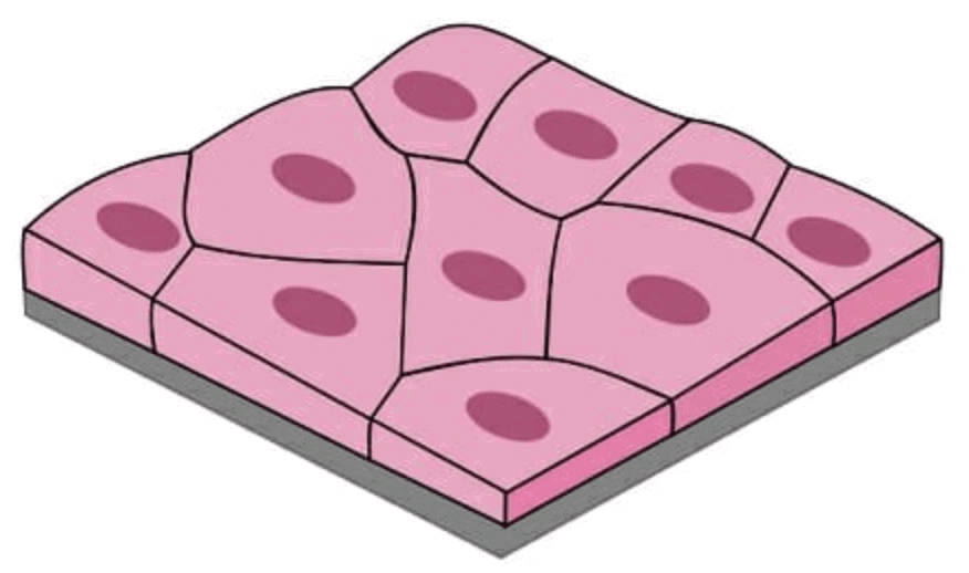
(i) What is the primary function of squamous epithelial cells in the body?
Ans: Squamous epithelial cells form a delicate lining and are involved in providing a selectively permeable surface for the transportation of substances.
(ii) Mention two locations in the body where the squamous epithelium is found and describe their specific functions.
Ans: Squamous epithelium is found in the skin, acting as a protective barrier, and in the lining of the mouth, contributing to its protective function and facilitating movement.
(iii) How does the structure of squamous epithelial cells relate to their function?
Ans: Squamous epithelial cells are extremely thin and flat, allowing for efficient exchange of materials across their surface due to their thin structure.
(iv) Discuss the role of squamous epithelium in the respiratory tract and blood vessels.
Ans: Squamous epithelium in the respiratory tract forms a selectively permeable surface and aids in gas exchange. In blood vessels, it contributes to a smooth lining for efficient blood flow.
Q8: Answer the following questions based on the diagram given below:
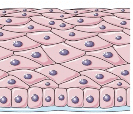
(i) What is the main purpose of the layered arrangement in stratified squamous epithelium?
Ans: The layered arrangement in stratified squamous epithelium provides protection against wear and tear due to its multiple layers.
(ii) Name two body parts where you would find stratified squamous epithelium and explain their protective roles.
Ans: Stratified squamous epithelium is found in the skin, providing protection against external factors, and in the esophagus, safeguarding against friction during swallowing.
(iii) How does the structure of stratified squamous epithelium support its protective function?
Ans: The stratified arrangement of cells provides strength and durability, making it suitable for protecting areas prone to mechanical stress.
(iv) Compare the structure and function of simple squamous epithelium with stratified squamous epithelium.
Ans: Simple squamous epithelium is a delicate lining facilitating material exchange, while stratified squamous epithelium is protective and has multiple layers to resist wear and tear.
Q9: Answer the following questions based on the diagram given below: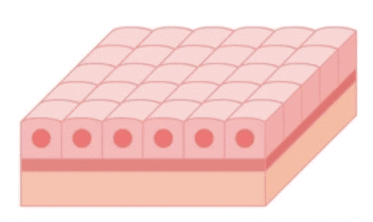
(i) What is the characteristic shape of cells in cuboidal epithelium, and where is it commonly found in the body?
Ans: The cells in the cuboidal epithelium are cube-shaped. It is found in the lining of kidney tubules and ducts of salivary glands.
(ii) How does cuboidal epithelium support its role in kidney tubules and salivary gland ducts?
Ans: The cuboidal shape provides mechanical support and aids in secretion and absorption in the kidney tubules and salivary gland ducts.
(iii) What additional specialization can cuboidal epithelial cells acquire, and how does this specialization benefit the body?
Ans: Cuboidal epithelial cells can specialize as gland cells, enabling the secretion of substances at the epithelial surface, and contributing to various physiological processes.
(iv) Discuss the importance of the lining provided by cuboidal epithelium in kidney tubules.
Ans: The lining of cuboidal epithelium in kidney tubules aids in the reabsorption of essential substances, maintaining proper balance and regulation in the body.
Q10: Answer the following questions based on the diagram given below:
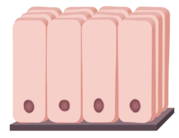
(i) Describe the structure of columnar epithelial cells, particularly focusing on their shape and unique features.
Ans: Columnar epithelial cells are tall and have hair-like projections called cilia on their outer surfaces, aiding in movement.
(ii) Where is ciliated columnar epithelium predominantly found, and what is its primary function?
Ans: Ciliated columnar epithelium is mainly found in the respiratory tract and helps in moving mucus forward to clear the airways.
(iii) Explain the significance of cilia in columnar epithelial cells within the respiratory tract.
Ans: The cilia in the respiratory tract's columnar epithelial cells move mucus, facilitating the removal of dust and particles, thus assisting in maintaining a clear airway.
(iv) Compare the functions of simple columnar epithelium and ciliated columnar epithelium.
Ans: Simple columnar epithelium facilitates absorption and secretion, whereas ciliated columnar epithelium primarily aids in movement and clearance of mucus in specific locations like the respiratory tract.
Q11: Answer the following questions based on the diagram given below:
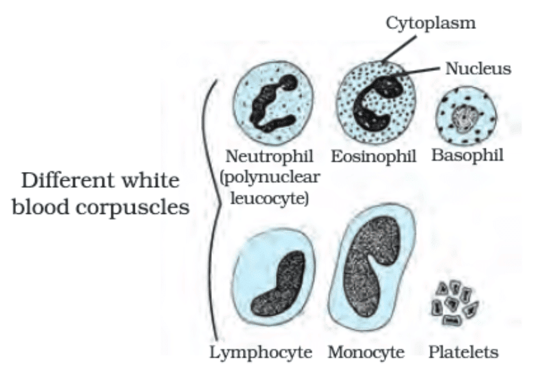
(i) What are the main types of blood cells found in the fluid matrix of blood called plasma?
Ans: The main types of blood cells found in the fluid matrix of blood called plasma are red blood corpuscles (RBCs), white blood corpuscles (WBCs), and platelets.
(ii) Which type of blood cell plays a crucial role in defending the body against infections and foreign invaders?
Ans: White blood corpuscles (WBCs), also known as leukocytes, play a crucial role in defending the body against infections and foreign invaders.
(iii) Among white blood corpuscles, which specific type is commonly known as polynuclear leucocyte?
Ans: Neutrophils, also referred to as polynuclear leucocytes, are a specific type of white blood corpuscle.
(iv) What is the primary function of eosinophils among the white blood corpuscles in the blood?
Ans: Eosinophils primarily function in immune responses and are particularly involved in combating parasitic infections and controlling allergic reactions.
(v) Which type of blood cell releases histamine and heparin, and is involved in allergic reactions and inflammation?
Ans: Basophils are the type of blood cells that release histamine and heparin and play a role in allergic reactions and inflammation.
(vi) What is the function of platelets in the blood, and what role do they play in maintaining the body's overall health?
Ans: Platelets are responsible for blood clotting and play a crucial role in preventing excessive bleeding when blood vessels are damaged, thus contributing to the body's overall health and wound healing.
Q12: Answer the following questions based on the diagram given below:
(i) What is the primary function of bone in the human body?
Ans: The primary function of bone is to provide a framework that supports the body, anchors muscles, and protects organs.
(ii) How does the composition of bone contribute to its strength and non-flexibility?
Ans: Bone's composition, particularly calcium and phosphorus compounds, provides strength and non-flexibility, ensuring a sturdy framework for the body.
(iii) Differentiate between ligaments and tendons based on their structure and function.
Ans: Ligaments connect bones to bones, are elastic, and have considerable strength with minimal matrix. Tendons connect muscles to bones, are fibrous, and strong, but have limited flexibility.
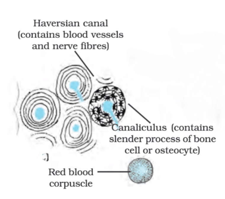
(iv) What is the role of Haversian canals in bones?
Ans: Haversian canals contain blood vessels and nerve fibres, facilitating the transportation of nutrients and oxygen to bone cells and the removal of waste products.
(v) Explain the significance of Canaliculus in bone structure.
Ans: Canaliculus contains slender processes of bone cells or osteocytes, allowing for communication and nutrient exchange between adjacent osteocytes, vital for bone cell health and functionality.
Q13: Answer the following questions based on the diagram given below:
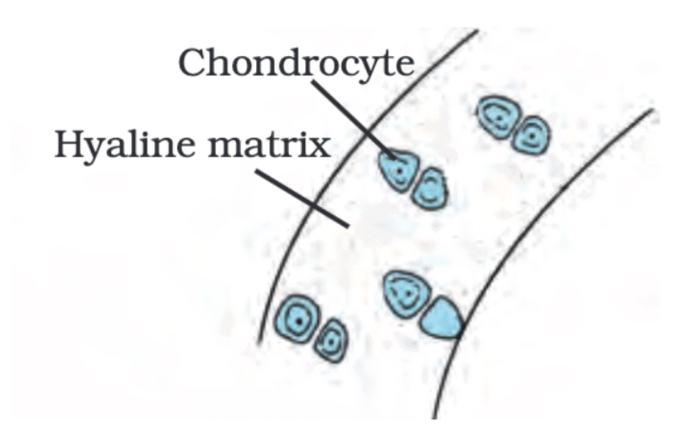
(i) Where is hyaline cartilage found in the body?
Ans: Hyaline cartilage is found in various locations such as joints, nose, ear, trachea, and larynx.
(ii) What is the composition of the solid matrix in hyaline cartilage?
Ans: The solid matrix of hyaline cartilage is composed of proteins and sugars.
(iii) How does hyaline cartilage contribute to joint function?
Ans: Hyaline cartilage smoothens bone surfaces at joints, allowing for smooth movement and reducing friction.
(iv) How does hyaline cartilage differ in structure compared to bone?
Ans: Unlike bones, hyaline cartilage has widely spaced cells and a solid, yet flexible, matrix.
(v) Mention one advantage of hyaline cartilage in terms of flexibility.
Ans: Hyaline cartilage's flexibility allows it to support various body parts without being rigid, enabling movement and flexibility in specific areas.
Q14: Answer the following questions based on the diagram given below:
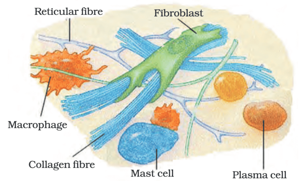
(i) Where is areolar connective tissue found in the body?
Ans: Areolar connective tissue is found between the skin and muscles, around blood vessels and nerves, in the bone marrow, and inside organs.
(ii) What is the main function of areolar tissue?
Ans: Areolar tissue supports internal organs, fills spaces inside organs, and aids in tissue repair.
(iii) Describe the characteristics of the matrix in areolar tissue.
Ans: The matrix of areolar tissue is loose and contains cells as well as fibres.
(iv) How does areolar tissue contribute to organ support?
Ans: Areolar tissue provides a supportive framework around organs and helps in maintaining their position within the body.
(v) What types of cells are typically found in areolar connective tissue?
Ans: Areolar tissue contains various cells, including fibroblasts, macrophages, and other connective tissue cells.
Q15: Answer the following questions based on the diagram given below:
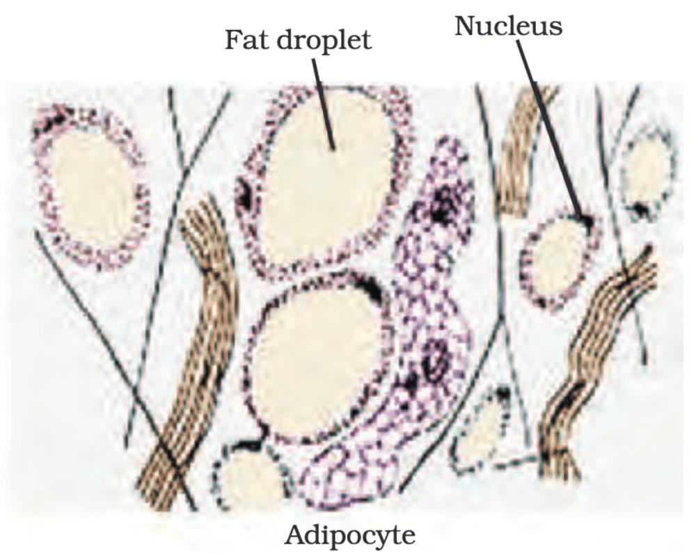
(i) Where is adipose tissue primarily located in the body?
Ans: Adipose tissue is primarily located below the skin and between internal organs.
(ii) What is the main function of adipose tissue in the body?
Ans: Adipose tissue acts as an insulator and stores fats for energy.
(iii) Describe the appearance of cells in adipose tissue.
Ans: Adipose tissue cells are filled with fat globules.
(iv) How does the structure of adipose tissue contribute to its insulating function?
Ans: The storage of fats in adipose tissue provides insulation, helping to maintain body temperature.
(v) What is the purpose of storing fats in adipose tissue?
Ans: Storing fats in adipose tissue serves as an energy reserve and insulation, providing a cushion and protection for organs.
Q16: Answer the following questions based on the diagram given below:
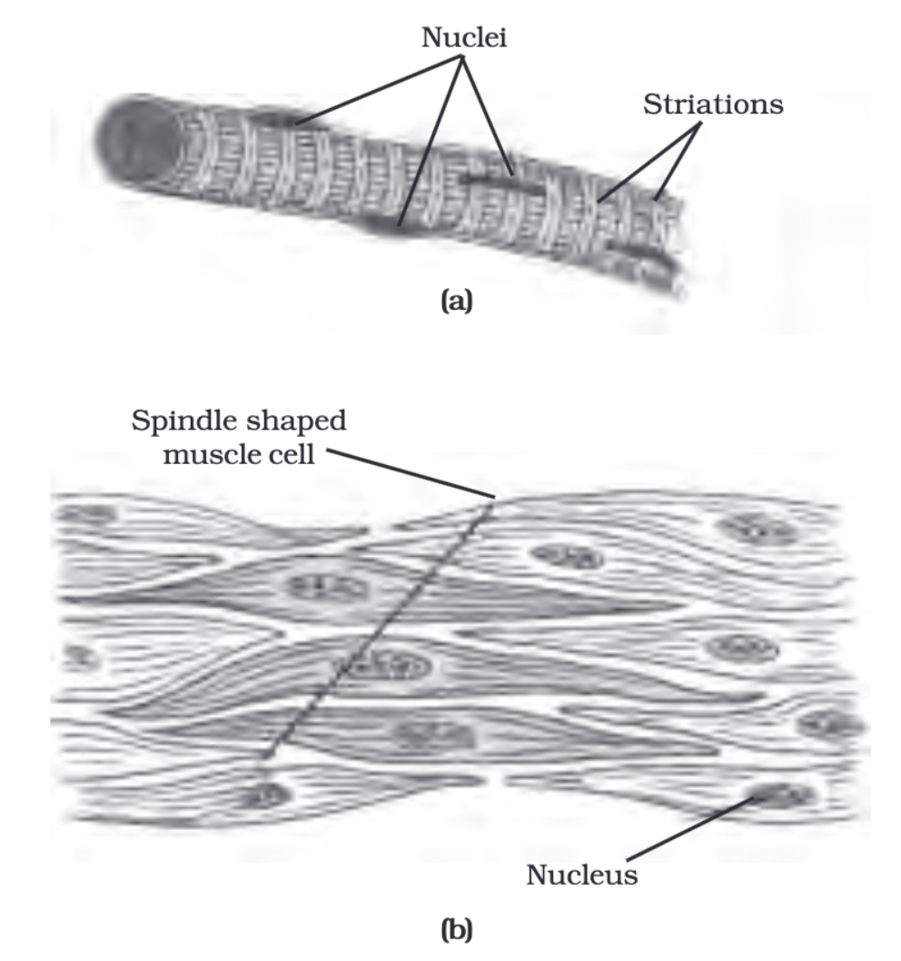
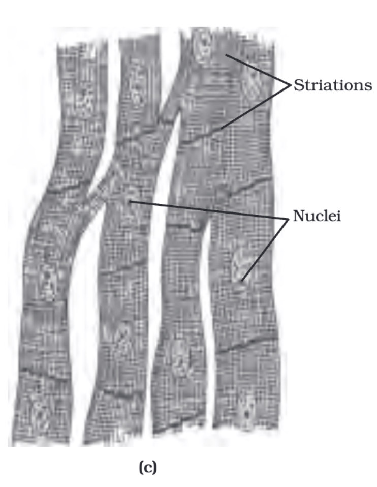
(i) Describe the appearance of striated muscle fibres under a microscope.
Ans: Striated muscle fibres appear to have alternate light and dark bands or striations when stained appropriately.
(ii) What is another name for skeletal muscles? Why are they called so?
Ans: Skeletal muscles are also known as voluntary muscles. They are called skeletal muscles because they are mostly attached to bones and help in body movement.
(iii) How are smooth muscles different from striated muscles in terms of shape and structure?
Ans: Smooth muscles have long, spindle-shaped cells with pointed ends and are uninucleate (have a single nucleus), while striated muscles have long, cylindrical, unbranched cells that are multinucleate (have many nuclei).
(iv) What are the involuntary functions controlled by smooth muscles?
Ans: Smooth muscles control involuntary movements like the movement of food in the alimentary canal, contraction and relaxation of blood vessels, and actions in the iris of the eye, ureters, and bronchi of the lungs.
(v) How do cardiac muscle cells differ from skeletal muscle cells?
Ans: Cardiac muscle cells are cylindrical, branched, and uninucleate (have a single nucleus), whereas skeletal muscle cells are long, cylindrical, unbranched, and multinucleate (have many nuclei). Cardiac muscles show rhythmic contraction and relaxation throughout life.
Q17: Answer the following questions based on the diagram given below:
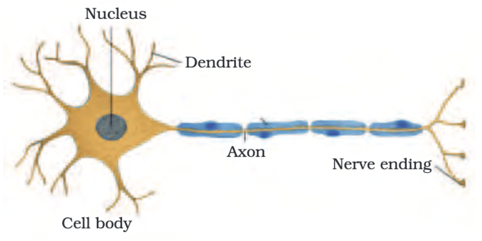
(i) What is the main function of a neuron in the nervous system?
Ans: A neuron transmits nerve impulses and helps in the communication of information throughout the body.
(ii) Name the three main parts of a neuron and briefly describe their functions.
Ans: The three main parts of a neuron are the dendrites (receive signals), the cell body (process signals), and the axon (transmits signals).
(iii) What is the role of the myelin sheath in a neuron?
Ans: The myelin sheath is a fatty layer that insulates the axon, allowing for faster and more efficient transmission of nerve impulses.
(iv) Explain how a nerve impulse is transmitted from one neuron to another.
Ans: A nerve impulse travels down the axon of a neuron and is transmitted to another neuron through a small gap called a synapse, where neurotransmitters carry the signal to the next neuron.
(v) Describe the importance of neurotransmitters in the functioning of a neuron.
Ans: Neurotransmitters are chemical messengers that transmit signals across synapses, facilitating communication between neurons and ensuring the proper functioning of the nervous system.
|
84 videos|384 docs|61 tests
|
FAQs on Important Diagrams: Tissues - Science Class 9
| 1. What are the four main types of tissues in the human body? |  |
| 2. How do epithelial tissues differ in structure and function? |  |
| 3. What is the role of connective tissue in the body? |  |
| 4. What are the characteristics of muscle tissue? |  |
| 5. How does nervous tissue function in the body? |  |


