NCERT Based Activity: The Fundamental Unit of Life | Science Class 9 PDF Download

Activity 5.1: Observation of Onion Epidermal Cells under a Compound Microscope
- Take a small piece from an onion bulb. Using forceps, peel off the epidermis from the concave side (inner layer) of the onion and place it in a watch-glass containing water to prevent folding or drying.
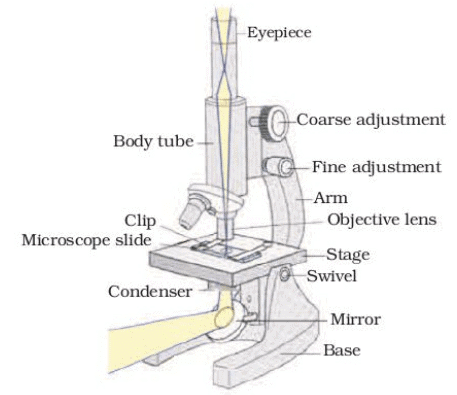 Fig. 5.1: Compound microscope
 - Place a drop of water on a glass slide, transfer a small piece of the peel to the slide, ensuring it is flat. Use a thin camel hair paintbrush if needed.
- Add a drop of safranin solution to the peel, then place a cover slip over it, avoiding air bubbles.
- Observe the prepared temporary mount of onion peel under low power and then high power of a compound microscope.
Q: What do we observe as we look through the lens?
Ans: Under the microscope, the onion peel reveals a pattern of small, rectangular or hexagonal compartments arranged in a brick-like structure. These compartments are the cells of the onion epidermis, with distinct cell walls and a darkly stained nucleus visible in each cell due to the safranin solution.
Q: Can we draw the structures that we are able to see through the microscope, on an observation sheet?
Ans: Yes, the structures can be drawn. The drawing should show rectangular or hexagonal cells with thick cell walls, a visible nucleus in each cell (stained dark by safranin), and possibly some cytoplasm. The arrangement resembles a honeycomb or brick wall pattern, similar to Fig. 5.2.
Q: Does it look like Fig. 5.2? 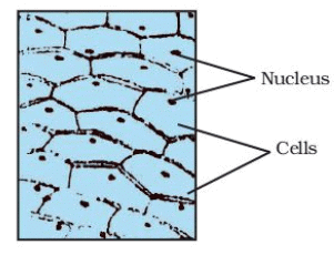 Fig. 5.2: Cells of an onion peelAns: Yes, the observed structure closely resembles Fig. 5.2, which depicts cells of an onion peel with clear cell walls, nuclei, and a regular, grid-like arrangement.
Q: Try preparing temporary mounts of peels of onions of different sizes. What do we observe? Do we see similar structures or different structures?
Ans: When peels from onions of different sizes are observed, the structures appear similar. The cells of the onion peel all look the same, regardless of the onion’s size, showing rectangular or hexagonal shapes, distinct cell walls, and a nucleus in each cell. The size of the onion does not alter the microscopic appearance of the epidermal cells.
Explanation: This activity demonstrates that the onion bulb is composed of cells, which are the basic structural units of living organisms. The similar appearance of cells across different onion sizes confirms that cells are consistent building blocks, supporting the cell theory (all plants and animals are composed of cells). The use of safranin stains the nucleus, making it easier to identify key cellular components like the cell wall, nucleus, and cytoplasm. This activity introduces students to microscopy and the concept of cells as the fundamental units of life.
Activity 5.2: Comparative Study of Plant Cells from Different Tissues
- Prepare temporary mounts of leaf peels, the tip of roots of onion, or peels of onions of different sizes.
Q: Do all cells look alike in terms of shape and size?
Ans: No, not all cells look alike in shape and size. For example:
- Onion peel cells (epidermal cells) are rectangular or hexagonal and relatively uniform in size.
- Leaf peel cells (e.g., from the lower epidermis) may include elongated epidermal cells and specialized guard cells (kidney-shaped) around stomata, which differ in shape and size from onion peel cells.
- Root tip cells may appear smaller, more rounded, or elongated, depending on the region (e.g., meristematic cells are small and cuboidal, while others are elongated).
Q: Do all cells look alike in structure?
Ans: No, cells differ in structure based on their function. For example:
- Onion peel cells have a prominent cell wall, nucleus, and cytoplasm but lack chloroplasts.
- Leaf peel cells may contain chloroplasts (especially in mesophyll cells) and stomata with guard cells, which are structurally distinct.
- Root tip cells may have denser cytoplasm and smaller vacuoles, especially in actively dividing meristematic regions.
Q: Could we find differences among cells from different parts of a plant body?
Ans: Yes, there are differences. For example:
- Onion peel cells are specialized for protection and storage, with large vacuoles and no chloroplasts.
- Leaf peel cells are adapted for photosynthesis (contain chloroplasts) or gas exchange (guard cells in stomata).
- Root tip cells are involved in growth and absorption, with structures suited for cell division or water uptake (e.g., root hair cells have extensions).
Q: What similarities could we find?
Ans: Similarities include:
- All cells have a cell wall (composed of cellulose), a plasma membrane, cytoplasm, and a nucleus (in non-dividing cells).
- All are eukaryotic plant cells with membrane-bound organelles.
- The basic organization (cell wall, plasma membrane, cytoplasm, nucleus) is consistent across different plant parts.
Explanation: This activity highlights the diversity and specialization of cells within a plant, demonstrating that while all cells share a basic structural organization (cell wall, plasma membrane, nucleus, cytoplasm), their shape, size, and internal structures vary depending on their function. This supports the concept of division of labor in multicellular organisms, where different cells perform specific roles (e.g., photosynthesis in leaves, absorption in roots, protection in epidermis). The activity reinforces the cell theory by showing that all plant parts are composed of cells, but it also introduces the idea of cellular differentiation.
Activity 5.3: Demonstration of Osmosis Using a De-shelled Egg
(a) Remove the shell of an egg by dissolving it in dilute hydrochloric acid (the shell, mostly calcium carbonate, dissolves, leaving a thin outer membrane). Place the de-shelled egg in pure water and observe after 5 minutes.
(b) Place a similar de-shelled egg in a concentrated salt solution and observe for 5 minutes.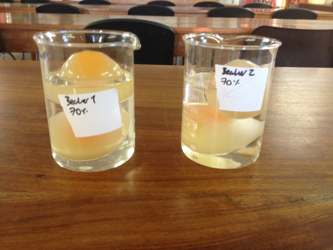
Ans: Observations:
- In pure water, the egg swells because water passes into it by osmosis.
- In concentrated salt solution, the egg shrinks because water passes out of the egg into the salt solution.
(a) Egg in pure water: The egg swells after 5 minutes. The egg’s contents (protected by the semi-permeable membrane) are hypertonic compared to pure water (hypotonic solution). Water moves into the egg by osmosis, causing it to swell.
(b) Egg in concentrated salt solution: The egg shrinks after 5 minutes. The concentrated salt solution is hypertonic compared to the egg’s contents. Water moves out of the egg into the salt solution by osmosis, causing the egg to shrink.
Explanation: This activity demonstrates osmosis, the movement of water across a selectively permeable membrane from a region of higher water concentration (lower solute concentration) to a region of lower water concentration (higher solute concentration). The egg’s membrane acts as a selectively permeable barrier, allowing water to pass but restricting larger solute molecules. In pure water (hypotonic), water enters the egg, increasing its volume. In a concentrated salt solution (hypertonic), water exits the egg, reducing its volume. This illustrates how osmosis regulates water movement in cells, critical for maintaining cell turgidity and function.
Activity 5.4: Osmosis in Plant Cells Using Raisins or Apricots
- Put dried raisins or apricots in plain water and leave them for some time. Then place them into a concentrated solution of sugar or salt. You will observe the following:
(a) Each gains water and swells when placed in water.
(b) However, when placed in the concentrated solution it loses water, and consequently shrinks.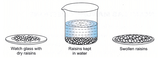
Ans: Observations:
(a) In plain water: The dried raisins or apricots swell after some time. Plain water is hypotonic compared to the concentrated contents of the raisins/apricots. Water enters the cells of the raisins/apricots by osmosis through their semi-permeable cell membranes, causing them to plump up.
(b) In concentrated sugar or salt solution: The raisins or apricots shrink. The concentrated solution is hypertonic compared to the cells’ contents. Water moves out of the cells into the surrounding solution by osmosis, causing the raisins/apricots to shrivel.
Explanation: Similar to Activity 5.3, this activity demonstrates osmosis using plant cells (in raisins or apricots). The semi-permeable cell membranes allow water to move based on the concentration gradient. In hypotonic plain water, water enters the cells, restoring turgidity. In hypertonic sugar/salt solution, water exits, causing plasmolysis (shrinkage). This activity reinforces the role of osmosis in water movement and its importance in plant cells, such as in root water absorption or maintaining turgor pressure.
Activity 5.5: Exploration of Electron Microscopes and Their Applications
- Find out about electron microscopes from resources in the school library or through the internet.
- Discuss it with your teacher.
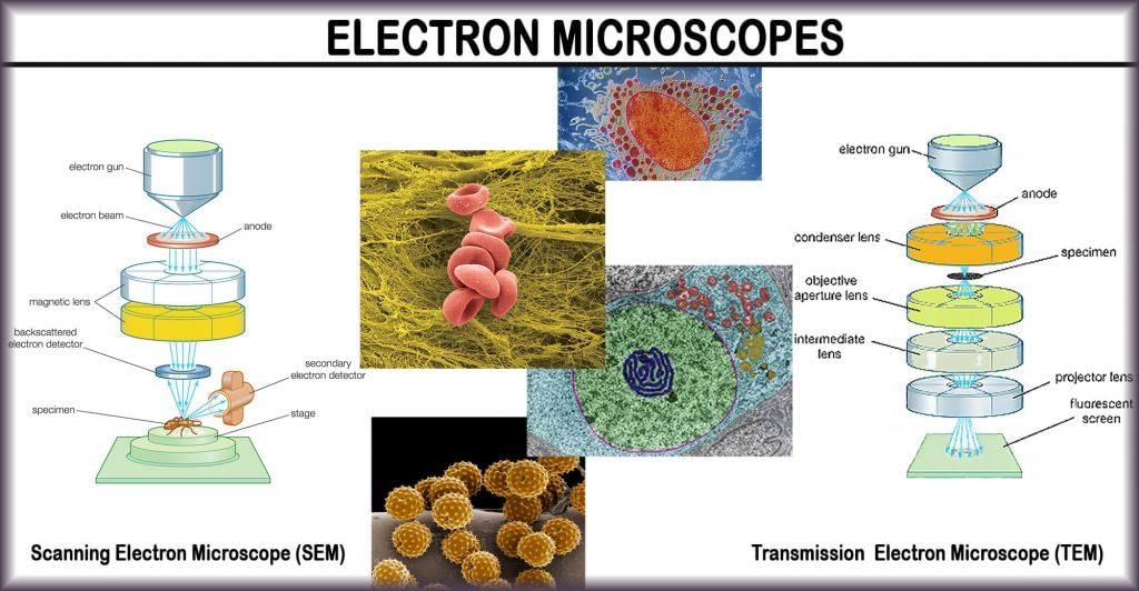
Ans: Observations/Expected Findings:
An electron microscope uses a beam of electrons instead of light to magnify specimens, achieving much higher resolution (up to 2 million times magnification) compared to light microscopes (up to 2000 times).
There are two main types:
- Transmission Electron Microscope (TEM): Electrons pass through a thin specimen, producing a 2D image of internal structures (e.g., organelles like mitochondria or ER).
- Scanning Electron Microscope (SEM): Electrons scan the surface of a specimen, producing a 3D image of surface details. Electron microscopes require specimens to be prepared in a vacuum, often dehydrated and coated with a conductive material (e.g., gold), and they can only view dead specimens due to the preparation process. They are critical for studying cell ultrastructure, such as the detailed organization of organelles (e.g., mitochondrial cristae, Golgi cisternae) and membrane systems, which are not visible under light microscopes.
Discussion Points with Teacher:
- Importance in cell biology: Electron microscopes revealed the complex internal structure of cells, confirming the presence of organelles like the endoplasmic reticulum, Golgi apparatus, and lysosomes.
- Comparison with light microscopes: Light microscopes are simpler, cheaper, and can view living cells, but they have lower resolution.
- Applications: Used in medical research, microbiology, and material science.
- Limitations: Expensive, requires specialized training, and cannot observe live cells.
Explanation: This activity encourages research and critical thinking about microscopy advancements. The electron microscope’s ability to reveal cell organelles’ fine details supports the understanding of eukaryotic cell complexity, as described in the chapter. It connects to the historical context of cell discovery, where the electron microscope (developed in 1940) expanded the cell theory by revealing intricate cellular structures.
Activity 5.6: Plasmolysis in Rhoeo Leaf Cells
- Mount the peel of a Rhoeo leaf in water on a slide and examine cells under the high power of a microscope. Note the small green granules, called chloroplasts. They contain a green substance called chlorophyll.
- Put a strong solution of sugar or salt on the mounted leaf on the slide. Wait for a minute and observe under a microscope.
Ans: Observations:
- Rhoeo leaf in water: Under the microscope, the cells appear turgid with distinct cell walls, cytoplasm, and a nucleus. Small green granules (chloroplasts) are visible, scattered in the cytoplasm, containing chlorophyll.
- After adding sugar/salt solution (living cells): After a minute, the cells show plasmolysis. The cytoplasm and plasma membrane shrink away from the cell wall due to water loss by osmosis into the hypertonic sugar/salt solution. The chloroplasts may appear clustered closer to the shrunken cytoplasm.
- Boiled Rhoeo leaf in water: The cells appear disorganized, with no turgidity. The cell membrane and organelles are damaged due to heat, and chloroplasts may appear clumped or faded. The cells are dead.
- After adding sugar/salt solution (dead cells): No plasmolysis occurs. The dead cells do not respond to the hypertonic solution because the plasma membrane is no longer functional, and osmosis requires a living, selectively permeable membrane.
Q: What do we see?
Ans: In living Rhoeo leaf cells, turgid cells with chloroplasts are visible initially, followed by plasmolysis (cytoplasm shrinking from the cell wall) after adding sugar/salt solution. In boiled (dead) cells, the structure is disrupted, and no plasmolysis occurs after adding the solution.
Q: What do we find? Did plasmolysis occur now?
Ans: Plasmolysis occurs in living cells but not in dead cells. The boiled cells, being dead, lack a functional plasma membrane, so osmosis (and thus plasmolysis) does not occur.
Q: What do we infer from this activity?
Ans: Only living cells with intact, selectively permeable membranes can undergo osmosis and plasmolysis. The cell wall in plant cells prevents bursting in hypotonic conditions and allows plasmolysis in hypertonic conditions, demonstrating the role of the cell wall and plasma membrane in osmoregulation.
Explanation: This activity illustrates plasmolysis, a phenomenon where living plant cells lose water in a hypertonic solution, causing the cytoplasm to pull away from the cell wall. It highlights the importance of the living plasma membrane in osmosis and the structural role of the cell wall in maintaining cell integrity. The contrast between living and dead cells emphasizes that osmosis is a process exclusive to living cells, reinforcing the concept of the cell as a dynamic, functional unit.
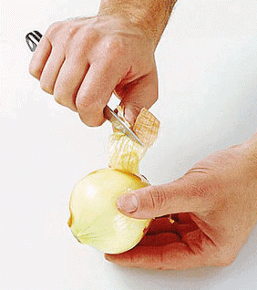
Activity 5.7: Observation of Human Cheek Cells under a Microscope
- Place a drop of water on a glass slide. Gently scrape the inside surface of the cheek using an ice-cream spoon. Check if any material gets stuck on the spoon.
- Transfer the material with a needle and spread it evenly on the glass slide.
- Add a drop of methylene blue solution to stain the material, then place a cover slip on it.
- Observe under a microscope.

Q: What do we observe?
Ans: Under the microscope, human cheek cells appear as flat, irregularly shaped (often polygonal or rounded) cells with a thin cell membrane. A darkly stained, spherical or oval nucleus is visible near the center of each cell due to the methylene blue stain. The cytoplasm is lightly stained, and no cell wall is present (unlike plant cells).
Q: What is the shape of the cells we see?
Ans: The cheek cells are irregular, often polygonal or rounded, with a flattened appearance, as they are epithelial cells from the inner lining of the mouth.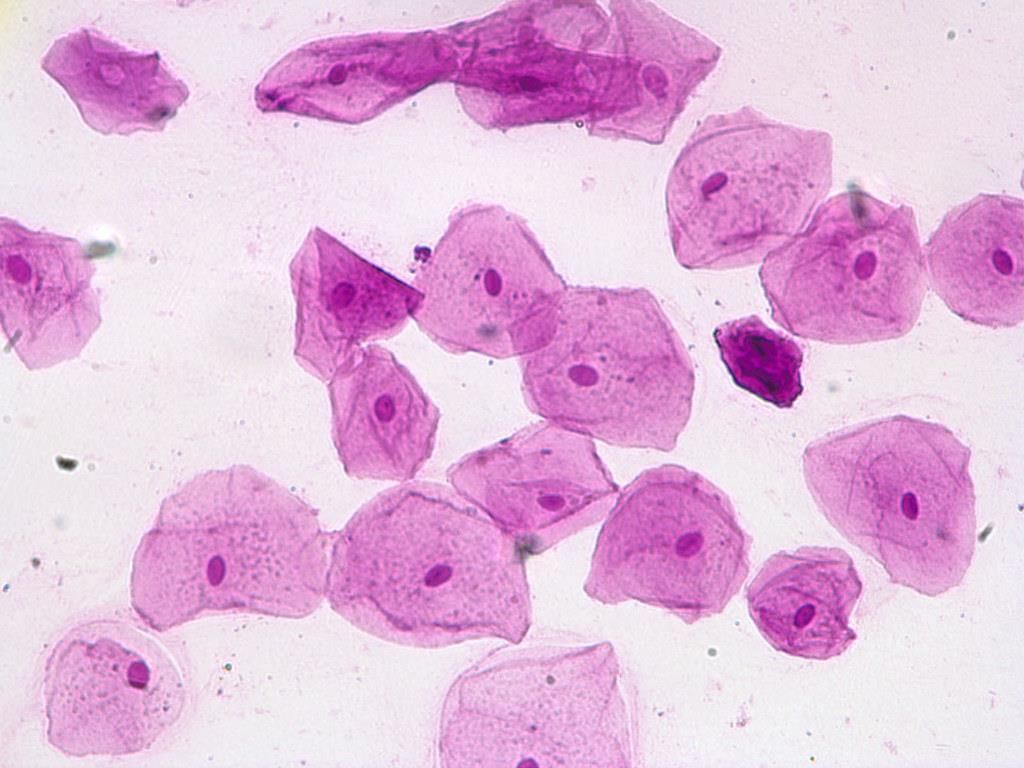
Q: Draw it on the observation sheet:
Ans: The drawing should depict flat, irregularly shaped cells with a thin cell membrane, a darkly stained nucleus (spherical or oval) near the center, and lightly stained cytoplasm. The cells lack a cell wall and chloroplasts, unlike onion cells.
Q: Was there a darkly coloured, spherical or oval, dot-like structure near the centre of each cell?
Ans: Yes, a darkly coloured, spherical or oval nucleus is visible in each cheek cell, stained by methylene blue.
Q: Were there similar structures in onion peel cells?
Ans: Yes, onion peel cells also have a nucleus, visible as a darkly stained, spherical or oval structure when stained with safranin (as in Activity 5.1). However, onion cells have a rigid cell wall and rectangular shape, unlike the flexible, irregular cheek cells.
Explanation: This activity compares animal (human cheek) and plant (onion peel) cells, highlighting similarities (presence of a nucleus, cytoplasm, and plasma membrane) and differences (animal cells lack a cell wall and chloroplasts, have irregular shapes). The nucleus, stained by methylene blue, is a key organelle in both cell types, controlling cellular activities and containing genetic material. The activity reinforces the cell theory by showing that both plant and animal organisms are composed of cells, with the nucleus as a universal feature in eukaryotic cells.
|
84 videos|478 docs|60 tests
|
FAQs on NCERT Based Activity: The Fundamental Unit of Life - Science Class 9
| 1. What is the fundamental unit of life? |  |
| 2. What are the main differences between prokaryotic and eukaryotic cells? | |
| 3. What is the structure and function of the cell membrane? | |
| 4. How do plant cells differ from animal cells? | |
| 5. Why are cells considered the basic unit of life? | |


practice quizzes
,NCERT Based Activity: The Fundamental Unit of Life | Science Class 9
,Important questions
,Exam
,Viva Questions
,shortcuts and tricks
,MCQs
,Extra Questions
,Free
,video lectures
,study material
,ppt
,Semester Notes
,past year papers
,Previous Year Questions with Solutions
,NCERT Based Activity: The Fundamental Unit of Life | Science Class 9
,Summary
,mock tests for examination
,Objective type Questions
,Sample Paper
,NCERT Based Activity: The Fundamental Unit of Life | Science Class 9
;


NCERT Based Activity: The Fundamental Unit of Life Free PDF Download
Importance of NCERT Based Activity: The Fundamental Unit of Life
NCERT Based Activity: The Fundamental Unit of Life Notes
NCERT Based Activity: The Fundamental Unit of Life Class 9 Questions
Study NCERT Based Activity: The Fundamental Unit of Life on the App
|
© EduRev
|
Education Revolution
|


|


